
What is keratin in skin
Keratin is a fibrous structural protein that forms the main building material of the outer skin layer (epidermis), as well as hair and nails. It is produced by keratinocytes, which make up about 90% of epidermal cells. As these cells mature and migrate toward the skin surface, they fill with keratin and eventually die, forming the tough, waterproof outer layer (stratum corneum) that protects the body.
Searching sources...
Reading File
Finding Sources
Searching the Web
keratin protein skin structure function
Finding Sources
Finding Sources
Reading File
Reading File
Reading File
Now I have enough rich textbook content to give a thorough answer. Let me also fetch the keratinocyte diagram from the histology textbook.
Loading Image
Here is a thorough explanation of keratin in skin, drawn from dermatology and histology textbooks.
Keratin in Skin
What Is Keratin?
Keratin is a family of fibrous structural proteins that form intermediate filaments (IFs) - one of the three main components of the cytoskeleton (alongside microtubules and actin filaments). In skin, keratin is the principal protein produced by keratinocytes, which make up roughly 90% of all epidermal cells. It gives the epidermis its mechanical toughness, chemical resistance, and waterproofing ability.
Types of Keratin
There are over 50 distinct keratin proteins encoded in the human genome, divided into two major classes:
| Type | Examples | Location |
|---|---|---|
| Type I (acidic) | KRT9, KRT10, KRT14, KRT15, KRT16, KRT17 | Skin, hair cortex |
| Type II (basic/neutral) | KRT1, KRT2, KRT5, KRT6a/b/c | Skin, hair cortex |
Keratins always work as obligate heterodimers - one type I and one type II protein pair together. In skin:
- Basal layer keratinocytes express the pair KRT5 + KRT14 (with KRT15 as a minor partner). This pair provides mechanical stability to the actively dividing cells anchored to the basement membrane.
- As cells differentiate and move suprabasally, they switch to KRT1 + KRT10, which are the keratins associated with the cornification process.
- KRT9 is uniquely expressed in the suprabasal layers of palmar and plantar skin, giving these areas their extra durability.
How Keratin Filaments Are Built
Assembly is a stepwise process (from Dermatology 2-Volume Set 5e):
- One type I + one type II keratin protein wind around each other in a coiled-coil to form a heterodimer.
- Two heterodimers associate antiparallelly to form a tetramer.
- Tetramers stack laterally into protofilaments.
- Protofilaments bundle into the mature 10-nm intermediate filament.
In light microscopy, these bundled filaments appear as tonofibrils, and they are anchored to desmosomes (cell-cell junctions) and hemidesmosomes (cell-basement membrane junctions), forming a mechanical network throughout the epidermis.
The Journey of Keratinocytes: Keratinization
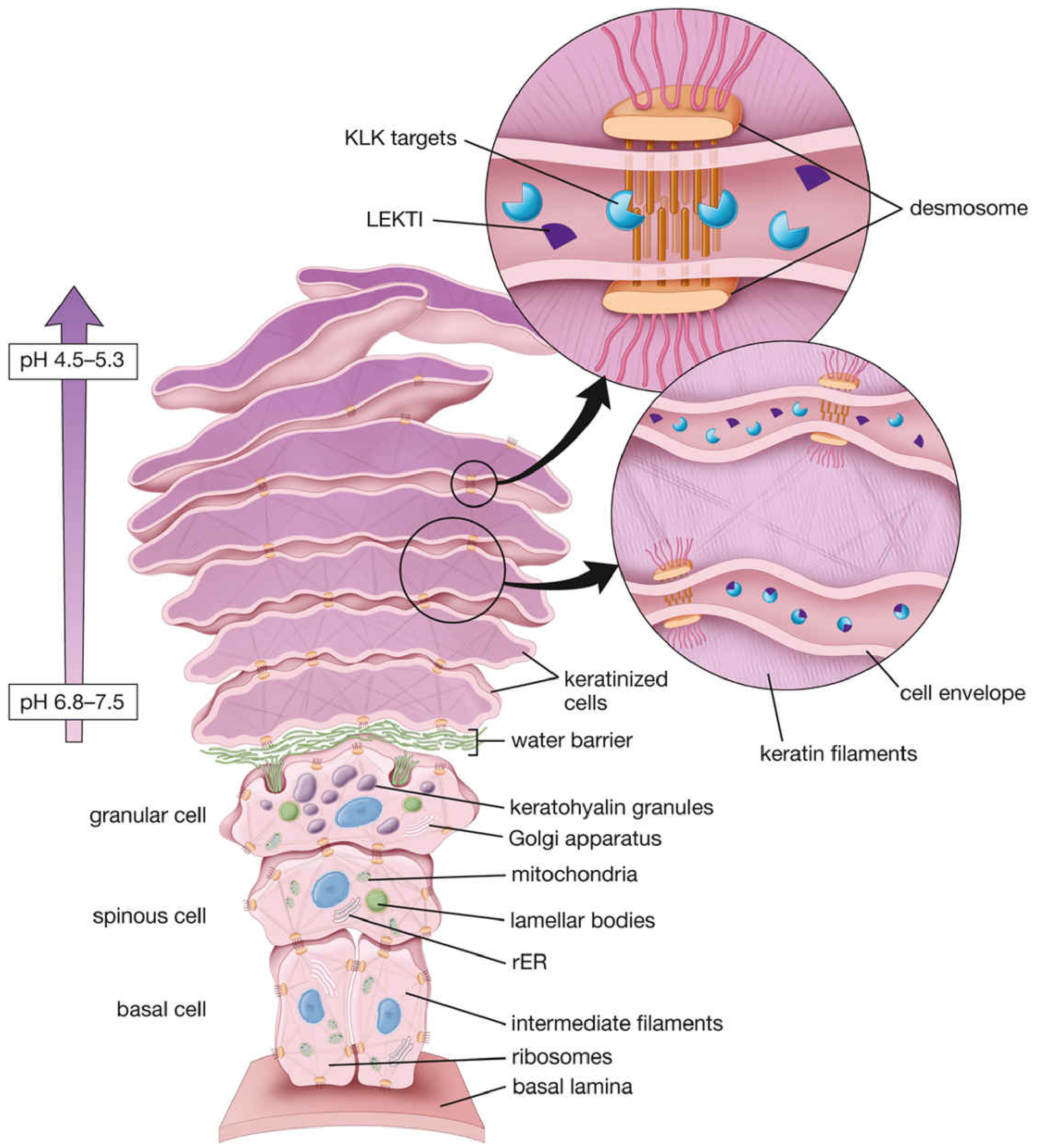
The diagram below illustrates the full life cycle from basal cell to desquamation:
| Layer | What happens to keratin |
|---|---|
| Stratum basale | KRT5/KRT14 filaments begin forming; cells are mitotically active |
| Stratum spinosum | Filament synthesis intensifies; KRT1/KRT10 replaces the basal pair; tonofibrils become prominent |
| Stratum granulosum | Keratohyalin granules appear - these contain filaggrin and trichohyalin, which aggregate keratin filaments into dense tonofibrils; lamellar bodies discharge lipids to form the water barrier |
| Stratum corneum | Full keratinization complete - cells (now called corneocytes) are dead, filled with keratin, surrounded by a cornified envelope; nucleus and organelles are gone |
This entire process from basal to cornified cell takes 2-6 hours for the granular-to-corneal transition, but weeks for the full journey from basal layer to surface.
"Soft" vs "Hard" Keratin
- Soft keratin - formed in skin epidermis; cells are loosely packed and eventually desquamate.
- Hard keratin - found in hair and nails; cells are tightly packed with high sulfur content (disulfide bonds via cysteine residues), making the structure rigid and permanent. Hair-specific keratins include KRT31-KRT40 (type I) and KRT81-KRT86 (type II).
Key Functions of Keratin in Skin
- Mechanical protection - resists shearing, compression, and abrasion.
- Water barrier - the cornified keratin layer prevents transepidermal water loss and blocks entry of external water/chemicals.
- Chemical resistance - insoluble in most solvents and resistant to proteases.
- Scaffold for cell architecture - anchors desmosomes and hemidesmosomes, distributing mechanical stress across the epithelium.
- Stress response - certain keratins (KRT6, KRT16, KRT17) are upregulated in wound healing and inflammation, helping coordinate the repair response.
Desquamation: Shedding the Old Keratin
Surface keratin does not accumulate indefinitely. The process of shedding (desquamation) is regulated by a pH-dependent protease system:
- At deeper layers, pH is near-neutral (~6.8-7.5), which allows the inhibitor LEKTI to suppress kallikrein-related serine peptidases (KLKs).
- As cells move to the surface, pH drops to 4.5-5.3, causing LEKTI to dissociate, freeing KLK5, KLK7, and KLK14 to digest desmosomal proteins and release the outermost corneocytes.
When Keratin Goes Wrong: Keratin Disorders
Mutations in keratin genes cause a range of genetic skin diseases, usually in autosomal dominant fashion because mutant keratin disrupts the normal filament assembly in a dominant-negative way. Examples:
| Disease | Mutated Keratin | Effect |
|---|---|---|
| Epidermolysis Bullosa Simplex (EBS) | KRT5 or KRT14 | Basal cell fragility, blistering |
| Epidermolytic Ichthyosis | KRT1 or KRT10 | Suprabasal cytolysis, scaling |
| Palmoplantar Keratoderma (PPK) | KRT9 (palmoplantar) | Thickened palms/soles |
| Pachyonychia Congenita | KRT6a, KRT16, KRT17 | Nail dystrophy, focal PPK |
Over 90% of pathogenic keratin mutations are missense mutations, with "hotspot" arginine residues in the helix initiation and termination motifs being especially common targets.
Sources:
- Dermatology 2-Volume Set, 5th edition - Biology of Keratinocytes, Keratin Disorders
- Histology: A Text and Atlas with Correlated Cell and Molecular Biology - Keratinocytes
- Junqueira's Basic Histology - Text and Atlas, 17th edition - Covering Epithelia
This is a shared conversation. Sign in to Orris to start your own chat.