
COPD
I'll start by reading the medical library skill, then research COPD comprehensively.
Searching sources...
Reading File
Now I'll search the library database and PubMed in parallel for comprehensive COPD coverage.
Finding Sources
Searching PubMed
"COPD" AND "management"
Searching Images
COPD emphysema lung histology pathology
Now let me read the key COPD sections from Murray & Nadel's textbook in parallel.
Reading File
Reading File
Reading File
Finding Sources
Excellent. Now I'll read the most comprehensive COPD sections - the main Murray & Nadel block27 section (longest, 9 paragraphs) and the Robbins pathology section in parallel.
Reading File
Reading File
Reading File
Now let me get the main Murray & Nadel dedicated COPD section and Robbins continued pathology, plus Goldman-Cecil management section.
Reading File
Reading File
Reading File
I now have rich content from multiple authoritative textbooks. Let me also fetch some key images and get management details from Goldman-Cecil.
Reading File
Loading Image
Loading Image
Excellent - I now have rich, comprehensive information from multiple authoritative sources. Let me also fetch the GOLD classification figure and get the detailed management content.
Loading Image
Reading File
I now have comprehensive content covering definition, epidemiology, pathology, pathogenesis, classification, and management. I have enough material to deliver a thorough, well-structured answer.
Chronic Obstructive Pulmonary Disease (COPD)
Definition
COPD is an umbrella term for conditions resulting in fixed (incompletely reversible) airway obstruction and dyspnea on exertion. The WHO defines it as "a common, preventable and treatable disease characterized by persistent respiratory symptoms and airflow limitation due to airway and/or alveolar abnormalities caused by exposure to noxious particles or gases."
Airflow limitation is confirmed spirometrically by a post-bronchodilator FEV1/FVC ratio < 0.7. Nearly all patients have overlapping components of emphysema (parenchymal destruction) and chronic bronchitis (airway inflammation/fibrosis), though the proportions vary.
- Goldman-Cecil Medicine, p. 892
- Robbins & Kumar Basic Pathology, p. 447
Epidemiology
| Statistic | Figure |
|---|---|
| Global prevalence | ~175 million |
| Annual deaths worldwide | ~3.2 million (3rd leading cause of death) |
| U.S. adult population affected (>40 yrs) | >10% |
| Attributable to smoking | ~80% |
| Annual U.S. cost | ~$50 billion |
Heavy smokers (35-50%) develop COPD; women appear more susceptible than men. Other risk factors include biomass fuel combustion, occupational dust (mines, grain handling, cotton mills), impaired lung development in childhood, airway hyperresponsiveness, and genetic polymorphisms (notably alpha-1 antitrypsin deficiency).
- Goldman-Cecil Medicine, p. 892
The Spectrum of Obstructive Lung Disease
The three major obstructive diseases overlap considerably:
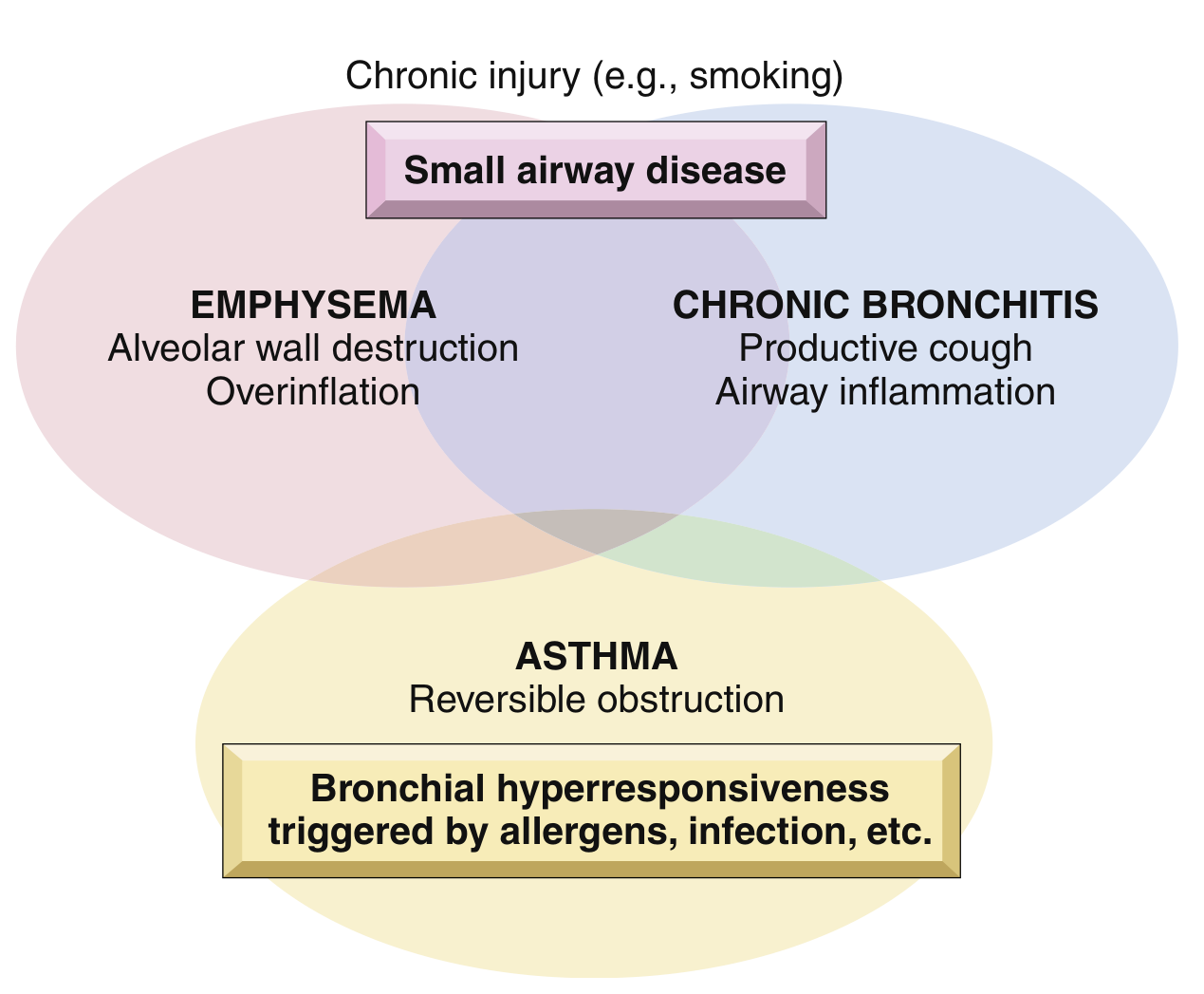
FIG. 11.4 - Schematic representation of overlap between chronic obstructive lung diseases. (Robbins & Kumar Basic Pathology)
Pathology
Emphysema
Emphysema is defined as permanent enlargement of air spaces distal to the terminal bronchioles with destruction of alveolar walls, without significant fibrosis. There are four anatomic patterns:
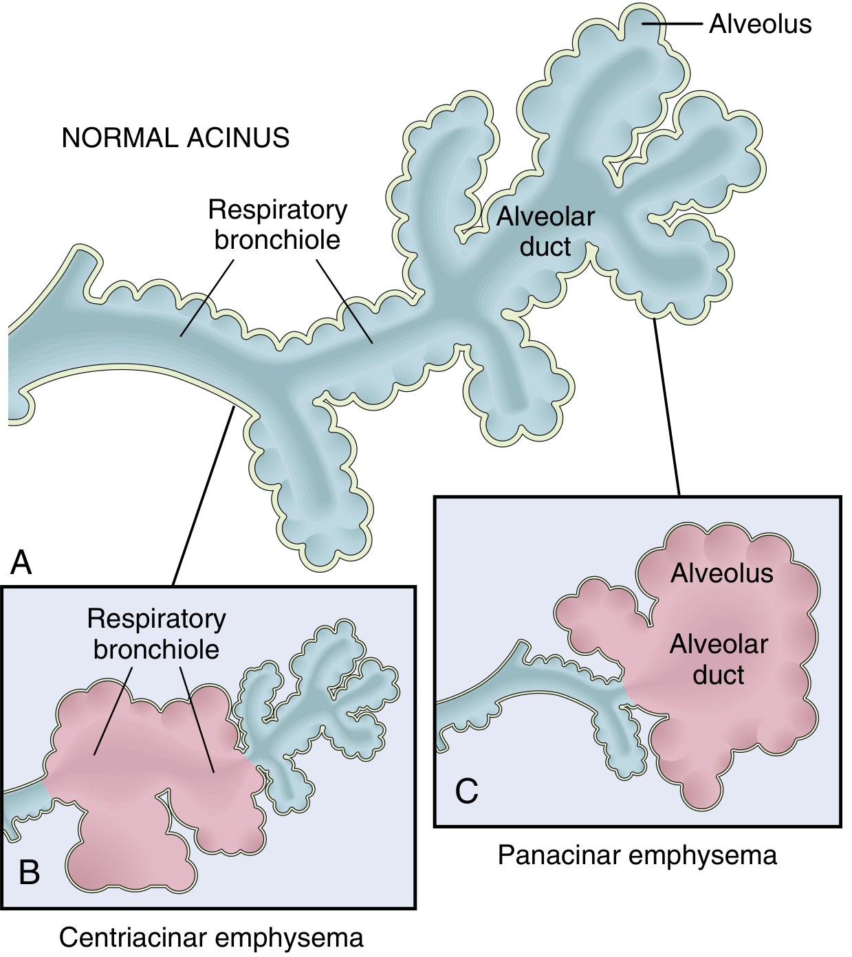
FIG. 11.5 - Major patterns of emphysema. (Robbins & Kumar Basic Pathology)
| Type | Location of Destruction | Association |
|---|---|---|
| Centriacinar (centrilobular) | Central/proximal acinus (respiratory bronchioles), sparing distal alveoli | Smoking; upper lobes; 20x more common than panacinar |
| Panacinar (panlobular) | Entire acinus uniformly affected | Alpha-1 antitrypsin deficiency; lower lobes |
| Distal acinar (paraseptal) | Distal alveolar ducts and sacs; subpleural | Spontaneous pneumothorax in young adults |
| Irregular | Irregular, periscarring distribution | Scar tissue; no functional significance |
Chronic Bronchitis
Defined clinically as productive cough for at least 3 months in each of 2 consecutive years (in the absence of another explanation).
Pathologically:
-
Hypertrophy and hyperplasia of bronchial submucosal mucous glands
-
Reid index (ratio of gland layer thickness to bronchial wall thickness) increased to >0.4
-
Goblet cell metaplasia, mucus plugging, luminal inflammatory infiltrate (lymphocytes, macrophages, neutrophils)
-
Small airway fibrosis (bronchiolitis obliterans) in severe cases
-
Robbins & Kumar Basic Pathology, p. 447-448
Pathogenesis
Protease-Antiprotease Imbalance (Primary Mechanism)
Cigarette smoke triggers an influx of neutrophils and macrophages into the lung. These cells release elastolytic proteases (neutrophil elastase, macrophage metalloproteinases) that degrade elastin, producing emphysema when they exceed antiprotease defenses. Alpha-1 antitrypsin (alpha-1 antiprotease) is the major inhibitor of neutrophil elastase; its deficiency (especially PiZZ phenotype) dramatically increases COPD risk.
Inflammation
- CD8+ lymphocytes and neutrophils are key effectors
- Unlike asthma, eosinophils are a minor component in most COPD (though a subgroup shows sputum/blood eosinophilia)
- Inflammation persists after smoking cessation due to microbiome changes and autoimmunity - explaining continued disease progression despite quitting
- Oxidative stress and reactive oxygen species amplify the inflammatory cascade
Vascular and Apoptotic Mechanisms
Cigarette smoke induces intimal thickening, smooth muscle proliferation, reduced VEGF expression, and endothelial cell apoptosis in septal vessels. Chronic hypoxemia causes pulmonary vasoconstriction, leading to pulmonary hypertension and eventually cor pulmonale in 20-30% of patients.
Small Airway Disease
IL-13 from T cells, elevated mucin production, and neutrophil elastase collectively damage the bronchial epithelium. Microbial colonization maintains inflammation and triggers exacerbations.
- Goldman-Cecil Medicine, p. 893-894
GOLD Classification
The GOLD (Global Initiative for Chronic Obstructive Lung Disease) classification uses two dimensions:
Spirometric severity (FEV1 % predicted, post-bronchodilator FEV1/FVC < 0.7):
| GOLD Grade | FEV1 % Predicted |
|---|---|
| GOLD 1 (Mild) | ≥80% |
| GOLD 2 (Moderate) | 50-79% |
| GOLD 3 (Severe) | 30-49% |
| GOLD 4 (Very Severe) | <30% |
Combined ABCD assessment adds symptom burden and exacerbation history:
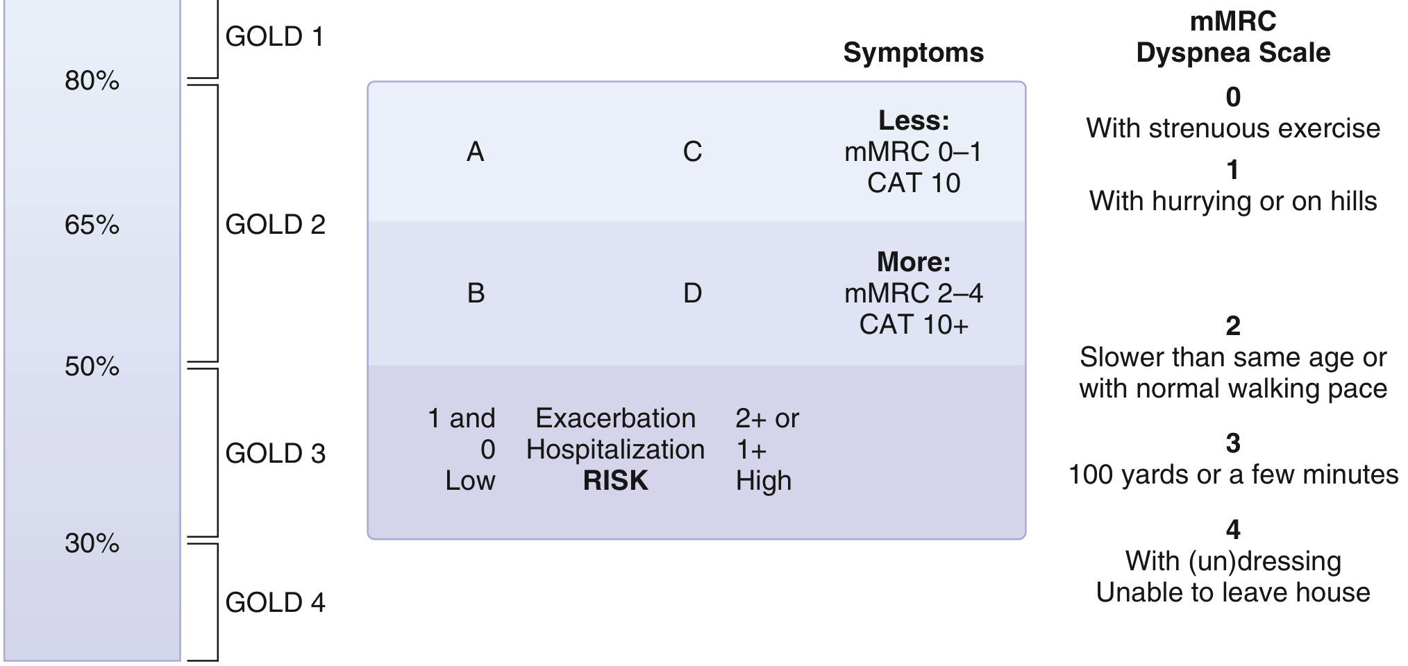
GOLD Classification: Groups A-D are determined by symptom burden (mMRC ≥2 or CAT ≥10 = more symptoms) and exacerbation risk (≥2 exacerbations or ≥1 hospitalization = high risk). (Goldman-Cecil Medicine)
Clinical Features
Symptoms
- Dyspnea - usually the first symptom; begins insidiously, progressively worsens
- Chronic productive cough with mucoid or mucopurulent sputum
- Wheezing, chest tightness
- Weight loss (can be marked, suggesting occult malignancy in the differential)
Classic Phenotypes
| Feature | "Pink Puffer" (Pure Emphysema) | "Blue Bloater" (Chronic Bronchitis Predominant) |
|---|---|---|
| Build | Thin, cachectic | Overweight |
| Breathing | Barrel-chested, pursed-lip breathing, prolonged expiration, hunched posture | Less prominent dyspnea |
| Gas exchange | Preserved until late; normocapnic | CO2 retention, hypoxia, cyanosis |
| Pulmonary HTN | Later onset | Earlier, more severe |
| OSA risk | Lower (lower BMI, hyperinflation protects upper airway) | Higher (higher BMI, lower respiratory drive) |
Pulmonary Function Tests
- Reduced FEV1 with normal or near-normal FVC → reduced FEV1/FVC ratio
- Reduced DLCO (reflects loss of alveolar-capillary surface)
- Increased TLC and RV (air trapping)
Complications
Acute Exacerbations of COPD (AECOPD)
Defined by increased dyspnea, increased cough, and increased/changed sputum - often with fever. The pathophysiology involves inability to maintain adequate alveolar ventilation against:
- High intrinsic PEEP (auto-PEEP)
- High airway resistance
- Flattened, mechanically disadvantaged diaphragm
- Respiratory muscle fatigue
This drives rising PaCO2 (hypercapnic respiratory failure) and hypoxemia.
Management of AECOPD:
- Short-acting bronchodilators (SABA ± SAMA)
- Systemic corticosteroids (5-day course)
- Antibiotics (if purulent sputum, increased CRP, or hospitalized)
- Controlled oxygen (target SpO2 88-92% to avoid CO2 retention)
- Non-invasive ventilation (NIV/BPAP) - strong recommendation when hypercapnic respiratory failure is present; reduces need for intubation and improves survival
- Invasive mechanical ventilation for NIV failure or contraindications
Pulmonary Hypertension (COPD-PH)
- COPD accounts for >80% of all chronic lung disease-related PH
- Mild PH (mPAP > 20-25 mmHg) is common; severe PH (mPAP ≥ 40 mmHg) occurs in only ~1-2.7%
- Driven by hypoxic vasoconstriction, vascular remodeling, and loss of capillary bed
OSA-COPD Overlap Syndrome
-
Up to 66% of COPD patients also have OSA
-
Higher risk of hospitalization and death than COPD alone
-
CPAP improves survival and reduces AECOPD hospitalizations
-
High-intensity BPAP is preferred when persistent hypercapnia is present
-
The "blue bloater" phenotype is at higher OSA risk; the "pink puffer" phenotype may be relatively protected
-
Murray & Nadel's Textbook of Respiratory Medicine, p. 2747, 2833
Management
Non-Pharmacologic (Foundation of Care)
- Smoking cessation - single most effective intervention; slows FEV1 decline
- Pulmonary rehabilitation - improves exercise tolerance and quality of life
- Long-term oxygen therapy (LTOT) - indicated when PaO2 ≤55 mmHg (or ≤60 with cor pulmonale/polycythemia); improves survival
- Vaccinations - influenza annually; pneumococcal; COVID-19
Pharmacologic (Inhaled Therapies - Primary Route)
| Class | Drugs | Role |
|---|---|---|
| Short-acting beta-2 agonists (SABA) | Salbutamol, terbutaline | Rescue (PRN) |
| Short-acting muscarinic antagonists (SAMA) | Ipratropium | Rescue or add-on |
| Long-acting beta-2 agonists (LABA) | Salmeterol, formoterol, indacaterol | Maintenance |
| Long-acting muscarinic antagonists (LAMA) | Tiotropium, umeclidinium, glycopyrronium | Maintenance (preferred first-line) |
| LABA + LAMA | Dual bronchodilation | More effective than either alone |
| Inhaled corticosteroids (ICS) | Budesonide, fluticasone | Add to LABA/LAMA when eosinophils ≥300 or recurrent exacerbations |
| ICS + LABA | Fixed combinations | For Group C/D with eosinophilic phenotype |
| Triple therapy (LAMA + LABA + ICS) | Trelegy, Trimbow | Severe disease (Group D) |
Note: ICS alone are not recommended as monotherapy. Their primary benefit is reducing exacerbation frequency, not improving FEV1. Eosinophil count guides ICS use.
Other agents:
- Roflumilast (PDE4 inhibitor) - for severe COPD with chronic bronchitis phenotype and frequent exacerbations
- Azithromycin (long-term, low-dose) - reduces exacerbation frequency in selected patients
- Alpha-1 antitrypsin augmentation - for confirmed severe deficiency
Interventional/Surgical
-
Lung volume reduction surgery (LVRS) - for upper-lobe predominant emphysema with low exercise capacity
-
Bronchoscopic lung volume reduction (endobronchial valves, coils) - less invasive alternatives
-
Lung transplantation - end-stage disease; improves quality of life; survival benefit less certain
-
Bullectomy - for giant bullae causing compression
-
Goldman-Cecil Medicine, p. 892-895
Key Points Summary
- COPD = fixed airflow obstruction (FEV1/FVC < 0.7 post-bronchodilator), caused by emphysema + small airway disease
- Major risk factor: cigarette smoking (80% of cases); also biomass fuels, occupational dust, alpha-1 antitrypsin deficiency
- Pathogenesis: protease-antiprotease imbalance + oxidative stress + persistent inflammation
- Diagnosis requires spirometry; supplemented by GOLD classification (grades 1-4 + groups A-D)
- Management is stepwise: smoking cessation → LAMA → LABA + LAMA → add ICS based on symptoms and exacerbation risk
- Exacerbations drive morbidity/mortality; NIV is strongly recommended for hypercapnic AECOPD
- Comorbidities are the rule: pulmonary hypertension, OSA, cardiovascular disease, anxiety/depression, lung cancer
Recent evidence note: A 2026 systematic review (PMID 41553852) confirms that integrated care models targeting COPD with comorbidities improve hospitalization outcomes and quality of life, supporting the current move toward multidisciplinary team-based management.
This is a shared conversation. Sign in to Orris to start your own chat.