


+16 more
So I'm , a second year BSc Cardiac Technology student I want a notes for my university as well as for my higher studies , so give me in such a way it should start from scratch and with diagram. flowcharts and table
I can see your study plan images clearly. Before I dive into building your notes, let me quickly clarify one thing to make sure I give you exactly what you need.
Asking for Preferences
Which topic/week should I start with for your notes? · What format do you want the notes in?
Starting with Week 1 - Heart Structure & Vessels. Let me pull from the medical library and build your full notes right now.
Searching sources...
Reading File
Finding Sources
Finding Sources
Finding Sources
Finding Sources
Finding Sources
Finding Sources
Finding Sources
Finding Sources
Reading File
Reading File
Reading File
Reading File
I now have excellent textbook content. Let me fetch the conduction system diagram and compile the full notes.
Loading Image
Finding Sources
Right now give day I notes - with detailed explanation with subtopics, diagram flow chart and table
Finding Sources
Finding Sources
Finding Sources
Reading File
Reading File
Finding Sources
Reading File
Reading File
Loading Image
Loading Image
I now have everything needed. Here are your complete Day 1 notes.
📚 BSc Cardiac Technology - WEEK 1, DAY 1
Gross Anatomy of the Heart: Chambers, Valves, Great Vessels & Pericardium
Study Plan Context: This is your foundation. Every disease topic - valve disease, heart failure, CAD, arrhythmias - builds on today's anatomy. Learn it structurally, not by rote.
1. OVERVIEW - THE HEART AT A GLANCE
The human adult heart weighs approximately 325 g (men) and 275 g (women). It is roughly the size of a clenched fist, located in the mediastinum (between the lungs), resting on the diaphragm, and enclosed within a protective sac called the pericardium.
📍 POSITION OF THE HEART
┌─────────────────────────────────────┐
│ THORACIC CAVITY │
│ ┌──────┐ ┌──────────┐ ┌──────┐ │
│ │Left │ │ HEART │ │Right │ │
│ │Lung │ │(mediastin│ │Lung │ │
│ │ │ │ -um) │ │ │ │
│ └──────┘ │ On diaphr│ └──────┘ │
│ │ -agm │ │
│ └──────────┘ │
└─────────────────────────────────────┘
Axis: base (top-right) → apex (bottom-left)
2. THE PERICARDIUM
2.1 What is it?
The pericardium is a fibroserous sac surrounding the heart and the roots of the great vessels. It has two main components.
2.2 Layers of the Pericardium
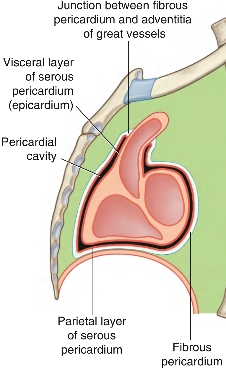
From Gray's Anatomy for Students - Sagittal Section of the Pericardium
PERICARDIUM - LAYER DIAGRAM
═══════════════════════════════════════════════
OUTERMOST
│
▼
┌─────────────────────────────────────────────┐
│ FIBROUS PERICARDIUM │
│ • Tough connective tissue outer layer │
│ • Cone-shaped; base on diaphragm │
│ • Attached to sternum (sternopericardial │
│ ligaments) → keeps heart in position │
│ • Limits cardiac over-distension │
│ • Innervated by phrenic nerve (C3-C5) │
└──────────────┬──────────────────────────────┘
│ Lines inner surface
▼
┌─────────────────────────────────────────────┐
│ SEROUS PERICARDIUM │
│ ┌─────────────────────────────────────┐ │
│ │ PARIETAL LAYER │ │
│ │ Lines inner surface of fibrous peri-│ │
│ │ cardium │ │
│ └────────────────┬────────────────────┘ │
│ │ Pericardial cavity │
│ │ (contains ~15-50 mL │
│ │ serous fluid) │
│ ┌────────────────▼────────────────────┐ │
│ │ VISCERAL LAYER = EPICARDIUM │ │
│ │ Adheres directly to heart surface │ │
│ └─────────────────────────────────────┘ │
└─────────────────────────────────────────────┘
INNERMOST (touching heart muscle)
═══════════════════════════════════════════════
| Layer | Location | Key Feature |
|---|---|---|
| Fibrous pericardium | Outermost | Tough, protective, non-distensible |
| Parietal serous pericardium | Inner face of fibrous layer | Smooth, secretes fluid |
| Pericardial cavity | Space between parietal & visceral | Contains 15-50 mL lubricating fluid |
| Visceral serous pericardium (epicardium) | On the heart surface | Outermost layer of heart wall |
⚠️ Clinical Link (Preview): When excess fluid fills the pericardial cavity (pericardial effusion), it can compress the heart - this is cardiac tamponade (covered in Week 5). Beck's triad = hypotension + muffled heart sounds + raised JVP.
3. THE THREE LAYERS OF THE CARDIAC WALL
CARDIAC WALL (inside → outside)
════════════════════════════════
BLOOD IN CHAMBER
│
▼
┌──────────────────────────────┐
│ ENDOCARDIUM │
│ • Innermost layer │
│ • Thin, smooth lining │
│ • Continuous with blood │
│ vessel endothelium │
│ • Most vulnerable to ischemia│
│ (supplied by smallest │
│ vessels) │
└──────────────┬───────────────┘
▼
┌──────────────────────────────┐
│ MYOCARDIUM │
│ • Middle, thickest layer │
│ • Striated cardiac muscle │
│ • Fibers 10-15 mm dia, │
│ 30-60 mm length │
│ • Contains: actin, myosin, │
│ troponins, CK, LDH, │
│ myoglobin │
└──────────────┬───────────────┘
▼
┌──────────────────────────────┐
│ EPICARDIUM │
│ • Outermost layer │
│ • = Visceral serous pericard │
│ • Coronary arteries run here │
└──────────────────────────────┘
│
▼
PERICARDIAL CAVITY
| Layer | Alternate Name | Function | Clinical Note |
|---|---|---|---|
| Endocardium | - | Smooth blood-contact surface | Site of infective endocarditis |
| Myocardium | - | Contracts, generates pressure | Damaged in MI; CK-MB, Troponin released |
| Epicardium | Visceral pericardium | Outer covering | Coronary arteries run here |
4. THE FOUR CHAMBERS
4.1 Overview Flowchart - Blood Flow Through the Heart
SYSTEMIC CIRCULATION → PULMONARY CIRCULATION FLOWCHART
═══════════════════════════════════════════════════════════
Deoxygenated blood Oxygenated blood
from body to body
│ ▲
▼ │
┌──────────────┐ ┌──────────────┐
│ RIGHT ATRIUM │ │ LEFT ATRIUM │
│ (thin wall) │ │ (thin wall) │
│ Receives: │ │ Receives: │
│ • SVC │ │ • Pulmonary │
│ • IVC │ │ veins (x4) │
│ • Coronary │ └──────┬───────┘
│ sinus │ │
└──────┬───────┘ MITRAL VALVE (bicuspid)
│ │
TRICUSPID VALVE ▼
│ ┌──────────────┐
▼ │ LEFT │
┌──────────────┐ │ VENTRICLE │
│ RIGHT │ │ (THICK wall │
│ VENTRICLE │ │ ~9-11 mm) │
│ (banana- │ │ Pumps to │
│ shaped; │ │ systemic │
│ thinner │ │ circulation │
│ wall ~3 mm) │ └──────┬───────┘
│ Pumps to │ │
│ pulmonary │ AORTIC VALVE
│ circulation │ │
└──────┬───────┘ ▼
│ AORTA → Body
PULMONARY VALVE
│
▼
PULMONARY ARTERY → Lungs
│
(Gas exchange)
│
PULMONARY VEINS → Left Atrium (above)
4.2 Right Side vs Left Side - Comparison Table
| Feature | Right Side | Left Side |
|---|---|---|
| Function | Receives deoxygenated blood; pumps to lungs | Receives oxygenated blood; pumps to body |
| Pressure | Low (~25/5 mmHg RV systolic/diastolic) | High (~120/80 mmHg LV) |
| Wall thickness - Atrium | Thin (~2 mm) | Thin (~3 mm) |
| Wall thickness - Ventricle | Thin (~3 mm); banana-shaped | Thick (~9-11 mm); conical |
| AV Valve | Tricuspid (3 cusps) | Mitral / Bicuspid (2 cusps) |
| Semilunar Valve | Pulmonary valve | Aortic valve |
| Inflow vessels | SVC, IVC, coronary sinus | 4 pulmonary veins |
| Outflow vessel | Pulmonary artery (pulmonary trunk) | Aorta |
💡 Why is the LV wall thicker? The left ventricle must generate enough pressure to push blood through the entire systemic circulation (aortic pressure ~80-120 mmHg). The right ventricle only needs to push against pulmonary arterial pressure (~10-25 mmHg). Pressure determines wall thickness - Laplace's Law.
5. THE FOUR HEART VALVES
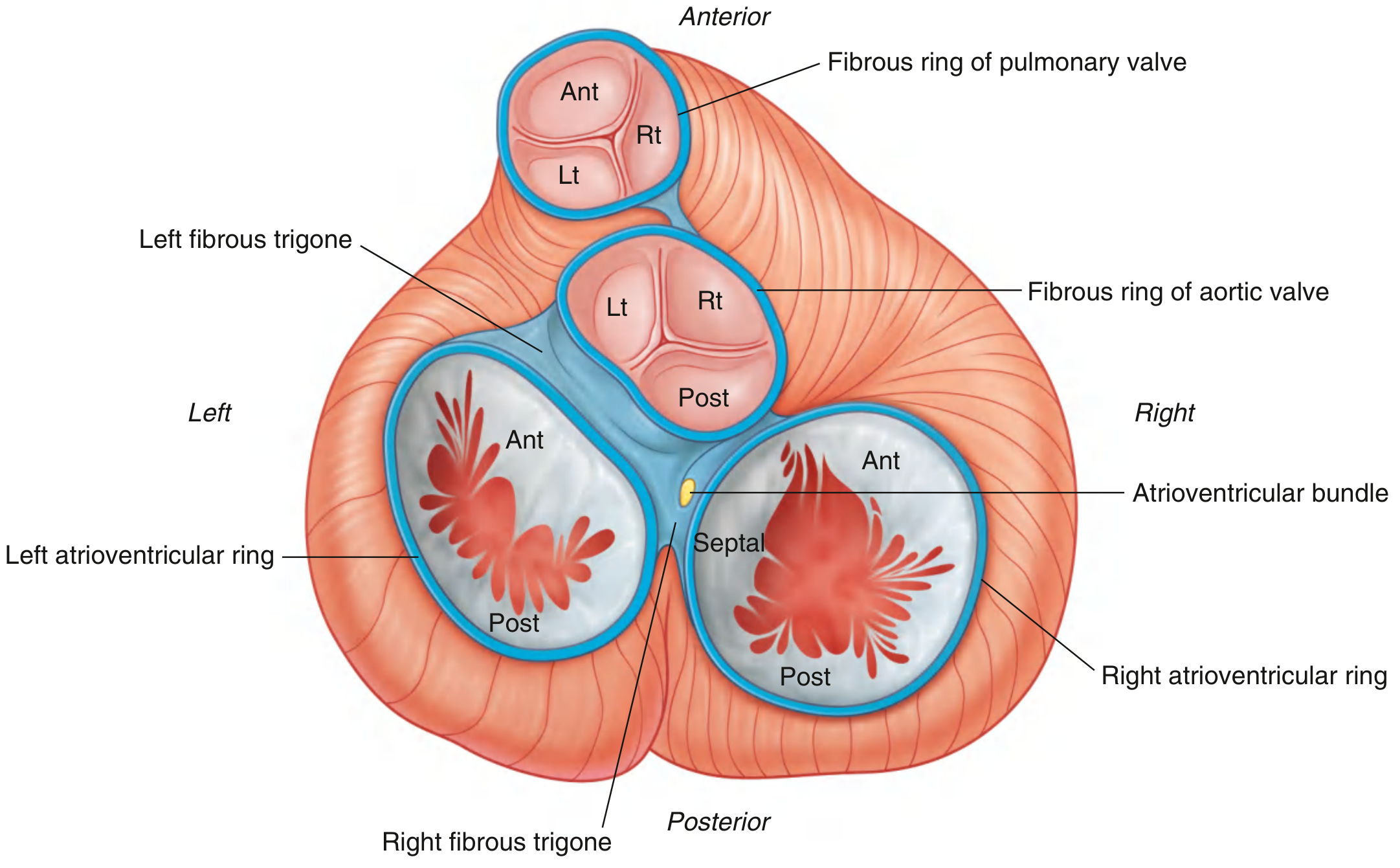
From Gray's Anatomy for Students - Cardiac Skeleton (Atria Removed). All four valves + their fibrous rings visible.
5.1 Classification
HEART VALVES
│
├── ATRIOVENTRICULAR (AV) VALVES
│ [between atria and ventricles]
│ │
│ ├── TRICUSPID VALVE (Right side)
│ │ • 3 cusps: anterior, posterior, septal
│ │ • Attached to papillary muscles via
│ │ chordae tendineae
│ │ • Prevents backflow RA → RV during systole
│ │
│ └── MITRAL VALVE (Left side)
│ • 2 cusps: anterior (large), posterior
│ • Also called bicuspid valve
│ • Also anchored by chordae tendineae
│ • Prevents backflow LA → LV during systole
│
└── SEMILUNAR VALVES
[between ventricles and great vessels]
│
├── PULMONARY VALVE (RV → Pulmonary artery)
│ • 3 cusps: anterior, right, left
│ • No chordae tendineae
│ • Opens during RV systole
│
└── AORTIC VALVE (LV → Aorta)
• 3 cusps: left, right, posterior
• Coronary arteries arise from left
& right aortic sinuses (cusps)
• No chordae tendineae
5.2 Valve Summary Table
| Valve | Type | Cusps | Location | Opens During | Closes During | Sound on Closure |
|---|---|---|---|---|---|---|
| Tricuspid | AV | 3 | Right AV junction | Diastole (filling) | Systole (RV contracts) | S1 (component) |
| Mitral (Bicuspid) | AV | 2 | Left AV junction | Diastole (filling) | Systole (LV contracts) | S1 (dominant) |
| Pulmonary | Semilunar | 3 | RV outflow | Systole (ejection) | Diastole | S2 (P2 component) |
| Aortic | Semilunar | 3 | LV outflow | Systole (ejection) | Diastole | S2 (A2 dominant) |
💡 Memory trick for valves opening/closing:
- AV valves are like inlet valves - they open to let blood IN to ventricles (diastole), close when ventricles squeeze (systole)
- Semilunar valves are outlet valves - they open when ventricles squeeze (systole), close when ventricles relax (diastole)
6. THE GREAT VESSELS
GREAT VESSELS ENTERING/LEAVING THE HEART
══════════════════════════════════════════════════════════
ENTERING THE HEART: LEAVING THE HEART:
┌───────────────────────┐ ┌────────────────────────┐
│ TO RIGHT ATRIUM: │ │ FROM RIGHT VENTRICLE: │
│ • Superior Vena Cava │ │ • Pulmonary Trunk │
│ (SVC) - upper body │ │ → splits into: │
│ • Inferior Vena Cava │ │ Right pulm. artery│
│ (IVC) - lower body │ │ Left pulm. artery │
│ • Coronary Sinus │ └────────────────────────┘
│ (heart's own venous│
│ drainage) │ ┌────────────────────────┐
└───────────────────────┘ │ FROM LEFT VENTRICLE: │
│ • AORTA │
┌───────────────────────┐ │ → Ascending aorta │
│ TO LEFT ATRIUM: │ │ → Aortic arch │
│ • 4 Pulmonary Veins │ │ → Descending aorta │
│ (carry oxygenated │ └────────────────────────┘
│ blood from lungs) │
└───────────────────────┘
| Vessel | Carries | Direction | Connects |
|---|---|---|---|
| Superior Vena Cava (SVC) | Deoxygenated | Into RA | Upper body → Heart |
| Inferior Vena Cava (IVC) | Deoxygenated | Into RA | Lower body → Heart |
| Coronary Sinus | Deoxygenated | Into RA | Heart muscle venous drainage |
| Pulmonary Trunk/Arteries | Deoxygenated | Out of RV | Heart → Lungs |
| Pulmonary Veins (x4) | Oxygenated | Into LA | Lungs → Heart |
| Aorta | Oxygenated | Out of LV | Heart → Entire body |
⚠️ Important exception to memorize: Pulmonary arteries carry deoxygenated blood. Pulmonary veins carry oxygenated blood. This is the REVERSE of the usual artery/vein rule and is a common exam question.
7. THE CORONARY ARTERIES (Introduction)
The coronary arteries supply blood to the heart muscle itself. They arise from the aortic sinuses at the base of the aorta.
CORONARY ARTERY ORIGIN & DISTRIBUTION
═══════════════════════════════════════════════
AORTA
│
┌──────┴──────┐
│ │
▼ ▼
RIGHT CORONARY LEFT CORONARY
ARTERY (RCA) ARTERY (LCA)
│ │
│ ┌────┴────┐
│ │ │
│ LEFT ANTERIOR CIRCUMFLEX
│ DESCENDING ARTERY (LCx)
│ (LAD) │
│ │ │
▼ ▼ ▼
RA, RV Anterior LV, Lateral LV,
SA node, Anterior Posterior LV
AV node septum (dominant)
Inferior LV
(in 85%)
| Coronary Artery | Main Branches | Territory Supplied |
|---|---|---|
| Right Coronary Artery (RCA) | SA nodal branch, Right marginal, Posterior interventricular (in 85%) | RA, RV, SA node, AV node, inferior LV wall, posterior septum |
| Left Coronary Artery (LCA - short main trunk) | Splits into LAD + LCx | Majority of LV |
| Left Anterior Descending (LAD) | Diagonal branches, Septal perforators | Anterior LV wall, anterior 2/3 of septum, anterior papillary muscle |
| Left Circumflex (LCx) | Obtuse marginal | Lateral LV wall, posterior LV (in left-dominant systems) |
💡 Clinical link (Week 3 preview): LAD blockage = anterior MI (most common, most deadly). RCA blockage = inferior MI + risk of AV nodal block. LCx blockage = lateral MI.
8. MASTER SUMMARY FLOWCHART - DAY 1
COMPLETE HEART ANATOMY - ONE-PAGE MIND MAP
═══════════════════════════════════════════════════════════════════
┌─────────────────┐
│ THE HEART │
│ Fist-sized, │
│ ~300g, mediastin│
└────────┬────────┘
│
┌─────────────────┼───────────────────┐
▼ ▼ ▼
┌─────────────┐ ┌──────────────┐ ┌──────────────┐
│ PERICARDIUM │ │ 3-LAYER │ │ 4 CHAMBERS │
│ │ │ WALL │ │ │
│ 1.Fibrous │ │ │ │ RA → RV │
│ (outer) │ │ 1.Epicardium │ │ ↓ ↓ │
│ 2.Serous │ │ 2.Myocardium │ │ LA ← LV │
│ -Parietal │ │ 3.Endocardium│ │ │
│ -Visceral │ │ │ │ Separated by:│
│ 3.Pericardial│ │ │ │ Interatrial &│
│ cavity │ │ │ │ Intervenicular│
│ (15-50mL) │ │ │ │ septa │
└─────────────┘ └──────────────┘ └──────┬───────┘
│
┌────────────────┼──────────────────┐
▼ ▼ ▼
┌────────────┐ ┌───────────┐ ┌──────────────┐
│ 4 VALVES │ │ GREAT │ │ CORONARY │
│ │ │ VESSELS │ │ ARTERIES │
│ AV Valves: │ │ │ │ │
│ -Tricuspid │ │ IN: SVC, │ │ RCA: │
│ (R side) │ │ IVC, │ │ SA/AV node, │
│ -Mitral │ │ 4 Pulm │ │ Inferior LV │
│ (L side) │ │ Veins │ │ │
│ │ │ │ │ LCA → LAD: │
│ Semilunar: │ │ OUT:Pulm │ │ Anterior LV │
│ -Pulmonary │ │ Trunk, │ │ │
│ (R side) │ │ Aorta │ │ LCx: │
│ -Aortic │ │ │ │ Lateral LV │
│ (L side) │ │ │ │ │
└────────────┘ └───────────┘ └──────────────┘
9. QUICK-RECALL TABLE - EXAM READY
| Structure | Key Numbers/Facts | Clinical Importance |
|---|---|---|
| Heart weight | 325g (M), 275g (F) | Cardiomegaly = >500g |
| Pericardial fluid (normal) | 15-50 mL | >250 mL = effusion symptomatic |
| LV wall thickness | ~9-11 mm | >12 mm = LVH (hypertension, HCM) |
| RV wall thickness | ~3 mm | Thickens in pulmonary hypertension |
| Pulmonary artery pressure | ~25/10 mmHg | >25 mmHg = pulmonary hypertension |
| Aortic pressure | ~120/80 mmHg | Hypertension if consistently >140/90 |
| Coronary artery origin | Aortic sinuses | Site of atherosclerosis in CAD |
| Dominant coronary (85%) | RCA provides posterior IV branch | "Right dominant" circulation |
10. DRAW FROM MEMORY - PRACTICE TASK (Day 1, Day 6)
After reading, close your notes and draw:
- 4-chamber cross-section - Label: RA, LA, RV, LV, all 4 valves, IVS
- Pericardium layers - Fibrous → Parietal serous → Cavity → Visceral serous → Myocardium
- Great vessels - Where each one enters/exits which chamber
11. SELF-QUIZ (Test yourself before Day 2)
- What are the three layers of the pericardium, from outside to inside?
- Which valve separates the LA from LV? How many cusps does it have?
- Why is the left ventricular wall thicker than the right?
- Name the vessel that carries oxygenated blood back to the heart from the lungs.
- Which coronary artery supplies the SA node in 85% of people?
- The endocardium is the layer most susceptible to ischemia - why?
- What is the normal volume of pericardial fluid?
Answers: 1. Fibrous, parietal serous, visceral serous (+ pericardial cavity between parietal & visceral). 2. Mitral/bicuspid valve, 2 cusps. 3. LV generates higher pressure (systemic vs pulmonary). 4. Pulmonary veins. 5. RCA. 6. Smallest vessels supply it; terminal arterial supply. 7. 15-50 mL.
Sources: Tietz Textbook of Laboratory Medicine 7th Ed | Gray's Anatomy for Students | Costanzo Physiology 7th Ed | Braunwald's Heart Disease | Medical Physiology (Boron & Boulpaep)
📅 Next up - Day 2: Coronary arteries in detail + Cardiac conduction system (SA node → AV node → Bundle of His → Purkinje fibres) + your first flowchart to draw from memory. Let me know when you're ready!
Please respond
Gross anatomy of Heart chambers valve great vessels and pericardium
Your Day 1 notes are already fully written above! Here they are again in a clean, complete format - everything on Gross Anatomy of the Heart: Chambers, Valves, Great Vessels & Pericardium.
Searching sources...
Finding Sources
Finding Sources
Reading File
Reading File
Loading Image
Loading Image
Here are your complete, detailed Day 1 notes:
📚 BSc CARDIAC TECHNOLOGY - WEEK 1, DAY 1
Gross Anatomy of the Heart: Chambers, Valves, Great Vessels & Pericardium
From scratch - University + Higher Studies Level
PART 1 - INTRODUCTION & POSITION OF THE HEART
The heart is a hollow, muscular, cone-shaped organ that functions as a dual pump - simultaneously driving blood through the pulmonary (lung) and systemic (body) circulations.
Key Facts:
| Parameter | Value |
|---|---|
| Weight (male) | ~325 g |
| Weight (female) | ~275 g |
| Size | Approximately the size of a clenched fist |
| Length | ~12 cm |
| Location | Middle mediastinum, between the lungs |
| Rests on | Diaphragm |
| Enclosed in | Pericardium |
| Apex points toward | Left, downward (5th intercostal space, mid-clavicular line) |
| Base faces | Upward and to the right |
POSITION IN THORAX
══════════════════════════════════════════════
Sternum
│
┌────────┴────────┐
│ Right Lung │ Left Lung │
│ │ │
│ ┌─────┴──────┐ │
│ │ HEART │ │
│ │ (mediastin │ │
│ │ -um) │ │
│ │ Base ─► ─►│ │
│ │ │ │ │
│ │ Apex │ │
│ └─────┬──────┘ │
└─────────────────┴───────────────┘
│
Diaphragm
PART 2 - THE PERICARDIUM
2.1 Definition
The pericardium is a fibroserous sac that encloses the heart and the proximal roots of the great vessels. It has two major layers.
2.2 Textbook Diagram - Layers of the Heart & Pericardium
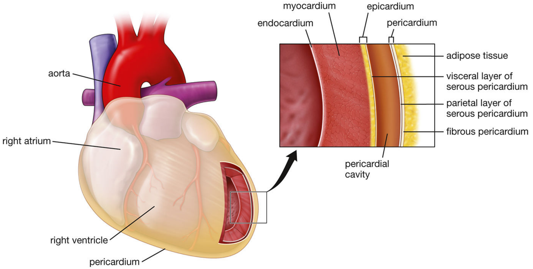
Histology: A Text and Atlas - Layers of the heart and pericardium. The inset shows the endocardium, myocardium, epicardium, pericardial cavity, serous layers, and fibrous pericardium from inside to outside.
2.3 Layer-by-Layer Flowchart
PERICARDIUM - FROM OUTSIDE IN
════════════════════════════════════════════════════
OUTSIDE (most superficial)
│
▼
┌───────────────────────────────────────────────┐
│ FIBROUS PERICARDIUM │
│ │
│ • Tough, dense connective tissue │
│ • Cone-shaped bag; base = diaphragm │
│ • Apex = continuous with adventitia of │
│ great vessels │
│ • Anteriorly attached to sternum via │
│ sternopericardial ligaments │
│ • Prevents over-distension of heart │
│ • Innervated by phrenic nerve (C3, C4, C5) │
│ • Blood supply: pericardiacophrenic vessels │
└─────────────────┬─────────────────────────────┘
│ Inner surface lined by...
▼
┌───────────────────────────────────────────────┐
│ SEROUS PERICARDIUM - PARIETAL LAYER │
│ │
│ • Thin, smooth membrane │
│ • Lines inner surface of fibrous pericardium │
│ • Secretes serous fluid │
└─────────────────┬─────────────────────────────┘
│
┌────────┴────────┐
│ PERICARDIAL │
│ CAVITY │
│ 15-50 mL fluid │
│ (lubricant) │
└────────┬────────┘
│
▼
┌───────────────────────────────────────────────┐
│ SEROUS PERICARDIUM - VISCERAL LAYER │
│ = EPICARDIUM │
│ │
│ • Adheres to heart surface │
│ • Single layer of mesothelial cells + │
│ underlying connective & adipose tissue │
│ • Coronary arteries & nerves run here │
│ • = Outermost layer of heart wall │
└─────────────────┬─────────────────────────────┘
│
▼
HEART MUSCLE (Myocardium)
INSIDE (deepest)
2.4 Pericardium Summary Table
| Layer | Type | Key Feature | Clinical Point |
|---|---|---|---|
| Fibrous pericardium | Fibrous | Tough, inelastic, protective | Limits distension; pain via phrenic nerve |
| Parietal serous pericardium | Serous | Smooth inner surface | Inflamed in pericarditis → friction rub |
| Pericardial cavity | Space | Contains 15-50 mL fluid | Effusion if >50 mL; tamponade if rapid fill |
| Visceral serous pericardium (Epicardium) | Serous | Adheres to heart | Coronary arteries run in adipose here |
⚠️ Clinical Link - Cardiac Tamponade (Week 5 preview): Rapid accumulation of fluid in the pericardial cavity (blood from trauma, or pericardial effusion from pericarditis) compresses the heart and stops it filling. This is Beck's Triad: hypotension + muffled heart sounds + raised JVP. Treatment = pericardiocentesis (needle drainage).
PART 3 - THREE LAYERS OF THE CARDIAC WALL
CARDIAC WALL CROSS-SECTION
════════════════════════════════════════════
BLOOD inside chamber
│
▼
┌──────────────────────────────────────┐
│ ENDOCARDIUM │
│ • Innermost layer │
│ • Single layer of endothelial cells │
│ + thin connective tissue │
│ • Smooth → prevents clot formation │
│ • Continuous with vessel endothelium│
│ • MOST vulnerable to ischemia │
│ (terminal, smallest vessels) │
└─────────────┬────────────────────────┘
│
▼
┌──────────────────────────────────────┐
│ MYOCARDIUM │
│ • Thickest, middle layer │
│ • Cardiac striated muscle fibers: │
│ - Diameter: 10-15 µm │
│ - Length: 30-60 µm │
│ • Proteins: Actin, Myosin, │
│ Troponin I, Troponin T │
│ • Enzymes: CK-MB, LDH, Myoglobin │
│ • Atrial myocardium = thinner │
│ • Ventricular myocardium = thicker │
│ (LV thickest - high pressure) │
└─────────────┬────────────────────────┘
│
▼
┌──────────────────────────────────────┐
│ EPICARDIUM │
│ • Outermost layer of heart │
│ • = Visceral layer of serous peric. │
│ • Mesothelial cells + adipose tissue│
│ • Coronary arteries run here │
│ • Cushions heart in pericardial │
│ cavity │
└──────────────────────────────────────┘
│
▼
Pericardial cavity
3.1 Wall Layers - Comparison Table
| Layer | Position | Composition | Function | Clinical Relevance |
|---|---|---|---|---|
| Endocardium | Innermost | Endothelium + connective tissue | Blood-contact surface, prevents thrombosis | Infective endocarditis; most ischemia-prone |
| Myocardium | Middle | Cardiac muscle (actin, myosin, troponin) | Generates contractile force | MI - troponin/CK-MB released into blood |
| Epicardium | Outermost | Mesothelium + adipose + connective tissue | Coronary artery support, lubrication | Coronary artery disease; pericarditis |
PART 4 - THE FOUR CHAMBERS
4.1 Overview
The heart has four chambers - two thin-walled atria (upper, receiving) and two thick-walled ventricles (lower, pumping).
4-CHAMBER HEART CROSS-SECTION
══════════════════════════════════════════════════════
SUPERIOR (Base)
│
┌──────────┴──────────┐
│ │
│ RIGHT ATRIUM (RA) │ LEFT ATRIUM (LA) │
│ • Receives: │ • Receives: │
│ - SVC (upper) │ - 4 pulm. veins │
│ - IVC (lower) │ • Thin wall (~3 mm) │
│ - Coronary sinus │ • Posterior chamber │
│ • Thin wall (~2 mm)│ • Largest atrium │
│ • Has: crista │ • Has: left auricle │
│ terminalis & │ (appendage) │
│ right auricle │ │
│ │ │
│ ─── TRICUSPID ─── │ ─── MITRAL ──── │
│ VALVE │ VALVE │
│ │ │
│ RIGHT VENTRICLE │ LEFT VENTRICLE (LV) │
│ (RV) │ │
│ • Banana-shaped │ • Conical/bullet │
│ • Wall: ~3 mm │ shaped │
│ • Lower pressure │ • Wall: 9-11 mm │
│ • Pulmonary │ • High pressure │
│ circulation │ • Systemic │
│ │ circulation │
└──────────┬──────────┘ │
│ │
PULMONARY AORTIC
VALVE VALVE
│ │
▼ ▼
Pulmonary artery Aorta
│
INFERIOR (Apex)
══════════════════════════════════════════════════════
Interventricular septum separates RV and LV
Interatrial septum separates RA and LA
4.2 Right Atrium (RA) - Detailed
- Receives deoxygenated blood from SVC (upper body), IVC (lower body), and coronary sinus (heart's own venous return)
- Inner wall has pectinate muscles (rough, comb-like ridges)
- The crista terminalis is a smooth muscular ridge separating the rough and smooth portions
- Contains the fossa ovalis (remnant of the foramen ovale from fetal life)
- Has a small pouch called the right auricle (appendage)
4.3 Left Atrium (LA) - Detailed
- Receives oxygenated blood from the 4 pulmonary veins (2 from each lung)
- Mostly smooth-walled (except the left auricle)
- Most posterior chamber of the heart
- Enlarged LA compresses the esophagus (seen on barium swallow in mitral stenosis)
4.4 Right Ventricle (RV) - Detailed
- Banana/crescent shaped when viewed in cross-section (wrapped around the LV)
- Wall thickness ~3 mm
- Contains trabeculae carneae (muscular ridges) and a moderator band (carries part of the right bundle branch)
- Pumps to the pulmonary circulation (low resistance) at ~25/5 mmHg
4.5 Left Ventricle (LV) - Detailed
- Circular in cross-section, conical/bullet shape
- Wall thickness 9-11 mm (3x thicker than RV)
- Contains trabeculae carneae and 2 papillary muscles (anterolateral & posteromedial)
- Pumps to the systemic circulation at ~120/80 mmHg
- Why thicker? Must generate much higher pressure - follows Laplace's Law: Wall stress ∝ Pressure × Radius / (2 × Wall thickness)
4.6 Right vs Left - Full Comparison Table
| Feature | Right Atrium | Left Atrium | Right Ventricle | Left Ventricle |
|---|---|---|---|---|
| Location | Upper right | Upper left/posterior | Lower right/anterior | Lower left |
| Shape | Oval | Oval | Banana/crescent | Bullet/conical |
| Wall thickness | ~2 mm | ~3 mm | ~3 mm | 9-11 mm |
| Inflow vessels | SVC, IVC, Coronary sinus | 4 Pulmonary veins | From RA via tricuspid | From LA via mitral |
| Outflow | To RV via tricuspid | To LV via mitral | Pulmonary trunk/artery | Aorta |
| AV Valve | Tricuspid (3 cusps) | Mitral (2 cusps) | - | - |
| Semilunar Valve | - | - | Pulmonary (3 cusps) | Aortic (3 cusps) |
| Pressure | Low (~5-8 mmHg) | Low (~8-12 mmHg) | Systolic ~25 mmHg | Systolic ~120 mmHg |
| Special structure | Crista terminalis, Fossa ovalis, Right auricle | Left auricle, Smooth walls | Moderator band | 2 papillary muscles |
| Failure signs | Peripheral edema, raised JVP | Pulmonary congestion | Cor pulmonale features | Pulmonary edema, SOB |
PART 5 - THE FOUR HEART VALVES
5.1 Textbook Diagram - All 4 Valves (Top View)
Gray's Anatomy for Students - Cardiac skeleton with all 4 valve rings: Pulmonary (top), Aortic (centre), Mitral/Left AV ring (left), Tricuspid/Right AV ring (right)
5.2 Classification Flowchart
HEART VALVES - CLASSIFICATION TREE
════════════════════════════════════════════════════════
HEART VALVES (4 total)
│
┌────────────┴────────────┐
▼ ▼
ATRIOVENTRICULAR SEMILUNAR
(AV) VALVES VALVES
[Inlet valves] [Outlet valves]
Between atria & Between ventricles &
ventricles great arteries
│ │
┌─────┴─────┐ ┌─────┴──────┐
▼ ▼ ▼ ▼
TRICUSPID MITRAL PULMONARY AORTIC
VALVE VALVE VALVE VALVE
(Right) (Left) (Right) (Left)
│ │ │ │
3 cusps 2 cusps 3 cusps 3 cusps
│ │ │ │
Has Has No chordae No chordae
chordae chordae tendineae tendineae
tendineae tendineae │ │
& papillary & papillary Opens in Opens in
muscles muscles systole systole
5.3 Mitral & Tricuspid Valve - Subvalvular Apparatus
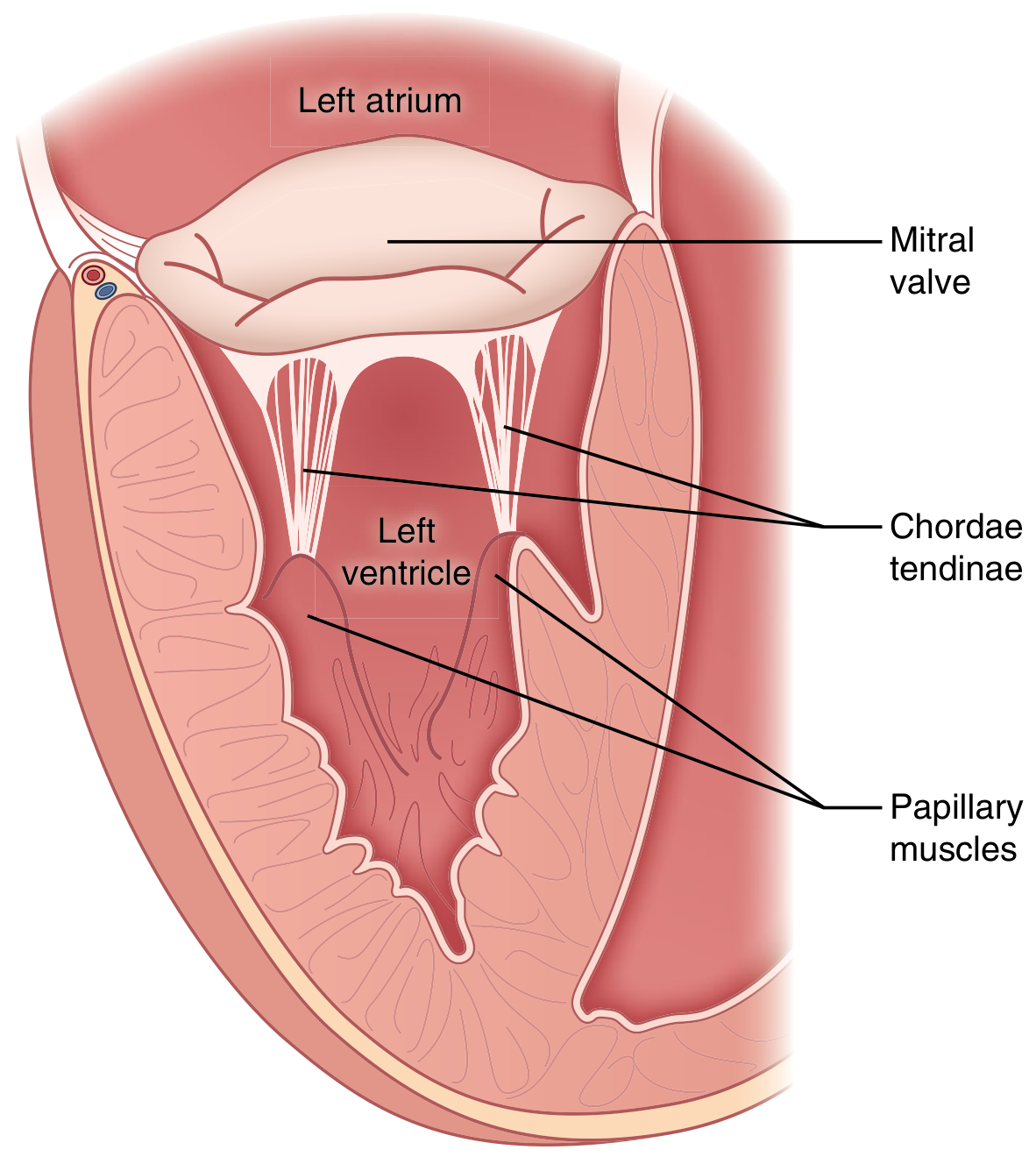
Sabiston Textbook of Surgery - The mitral valve's subvalvular apparatus: leaflets anchored by chordae tendinae to papillary muscles. This prevents leaflet prolapse back into the atrium during systole.
5.4 How AV Valves Work - Mechanism Flowchart
AV VALVE MECHANISM (Mitral & Tricuspid)
══════════════════════════════════════════════
DIASTOLE (ventricular filling):
┌─────────────────────────────────────┐
│ Ventricular pressure < Atrial press │
│ │ │
│ ▼ │
│ AV Valves OPEN │
│ Blood flows: Atrium → Ventricle │
│ Chordae tendineae - SLACK │
│ Papillary muscles - RELAXED │
└─────────────────────────────────────┘
│
▼
SYSTOLE (ventricular contraction):
┌─────────────────────────────────────┐
│ Ventricular pressure > Atrial press │
│ │ │
│ ▼ │
│ AV Valves attempt to CLOSE │
│ Blood pushes leaflets upward │
│ BUT: Papillary muscles CONTRACT │
│ pulling chordae tendineae │
│ TAUT │
│ → Prevents leaflet prolapse │
│ back into atrium │
│ AV Valves CLOSE completely │
│ → Produces HEART SOUND S1 │
└─────────────────────────────────────┘
⚠️ Clinical Link: If a papillary muscle ruptures (e.g., in inferior MI from RCA occlusion), chordae go slack and the mitral leaflet prolapses back into the LA during systole → Acute Mitral Regurgitation (medical emergency: sudden pulmonary edema).
5.5 Complete Valve Reference Table
| Valve | Type | Location | Cusps | Opens | Closes | Sound | Disease if Damaged |
|---|---|---|---|---|---|---|---|
| Tricuspid | AV | Right AV junction | 3 (ant., post., septal) | Diastole | Systole | S1 | Tricuspid stenosis/regurgitation |
| Mitral (Bicuspid) | AV | Left AV junction | 2 (ant., post.) | Diastole | Systole | S1 (dominant) | Mitral stenosis (rheumatic), MVP |
| Pulmonary | Semilunar | RV outflow | 3 | Systole | Diastole | S2 (P2) | Pulmonary stenosis (congenital) |
| Aortic | Semilunar | LV outflow | 3 (L, R, posterior) | Systole | Diastole | S2 (A2 dominant) | Aortic stenosis (calcific), AR |
5.6 Heart Sounds - Simple Table
| Sound | Name | Cause | Heard at |
|---|---|---|---|
| S1 | "Lub" | Closure of Mitral + Tricuspid valves at start of systole | Apex (mitral area) |
| S2 | "Dub" | Closure of Aortic + Pulmonary valves at end of systole | Left sternal edge (pulmonary), Right 2nd ICS (aortic) |
| S3 | Gallop | Rapid ventricular filling (early diastole) | Normal in young; abnormal in HF |
| S4 | Atrial gallop | Forceful atrial contraction against stiff LV | Aortic stenosis, LVH, hypertension |
PART 6 - THE GREAT VESSELS
6.1 Complete Flowchart - Pulmonary + Systemic Circuits
COMPLETE BLOOD FLOW CIRCUIT
════════════════════════════════════════════════════════════════
BODY TISSUES
(deoxygenated blood)
│
SVC (upper body) ──┐
IVC (lower body) ──┤──► RIGHT ATRIUM (RA)
Coronary Sinus ──┘ │
Tricuspid Valve
│
▼
RIGHT VENTRICLE (RV)
│
Pulmonary Valve
│
▼
PULMONARY TRUNK
│
┌────────────┴────────────┐
▼ ▼
Right Pulmonary Left Pulmonary
Artery Artery
│ │
└───────── LUNGS ──────────┘
│
(Gas exchange occurs)
O2 added, CO2 removed
│
┌────────────┴────────────┐
▼ ▼
Right pulm. Left pulm.
veins (x2) veins (x2)
│ │
└────────────┬────────────┘
▼
LEFT ATRIUM (LA)
│
Mitral Valve
│
▼
LEFT VENTRICLE (LV)
│
Aortic Valve
│
▼
AORTA
┌────────────┴────────────┐
▼ ▼
Ascending aorta Descending aorta
│
Aortic arch
(brachiocephalic,
L. common carotid,
L. subclavian branches)
│
▼
BODY TISSUES
(cycle repeats)
6.2 Great Vessels Summary Table
| Vessel | Blood Type | Direction | Connects |
|---|---|---|---|
| Superior Vena Cava (SVC) | Deoxygenated | INTO Right Atrium | Head, neck, upper limbs → Heart |
| Inferior Vena Cava (IVC) | Deoxygenated | INTO Right Atrium | Abdomen, pelvis, lower limbs → Heart |
| Coronary Sinus | Deoxygenated | INTO Right Atrium | Heart muscle (venous drainage) |
| Pulmonary Trunk | Deoxygenated | OUT of Right Ventricle | Heart → Lungs |
| Right Pulmonary Artery | Deoxygenated | INTO right lung | Heart → Right lung |
| Left Pulmonary Artery | Deoxygenated | INTO left lung | Heart → Left lung |
| Pulmonary Veins (x4) | OXYGENATED | INTO Left Atrium | Lungs → Heart |
| Aorta | OXYGENATED | OUT of Left Ventricle | Heart → Entire body |
💡 Key Exam Fact: Pulmonary arteries carry deoxygenated blood. Pulmonary veins carry oxygenated blood. This is the only exception to the rule that arteries carry oxygenated blood.
PART 7 - CORONARY ARTERIES (Gross Introduction)
7.1 Overview
The heart receives its own blood supply via the coronary arteries, which arise from the aortic sinuses (sinuses of Valsalva) at the root of the ascending aorta, just above the aortic valve cusps.
CORONARY ARTERY ANATOMY - FLOWCHART
═══════════════════════════════════════════════════════
AORTA
│
┌──────────┴──────────┐
│ │
▼ ▼
RIGHT CORONARY ARTERY LEFT CORONARY ARTERY
(RCA) (LCA) - short trunk
│ │
│ ┌──────┴──────┐
│ │ │
│ ▼ ▼
│ LEFT ANTERIOR CIRCUMFLEX
│ DESCENDING ARTERY (LCx)
│ (LAD) │
│ "the widow │
│ maker" │
│ │ │
▼ ▼ ▼
BRANCHES: SUPPLIES: SUPPLIES:
• SA nodal br. • Anterior • Lateral LV wall
• Right marginal LV wall • Posterior LV
branch • Anterior (if left-dominant)
• Posterior 2/3 of • Left atrium
interventricular septum
branch (in 85%)• Anterior
papillary
muscle
SUPPLIES:
• RA, RV
• SA node (85%)
• AV node (85%)
• Inferior LV wall
• Posterior septum
7.2 Coronary Artery Supply Table
| Artery | Territory | ECG Changes if Blocked | Clinical Significance |
|---|---|---|---|
| RCA | RA, RV, SA node, AV node, inferior LV | Inferior MI: ST elevation in II, III, aVF | Risk of bradycardia, AV block, RV infarct |
| LAD | Anterior LV, anterior septum, apex | Anterior MI: ST elevation in V1-V4 | Most common; causes large MI ("widow maker") |
| LCx | Lateral LV, posterior LV (left-dominant) | Lateral MI: ST elevation in I, aVL, V5-V6 | May be silent; posterior MI |
7.3 Coronary Dominance
- Right dominant (85%): RCA provides the posterior interventricular branch → supplies inferior LV + AV node
- Left dominant (8%): LCx provides the posterior interventricular branch
- Co-dominant (7%): Both contribute equally
PART 8 - MASTER INTEGRATIVE FLOWCHART
WEEK 1, DAY 1 - COMPLETE OVERVIEW
════════════════════════════════════════════════════════════════════
┌──────────────────────┐
│ THE HEART │
│ ~300 g, mediastinum│
│ Cone-shaped │
└──────────┬───────────┘
│
┌─────────────────────────┼──────────────────────────┐
▼ ▼ ▼
┌──────────────┐ ┌──────────────────┐ ┌──────────────────┐
│ PERICARDIUM │ │ CARDIAC WALL │ │ 4 CHAMBERS │
│ │ │ (3 layers) │ │ │
│ 1. Fibrous │ │ │ │ RA - RV (Right) │
│ (outer) │ │ 1. Epicardium │ │ LA - LV (Left) │
│ │ │ 2. Myocardium │ │ │
│ 2. Serous │ │ 3. Endocardium │ │ Separated by: │
│ - Parietal │ │ │ │ Interatrial & │
│ - Visceral │ │ (Outside→Inside)│ │ Interventricular│
│ │ │ │ │ Septa │
│ 3. Pericardial│ └──────────────────┘ │ │
│ cavity │ └────────┬─────────┘
│ (15-50 mL)│ │
└──────────────┘ │
┌─────────────────┼──────────────────┐
▼ ▼ ▼
┌─────────────┐ ┌─────────────┐ ┌──────────────┐
│ 4 VALVES │ │ GREAT VESSELS│ │ CORONARY │
│ │ │ │ │ ARTERIES │
│ AV Valves: │ │ IN to heart:│ │ │
│ - Tricuspid │ │ SVC, IVC, │ │ RCA: │
│ (3 cusps) │ │ Coron. sinus│ │ Inferior LV, │
│ - Mitral │ │ Pulm. veins │ │ SA+AV node │
│ (2 cusps) │ │ │ │ │
│ │ │ OUT of heart│ │ LAD: │
│ Semilunar: │ │ Pulm. trunk │ │ Anterior LV │
│ - Pulmonary │ │ Aorta │ │ │
│ - Aortic │ │ │ │ LCx: │
│ │ │ │ │ Lateral LV │
└─────────────┘ └─────────────┘ └──────────────┘
PART 9 - QUICK REVISION TABLE (Exam-Ready)
| Topic | Key Point | Number to Remember |
|---|---|---|
| Heart weight | 325g (M), 275g (F) | Cardiomegaly >500g |
| Pericardial fluid | Normal lubricating fluid | 15-50 mL |
| Pericardial layers | Fibrous → Parietal serous → Cavity → Visceral serous | 3 structural layers |
| Cardiac wall layers | Endocardium → Myocardium → Epicardium | 3 layers |
| LV wall thickness | 9-11 mm; thickest | >12 mm = LVH |
| RV wall thickness | ~3 mm; banana-shaped | Thickens in pulmonary HTN |
| Cardiac chambers | 4: RA, LA, RV, LV | Two atria + two ventricles |
| AV valves | Tricuspid (R, 3 cusps), Mitral (L, 2 cusps) | Open in diastole |
| Semilunar valves | Pulmonary (R), Aortic (L) - both 3 cusps | Open in systole |
| S1 sound | Mitral + Tricuspid closure | Start of systole |
| S2 sound | Aortic + Pulmonary closure | End of systole |
| Pulmonary veins | Carry oxygenated blood | 4 veins → Left Atrium |
| Pulmonary arteries | Carry deoxygenated blood | Arise from RV |
| Coronary arteries | Arise from aortic sinuses | 2 main (RCA + LCA) |
| Right dominance | RCA supplies posterior IV branch + AV node | 85% of people |
| LAD | "Widow maker" - supplies anterior LV + septum | Most common MI vessel |
PART 10 - DRAW FROM MEMORY (Practice)
After reading all the above, close your notes and try to draw:
- 4-chamber cross-section - Label RA, LA, RV, LV, all 4 valves, septum, inflow & outflow vessels
- Pericardium layers - from fibrous pericardium down to myocardium with the pericardial cavity shown
- Complete blood flow circuit - start from the body → RA → RV → lungs → LA → LV → body
- Coronary artery tree - from the aorta, RCA and LCA with their branches and territories
PART 11 - SELF-QUIZ
| # | Question | Answer |
|---|---|---|
| 1 | What are the 3 layers of the cardiac wall (outside to inside)? | Epicardium → Myocardium → Endocardium |
| 2 | How many layers does the pericardium have? Name them. | Fibrous + Parietal serous + Pericardial cavity + Visceral serous (epicardium) |
| 3 | Which valve separates LA from LV? How many cusps? | Mitral valve; 2 cusps |
| 4 | Why is LV wall thicker than RV? | LV pumps against systemic pressure (~120 mmHg); RV only against pulmonary pressure (~25 mmHg) |
| 5 | Pulmonary veins carry what type of blood? To where? | Oxygenated blood → Left Atrium |
| 6 | Which coronary artery supplies the SA and AV nodes in 85%? | Right Coronary Artery (RCA) |
| 7 | What is normal pericardial fluid volume? | 15-50 mL |
| 8 | S1 is caused by closure of which valves? | Mitral + Tricuspid (AV valves) at start of systole |
| 9 | What prevents AV valve leaflets from prolapsing into the atria? | Chordae tendineae + papillary muscles |
| 10 | What is cardiac dominance? Which is most common? | Which vessel supplies the posterior interventricular artery; Right dominant (85%) |
Sources: Histology: A Text and Atlas (Pawlina) 8th Ed | Gray's Anatomy for Students | Sabiston Textbook of Surgery 21st Ed | Tietz Textbook of Laboratory Medicine 7th Ed | Costanzo Physiology 7th Ed
📅 Ready for Day 2? Next topic: Coronary arteries in detail + Cardiac Conduction System (SA node → AV node → Bundle of His → Purkinje fibres) with flowcharts, ECG correlation preview, and your first pacemaker rate table. Just say "Day 2 notes" and I'll build them immediately!
This is a shared conversation. Sign in to Orris to start your own chat.