
IMG_20260518_104914.jpg
What are the finding abnormal here how to read if someone tell me to read this x ray reference orthopedcis standard book
Reading File
Finding Sources
Finding Sources
Finding Sources
Reading File
Finding Sources
Finding Sources
Reading File
Finding Sources
Finding Sources
Reading File
Reading File
Loading Image
Loading Image
Loading Image
I now have everything needed. Here is a comprehensive structured answer:
Elbow X-Ray — Findings & How to Read It
What Is This X-Ray?
This is an AP (anteroposterior) + Lateral view of the elbow — the two standard views required for any elbow assessment. The patient appears to be a child/adolescent based on visible open growth plates (physes).
Abnormal Findings in This X-Ray
1. Distal Humerus Fracture (Supracondylar Pattern)
On the AP view (left image), there is disruption and irregular density in the distal humeral metaphysis — the region just above the condyles is markedly abnormal with loss of normal trabecular architecture and bony irregularity. This is a supracondylar fracture of the humerus, the most common elbow fracture in children.
2. Posterior Fat Pad Sign (Lateral View)
On the lateral view (right image), there is a visible posterior fat pad — a radiolucent lucency posterior to the distal humerus. This is always pathological and indicates a haemarthrosis (blood in the joint) from an intra-articular fracture.
"Any posterior fat pad is pathological and indicates the presence of joint effusion. In the setting of trauma, patients with a radiological posterior fat pad sign are assumed to have an intra-articular skeletal injury." — Rosen's Emergency Medicine, 10th Ed.
3. Anterior Humeral Line Violation (Lateral View)
On the lateral view, a line drawn along the anterior cortex of the humerus (the "Anterior Humeral Line / AHL") should normally pass through the middle third of the capitellum. In an extension-type supracondylar fracture, this line passes anterior to the capitellum or through its anterior third — indicating posterior displacement of the distal fragment.
4. Significant Bony Displacement/Comminution
The distal humerus on the AP view shows marked fragmentation/irregularity consistent with a displaced fracture (Gartland Type II or III).
How to Systematically Read an Elbow X-Ray (Standard Method)
Step 1 — Confirm Two Views
Always read AP + true lateral together. Never report on a single view.
Step 2 — AP View: Draw Baumann's Angle
Draw a line along the humeral shaft axis and another along the lateral condyle physis (growth plate). Normal Baumann's angle = 70–75° (some texts say 64–81°). An increased angle means varus deformity (cubitus varus/gunstock deformity risk after fracture).
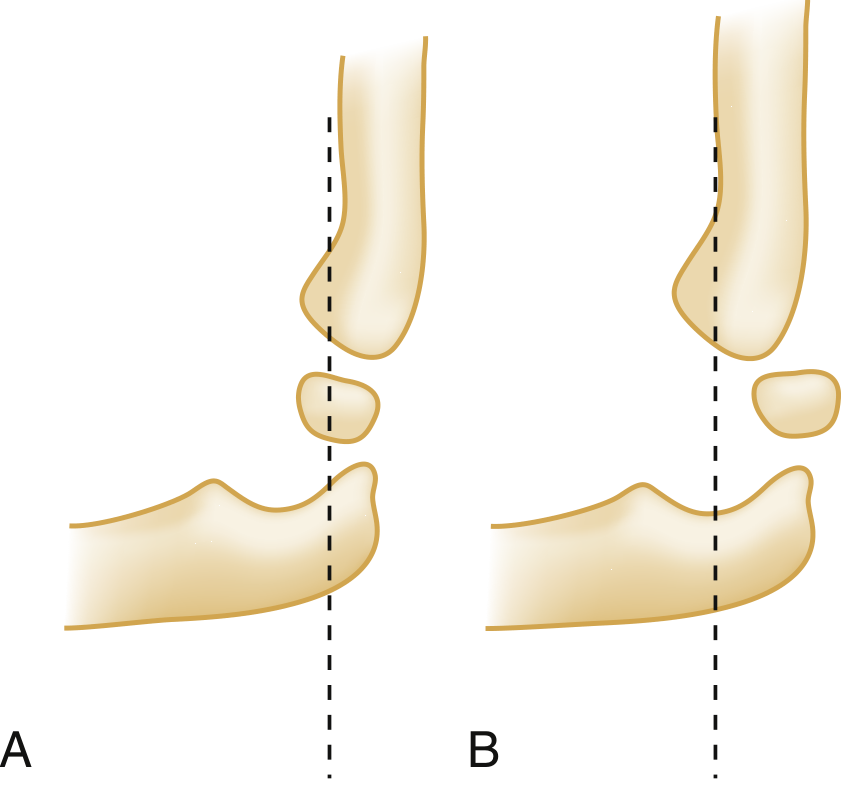
Step 3 — Lateral View: Draw the Anterior Humeral Line (AHL)
- Draw a line down the anterior cortex of the humerus
- It should bisect the middle third of the capitellum
- If the line passes through the anterior third or misses the capitellum → extension-type supracondylar fracture
Step 4 — Lateral View: Check Radiocapitellar Line
Draw a line through the long axis of the radial shaft — it must pass through the centre of the capitellum on all views. If not → suspect radial head dislocation, Monteggia fracture, or lateral condyle fracture.
Step 5 — Lateral View: Fat Pad Signs
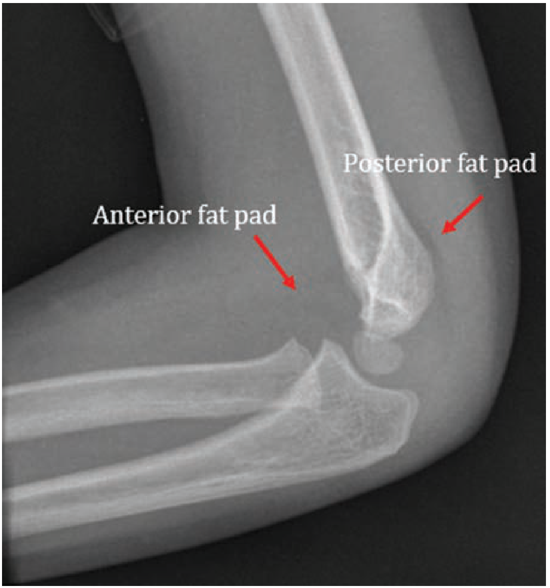
| Fat Pad | Appearance | Significance |
|---|---|---|
| Anterior (small, flat) | Normal — sits flush to anterior humerus | Normal |
| Anterior (convex/"sail sign") | Triangular, lifted away | Pathological — effusion |
| Posterior | Any visibility | Always pathological → haemarthrosis |
Step 6 — CRITOE Mnemonic (Paediatric Elbow Ossification Centers)
In children, use CRITOE to check ossification centres appear in order (age of appearance):
| Letter | Centre | Age (years) |
|---|---|---|
| C | Capitellum | 1 |
| R | Radial head | 3 |
| I | Internal (medial) epicondyle | 5 |
| T | Trochlea | 7 |
| O | Olecranon | 9 |
| E | External (lateral) epicondyle | 11 |
If a centre appears out of order or is displaced, suspect a fracture or avulsion.
Step 7 — Look for Specific Fracture Lines
Systematically trace every cortex: distal humerus, lateral condyle, medial epicondyle, olecranon, radial head, and proximal ulna.
Textbook Reference: Gartland Classification of Supracondylar Fractures
| Type | Description | Treatment |
|---|---|---|
| Type I | Undisplaced | Collar and cuff / backslab × 3 weeks |
| Type II | Displaced, posterior cortex intact | Closed reduction + above-elbow cast |
| Type III | Completely displaced, no cortical contact | Closed/open reduction + K-wire fixation |
| Type IV | Multi-plane instability | Operative fixation |
Critical complications to always check: Vascular (radial/brachial artery), Neurological (anterior interosseous nerve most common), Volkmann's ischaemic contracture.
Summary of This X-Ray
| Finding | Present? |
|---|---|
| Distal humeral fracture (supracondylar pattern) | ✅ Yes |
| Posterior fat pad sign | ✅ Yes (lateral view) |
| Anterior fat pad/sail sign | Likely present |
| AHL violation | ✅ Yes (extension type) |
| Growth plate disruption | ✅ Yes |
| Open fracture / soft tissue gas | Not visible |
Likely diagnosis: Displaced extension-type supracondylar fracture of the humerus (Gartland Type II–III) — requires urgent orthopaedic assessment, neurovascular examination, and likely closed reduction + K-wire fixation.
Sources: Bailey & Love's Short Practice of Surgery 28th Ed.; Rosen's Emergency Medicine 10th Ed.; Tintinalli's Emergency Medicine; Miller's Review of Orthopaedics 9th Ed.
It is a case of poster neglects elvow disslocation since 3 months
Finding Sources
Finding Sources
Finding Sources
Finding Sources
Finding Sources
Reading File
Reading File
Finding Sources
Finding Sources
Finding Sources
Loading Image
Searching PubMed
neglected posterior elbow dislocation treatment
Reading Article
PMID: 33996454
Reading Article
PMID: 42078978
Now I have all the information needed. Here is the complete structured answer:
Neglected Posterior Elbow Dislocation (3 Months Old)
Correcting the Previous Diagnosis
Now that you have provided the clinical context — 3 months untreated — the X-ray findings make complete sense. This is NOT a supracondylar fracture. Re-reading the X-ray with this context:
X-Ray Findings in This Case (Explained)
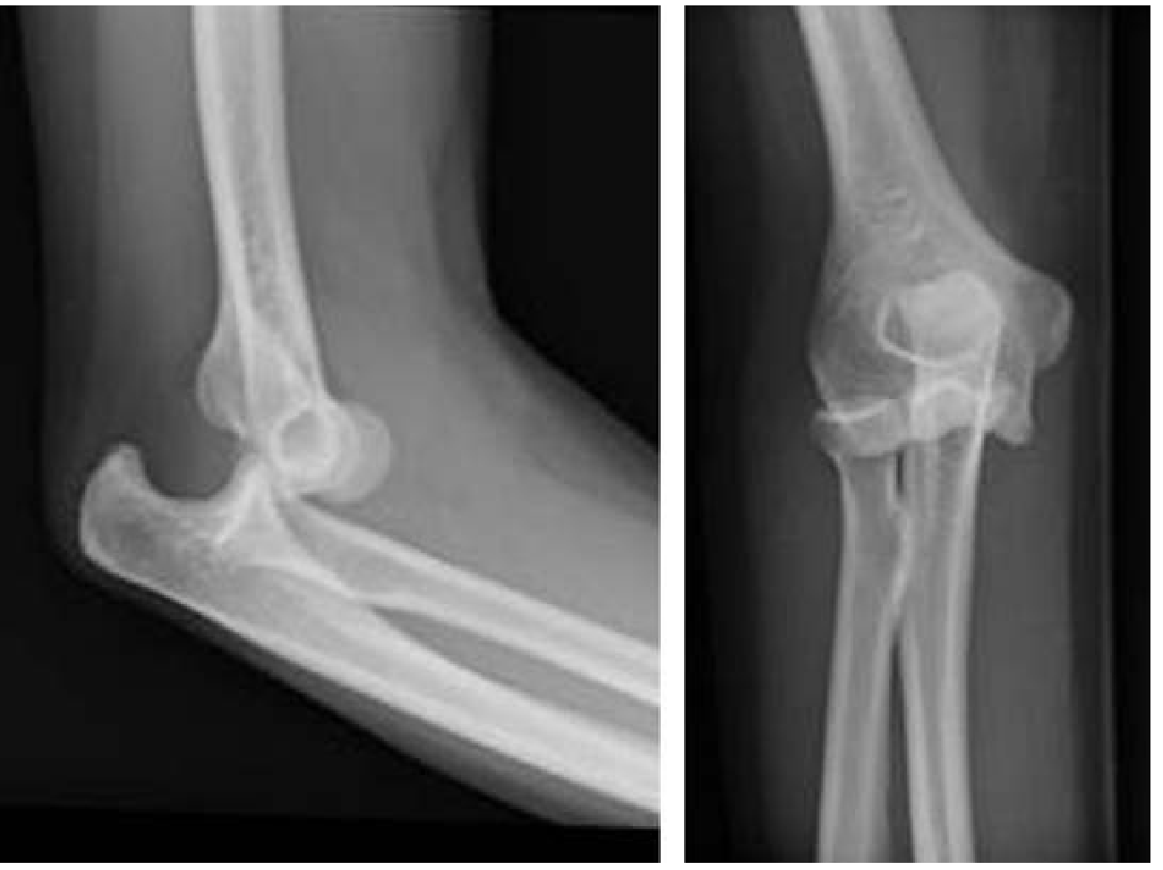
Here is a textbook reference image of an acute posterior elbow dislocation for comparison:
On the AP View (left image of your X-ray):
| Finding | Explanation |
|---|---|
| Olecranon displaced posteriorly and superiorly | The radius and ulna are dislocated posteriorly — the ulnohumeral articulation is lost |
| Disorganised, dense bone mass around the distal humerus | Heterotopic ossification / myositis ossificans — new bone formation in peri-articular soft tissues after 3 months of untreated dislocation |
| Loss of normal joint space | The trochlea-olecranon relationship is completely disrupted |
| Bony irregularity of distal humerus | Remodelling changes from chronic unreduced dislocation |
On the Lateral View (right image):
| Finding | Explanation |
|---|---|
| Posterior displacement of radius + ulna relative to humerus | Classic posterior dislocation — the forearm bones are posterior to the humerus |
| Capitellum visible anteriorly, uncovered | No longer articulating with the radial head |
| Heterotopic ossification / callus | Irregular new bone in posterior and peri-articular region (3 months of organisation) |
| Posterior fat pad obliterated/replaced | Chronic changes have replaced the normal soft tissue planes |
Definition & Classification
"Untreated traumatic posterior dislocation of the elbow joint, 3 weeks or older, is defined as 'neglected posterior dislocation of the elbow'. Approximately 90% are of posterolateral type." — Pal et al., J Clin Orthop Trauma, 2021 [PMID: 33996454]
At 3 months, this is a well-established neglected dislocation with significant secondary pathology.
Pathological Changes at 3 Months (Why It's Difficult)
By 3 months, multiple structures have undergone irreversible changes:
| Structure | Change |
|---|---|
| Joint capsule | Contracted, fibrotic, scarred |
| Collateral ligaments | Shortened, inelastic |
| Muscles (brachialis, triceps) | Fibrosed, shortened — brachialis is most affected |
| Peri-articular soft tissue | Heterotopic ossification / myositis ossificans |
| Articular cartilage | Degenerative changes underway |
| Neurovascular | Risk of ulnar nerve, median nerve, brachial artery adhesion/tethering |
Radiological Checklist for Neglected Elbow Dislocation
When reporting this X-ray, comment on:
- ✅ Direction of dislocation — posterior (olecranon/radius behind humerus)
- ✅ Radiocapitellar line disrupted — radial head not pointing to capitellum
- ✅ Anterior humeral line disrupted — capitellum uncovered
- ✅ Heterotopic ossification — peri-articular new bone (amount and location)
- ✅ Associated fractures — coronoid process? radial head? medial epicondyle avulsion?
- ✅ Degree of articular damage — joint space narrowing, subchondral changes
- ✅ Bony remodelling — pseudoarthrosis or false joint formation?
Treatment of Neglected Posterior Elbow Dislocation (3 Months)
"Goal of surgical treatment is to achieve a painless, stable and mobile elbow with a congruent joint space." — Pal et al., 2021
Why Closed Reduction Fails at 3 Months
Closed reduction is only feasible in the first 2–3 weeks. After that, soft tissue contractures, fibrous organisation, and heterotopic bone make it impossible without causing neurovascular injury.
Surgical Treatment: Open Reduction (ORIF)
Approach: Posterior approach (most commonly used)
Steps:
- Ulnar nerve identification and protection (compulsory)
- Release of posterior capsule and contractures
- Excision of heterotopic bone / fibrous tissue blocking reduction
- Release of medial and lateral collateral ligaments as needed (then repair)
- Reduction of the joint under direct vision
- Stabilisation — usually with hinged external fixator or K-wires if unstable
- Collateral ligament repair/reconstruction
Recent outcomes data:
"23 patients treated via posterior approach surgery — average time onset to surgery 7.83 months. Mean DASH score 15.32, MEPI score 90 (satisfactory functional outcomes)." — Hadinoto et al., Malaysian Orthop J, 2026 [PMID: 42078978]
Post-operative Protocol
- Hinged elbow brace with early active motion
- Aggressive physiotherapy — crucial for regaining range of motion
- NSAIDs / low-dose radiation to prevent recurrence of heterotopic ossification
Complications
| Complication | Notes |
|---|---|
| Heterotopic ossification | Already present; risk of recurrence post-op |
| Ulnar nerve injury | Pre-existing or intra-operative — must be assessed |
| Re-dislocation / instability | Due to ligamentous laxity |
| Limited range of motion | Most common residual problem; target 30–130° |
| Post-traumatic arthritis | Articular cartilage damage from 3 months of neglect |
| Volkmann's ischaemia | Less risk now, but vascular assessment mandatory |
Summary Card
| Item | Finding |
|---|---|
| Diagnosis | Neglected posterior elbow dislocation (3 months) |
| Direction | Posterior (posterolateral) |
| Secondary changes | Heterotopic ossification, capsular contracture, ligament fibrosis |
| Treatment | Open reduction via posterior approach |
| Key nerve to protect | Ulnar nerve |
| Prognosis | Good functional outcome possible with surgery + physio |
Sources: Pal et al. J Clin Orthop Trauma 2021 [PMID: 33996454]; Hadinoto et al. Malays Orthop J 2026 [PMID: 42078978]; Tintinalli's Emergency Medicine; Rockwood & Green's Fractures in Adults 10th Ed.
This is a shared conversation. Sign in to Orris to start your own chat.