

this face bone is too much paining not able to eat properly like chew etc
temporomandibular joint anatomy diagram
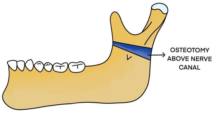
This medical illustration depicts a lateral view of the human mandible, highlighting a surgical modification for treating temporomandibular joint (TMJ) ankylosis. The diagram shows the primary anatomical features of the mandible, including the alveolar ridge with teeth, the mandibular body, and the ramus featuring the coronoid and condylar processes. A highlighted blue band across the upper portion of the mandibular ramus represents a 'Modified Osteotomy Above Nerve Canal.' This specific surgical plane is positioned superior to the inferior alveolar nerve (IAN) canal, which is indicated by a 'v' shaped marking on the ramus. The illustration demonstrates the Esmarch procedure variant where the bone cut is performed high on the ramus to separate the ankylosed condylar unit from the functional mandibular body while aiming to preserve nerve integrity. This anatomical diagram is used to explain surgical planning in maxillofacial surgery, specifically for patients with recurrent joint fusion or altered vascular anatomy.
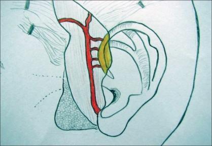
This medical anatomical diagram illustrates the surgical vascular anatomy for a vascularized auricular cartilage flap. The illustration shows a profile view of the human ear and the preauricular region. The superficial temporal artery (STA) is highlighted in red, coursing vertically in the preauricular plane. Several horizontal arterial branches are shown originating from the STA and extending toward the root of the helix. A specific yellow-shaded region identifies a cartilage flap being harvested from the helical root, demonstrating its relationship to the supplying vascular pedicle. Key anatomical structures depicted include the helix, antihelix, tragus, and concha. The diagram serves as a surgical planning aid for reconstructive procedures, such as temporomandibular joint (TMJ) ankylosis surgery, where vascularized cartilage is used for interpositional arthroplasty. It highlights the importance of preserving the delicate arterial supply from the STA to the helical cartilage to ensure flap viability.
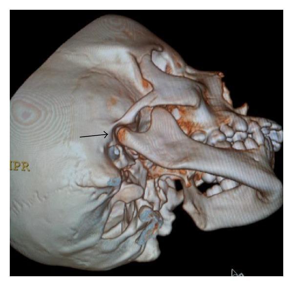
This diagnostic image is a 3D computed tomography (CT) reconstruction of a human skull, providing a lateral-oblique view of the right craniofacial region. The visualization highlights the normal anatomy of the temporomandibular joint (TMJ). A black arrow indicates the right mandibular condyle, which exhibits a smooth, rounded cortical surface and maintains a proper anatomical relationship with the glenoid fossa of the temporal bone. Key visible structures include the mandible, maxilla with developing mixed dentition, zygomatic bone, and the temporal bone. The image serves as a healthy baseline comparison for evaluating maxillofacial asymmetries or developmental pathologies like condylar aplasia. This anatomical diagram is highly relevant for dentistry and oral and maxillofacial surgery education, demonstrating the standard structural integrity and alignment of the TMJ components.
jaw pain near ear difficulty chewing causes TMJ vs dental abscess vs mumps when to see doctor
What this likely is
| Cause | Clues |
|---|---|
| TMJ dysfunction | Pain worse with chewing/yawning, clicking or popping, jaw stiffness, no fever |
| Dental problem (abscess, decaying/impacted tooth) | Throbbing pain, sensitivity to hot/cold, possible swelling, sometimes fever |
| Mumps/parotitis (salivary gland) | Swelling in front of/below ear, pain when chewing or with sour foods, may follow flu-like illness |
| Jaw trauma or fracture | Recent injury/blow to the face, visible swelling/bruising, jaw misaligned |
| Ear infection (referred pain) | Ear-specific symptoms, hearing change, discharge |
What to do now
- Stick to soft foods (soup, mashed food, yogurt) and avoid wide mouth-opening, gum, or hard/chewy foods until it settles - textbook guidance (Tintinalli's Emergency Medicine) specifically advises soft diet plus simple analgesics like NSAIDs (e.g., ibuprofen) for non-traumatic TMJ pain.
- Warm compress on the joint can ease muscle spasm.
- Avoid clenching/grinding teeth; try not to rest your chin on your hand (as in the photo) since that puts pressure on the joint.
When to get seen in person (don't just wait it out)
- Fever, visible swelling, or redness (suggests infection/abscess, not just TMD)
- History of recent injury/blow to the jaw or face (need to rule out fracture or dislocation)
- Jaw locking open or stuck, or visible asymmetry/deformity
- Numbness in the face or jaw
- Pain that's severe, not improving in a few days, or getting worse despite soft diet and NSAIDs
it is tmj
"temporomandibular joint disorders" AND therapy
TMJ jaw pain home exercises stretches self care relief
Immediate relief (next few days)
- Soft diet only - soups, khichdi, dal, mashed vegetables, yogurt, smoothies. Avoid anything that needs wide opening or hard chewing (bread crusts, meat, nuts, chewing gum, apples). This is the standard first recommendation in emergency medicine texts for non-traumatic TMJ pain.
- NSAIDs (ibuprofen or similar, if no contraindication) for pain and inflammation - simple analgesics are the first-line drug treatment per Tintinalli's Emergency Medicine.
- Heat or cold: warm compress for muscle tightness, or an ice pack for 10-15 min if there's acute swelling/inflammation.
- Rest the joint: avoid wide yawning, don't rest your chin on your hand or fist (like in your photo), avoid clenching or grinding teeth, don't chew gum.
Jaw exercises (once acute pain settles a bit)
- Relaxed jaw posture: keep teeth slightly apart, lips gently closed (the "uh" position) through the day to take pressure off the joint.
- Gentle active stretch: slowly open your jaw only as far as comfortable (no pain/clicking), hold a few seconds, release. Repeat a few times a day.
- Side-to-side and forward jaw movement: slow, small, controlled motions to loosen the joint - stop immediately if it causes sharp pain.
Who to see next
- If pain persists beyond a week or two despite soft diet + NSAIDs, see a dentist or oral-maxillofacial/ENT specialist for a proper exam - they may fit a night guard/occlusal splint, which has solid evidence for reducing TMJ pain, especially if you grind your teeth at night.
- A recent network meta-analysis of conservative treatments (Ferrillo et al., 2025, PMID 36148997) supports splints, physiotherapy, and manual therapy as effective conservative options - so this isn't just old-textbook advice, current evidence backs it.
- Go in sooner (don't just self-manage) if you notice: fever or visible facial swelling, the jaw locking open or shut, numbness, it followed an injury/blow to the face, or the pain keeps getting worse rather than better - these need in-person evaluation to rule out a fracture, dental abscess, or dislocation rather than plain TMD.
why this happens
"temporomandibular joint disorders" AND etiology
what causes TMJ disorder risk factors bruxism stress
Why TMJ pain happens
- Bruxism / teeth grinding and clenching - especially at night, often stress-related. This overloads the joint and chewing muscles.
- Emotional stress and anxiety - increases muscle tension and clenching, a well-documented driver (stress -> bruxism -> TMD is a recognized cycle in recent literature).
- Habits: excessive gum chewing, nail biting, resting the chin on your hand (like in your photo - that puts sustained pressure directly on the joint), wide yawning, or prolonged mouth opening (e.g., long dental procedures).
- Teeth malocclusion or jaw malalignment - uneven bite forces the joint to work asymmetrically.
- Disc displacement within the joint - causes clicking/popping and uneven movement.
- Degenerative joint disease (osteoarthritis) of the TMJ from chronic wear over time.
- Trauma - a blow to the jaw or face, even an old one.
- Systemic inflammatory disease - rheumatoid arthritis, lupus, or other autoimmune arthritis can involve the TMJ.
- Textbooks note that despite popular belief, studies have found no strong, consistent link between bruxism/occlusion alone and TMD - it's usually multifactorial (a combination of muscle overuse, stress, joint mechanics, and sometimes systemic disease) rather than one single cause - Tintinalli's Emergency Medicine.
actually i sleep good then when i get up in the morning this happens
TMJ jaw pain worse in morning after sleep causes sleeping position bruxism sleep apnea
Nocturnal bruxism (sleep clenching/grinding) - the most likely cause
- Teeth grinding/clenching while asleep puts sustained, repetitive force through the joint and chewing muscles all night. Since you're asleep, there's no pain signal registering - the damage/strain shows up only once you wake and start moving/using the joint. This is the classic presentation of sleep bruxism-related TMD, well documented across sleep medicine and psychiatry texts.
- Sleep bruxism is now classified as a sleep-related movement disorder, and morning jaw pain/stiffness with normal daytime comfort is its textbook hallmark.
Sleeping position
- If you sleep on your side or stomach, especially with your hand/arm tucked under your jaw or cheek, that applies direct sustained pressure on the TMJ for hours - similar to the chin-resting habit in your photo, just for 6-8 hours instead of a few minutes.
- Back sleeping is generally best for TMJ because it avoids any direct pressure on the joint and keeps head/neck alignment neutral.
Possible underlying driver: stress or sleep-disordered breathing
- Stress and anxiety are strongly linked to nocturnal clenching - your body may be tensing the jaw as a subconscious stress response overnight even if you're sleeping "well."
- Snoring or sleep apnea can also trigger jaw clenching/thrusting as the body tries to keep the airway open - worth considering if you also snore, wake up tired despite adequate hours, or have been told you stop breathing briefly at night.
What this means for you practically
- Try switching to back sleeping for a week or two and see if it changes things.
- Watch for morning signs of clenching: flattened/worn teeth edges, sore jaw muscles you can feel when you press on your cheek near the ear, or a partner noticing grinding sounds at night.
- If it keeps happening, a dentist can fit a night guard (occlusal splint) - this is the standard, evidence-backed fix specifically for grinding-related morning TMJ pain, since it protects the joint and teeth from the clenching forces overnight.
- If you also snore heavily or feel unrefreshed despite good sleep duration, mention that to a doctor too, since treating an underlying sleep-breathing issue can resolve the clenching that's driving the jaw pain.