
25 mark answer for surgical anatomy of fascial spaces and spread of infection from lower 3rd molar
Reading File
Now let me search the medical library for comprehensive content on fascial spaces and spread of infection from lower third molar.
Finding Sources
Finding Sources
Finding Sources
Finding Sources
Finding Sources
Finding Sources
Finding Sources
Finding Sources
Excellent - found strong sources. Let me read the most relevant sections in parallel.
Reading File
Reading File
Reading File
Excellent content found. Let me now read the key Cummings section on Mandibular Spaces and fetch the important diagrams.
Reading File
Loading Image
Reading File
Loading Image
Now let me read the detailed section on the individual spaces and their contents from the Cummings book, and also read the Scott-Brown section on submandibular space anatomy.
Reading File
Reading File
I now have rich, comprehensive content from authoritative sources. Let me compile a complete 25-mark answer.
Surgical Anatomy of Fascial Spaces and Spread of Infection from Lower Third Molar
Introduction
Odontogenic infections from the lower third molar (mandibular wisdom tooth) are among the most common and potentially life-threatening dental emergencies. The spread of such infections is governed by the anatomy of fascial spaces - potential spaces between layers of fascia, muscle, and bone that are filled with loose areolar tissue. Understanding these spaces, their boundaries, and their communications is the cornerstone of predicting, diagnosing, and managing deep cervicofacial infections.
I. Anatomy of Fascial Layers of the Head and Neck
The head and neck are invested by three layers of fascia:
-
Superficial cervical fascia - a thin layer beneath the skin containing the platysma, facial muscles, and superficial vessels/nerves. It does not form significant barriers to infection.
-
Deep cervical fascia - the main structural layer, divided into three layers:
- Superficial layer (investing layer): encircles the entire neck; splits to enclose the sternocleidomastoid, trapezius, submandibular gland, and parotid gland.
- Middle layer (pretracheal/visceral fascia): anterior portion enclosing the strap muscles, trachea, esophagus, and thyroid gland.
- Deep layer (prevertebral fascia): covers the prevertebral muscles; further divided into the alar fascia anteriorly and prevertebral fascia posteriorly - these create the critically important danger space.
II. Classification of Fascial Spaces
Fascial spaces are classified as primary (directly involved from a tooth apex) or secondary (involved by spread from primary spaces).
A. Primary Spaces of the Mandible
These receive direct spread from mandibular tooth apices through bone:
1. Submental Space
- Boundaries: Anterior - symphysis menti; Posterior - body of hyoid; Superiorly - mylohyoid muscle; Inferiorly - skin and platysma; Laterally - anterior bellies of digastric muscles.
- Contents: Submental lymph nodes, anterior jugular vein tributaries, fat.
- Source: Infected mandibular incisors (when root apices lie below the mentalis muscle attachment).
- Communications: Submandibular space bilaterally.
2. Sublingual Space
- Boundaries: Superior - oral mucosa of the floor of mouth; Inferior - mylohyoid muscle; Lateral - inner surface of mandible; Medial - genioglossus and geniohyoid muscles; Open posteriorly - communicates freely with submandibular space around the free posterior border of mylohyoid.
- Contents: Sublingual gland, deep part of submandibular gland and its duct (Wharton's duct), lingual nerve, hypoglossal nerve, lingual artery and veins.
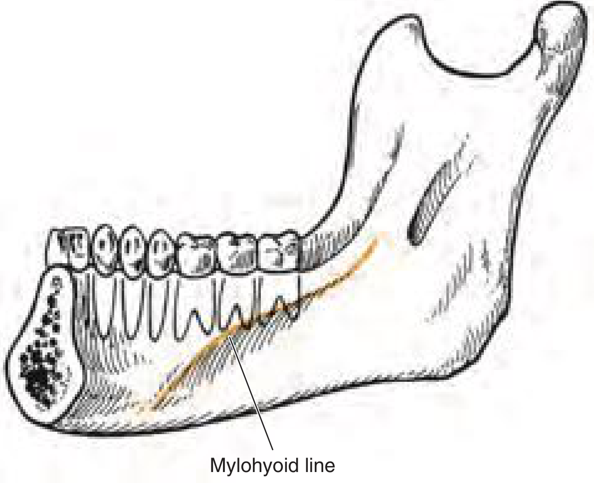
- Source: Mandibular premolars and first molar (apices lie above the mylohyoid line/ridge).
- Clinical feature: Intraoral swelling of the floor of mouth, tongue elevation; minimal extraoral swelling.
3. Submandibular Space
- Boundaries: Superior - mylohyoid muscle; Inferior - investing layer of deep cervical fascia; Anterior/medial - anterior belly of digastric; Open posteriorly - communicates with sublingual space and secondary spaces.
- Contents: Superficial lobe of submandibular gland, submandibular lymph nodes, facial artery and vein, lingual nerve, hypoglossal nerve.
- Source: Second and third mandibular molars - their root apices lie below the mylohyoid ridge (the key anatomical determinant).
- Clinical feature: Extraoral swelling below the mandible, expanding toward the digastric and hyoid.
The critical anatomical determinant: The position of a tooth's root apex relative to the mylohyoid line (mylohyoid ridge) determines whether infection enters the sublingual space (above the line) or the submandibular space (below the line). The apices of the lower 3rd molar lie consistently below this line, directing infection primarily into the submandibular space.
B. Secondary Spaces
These are involved by spread from primary spaces:
4. Masticator Space (Masseteric Space / Pterygomandibular Space)
- Boundaries: Enclosed by the superficial layer of the deep cervical fascia, split to enclose the masseter laterally and the medial pterygoid medially; extends from the zygomatic arch to the inferior mandible.
- Contents: Masseter muscle, medial and lateral pterygoid muscles, ramus of mandible, inferior alveolar nerve and vessels, lingual nerve.
- Source: Lower third molar - spread via lingual or buccal cortical perforation; also direct pericoronitis.
- Clinical features: Trismus (spasm of pterygoid/masseter muscles is pathognomonic), extraoral swelling over the angle of mandible.
- Communications: Parapharyngeal space, buccal space, temporal space.
5. Buccal Space
- Boundaries: Anterolateral - skin; Posteromedial - buccinator muscle; Superior - zygomatic arch; Inferior - lower border of mandible.
- Contents: Buccal fat pad, parotid duct, facial nerve branches, facial artery and vein.
- Source: Maxillary molars primarily (superior to buccinator attachment); lower molars can also be involved.
- Clinical feature: Dramatic facial swelling, trismus.
6. Parapharyngeal Space (Lateral Pharyngeal Space)
This is the most important secondary space - a hub communicating with virtually all other deep neck spaces.
- Shape: Inverted pyramid; from petrous temporal bone superiorly to the hyoid bone inferiorly.
- Boundaries: Medially - superior pharyngeal constrictor; Laterally - medial pterygoid muscle, parotid gland, and mandible; Posteriorly - prevertebral fascia; Anteriorly - pterygomandibular raphe.
- Divided by the styloid process and stylopharyngeus into:
- Pre-styloid (anterior) compartment: Contains fat, connective tissue, maxillary artery, inferior alveolar nerve, lingual nerve, auriculotemporal nerve.
- Post-styloid (posterior) compartment: Contains carotid sheath (internal carotid artery, internal jugular vein, vagus nerve), glossopharyngeal and hypoglossal nerves, sympathetic chain, lymph nodes.
- Significance: The APPS (anterior PPS) tends to form pus/abscess; the PPPS (posterior PPS) tends to cause lymphadenitis and is less prone to abscess.
- Communications: Submandibular space, retropharyngeal space, masticator space, peritonsillar space, carotid sheath (inferiorly to mediastinum).
7. Retropharyngeal Space
- Boundaries: Anterior - posterior pharyngeal wall (buccopharyngeal fascia); Posterior - alar fascia; Lateral - carotid sheaths; Superior - base of skull; Inferior - C6/T4 level.
- Contents: Retropharyngeal lymph nodes, loose areolar tissue.
- Communications: Parapharyngeal space, danger space.
8. Danger Space (Space 4 of Grodinsky and Holyoke)
- Boundaries: Anterior - alar fascia; Posterior - prevertebral fascia; Superior - base of skull; Inferior - diaphragm.
- Contents: Only loose areolar tissue - hence the name "danger."
- Critical significance: Provides a direct, unobstructed route for infection from the neck to the posterior mediastinum and even the pericardium/pleural cavity. This is the mechanism of descending necrotizing mediastinitis, one of the most lethal complications.
9. Prevertebral Space
- Boundaries: Anterior - prevertebral fascia; Posterior - vertebral bodies; Lateral - transverse processes; Superior - base of skull; Inferior - coccyx.
- Contents: Prevertebral muscles, vertebral artery and vein, brachial plexus, phrenic nerve.
III. Spread of Infection from Lower Third Molar - Step by Step
The lower third molar (mandibular wisdom tooth) is the most common source of serious odontogenic infection. Its unique anatomy makes it particularly dangerous:
- Roots are long, often curved, often close to the inferior alveolar nerve.
- Root apices consistently lie below the mylohyoid line on the lingual surface of the mandible.
- Pericoronal infection (pericoronitis) is common due to partial eruption.
- Lingual bone at this site is thin and easily perforated.
Pathway of Spread
Step 1 - Periapical/Pericoronitis → Infection begins at the apical region or around the crown of the impacted/erupting lower 3rd molar.
Step 2 - Bony perforation → Infection erodes through bone. At the lower 3rd molar:
- Lingual cortical plate is thinner and perforated more readily.
- Root apices of lower 3rd molar are below the mylohyoid attachment → infection enters the submandibular space (not the sublingual space).
- Buccal cortical perforation → buccal space or masticator space.
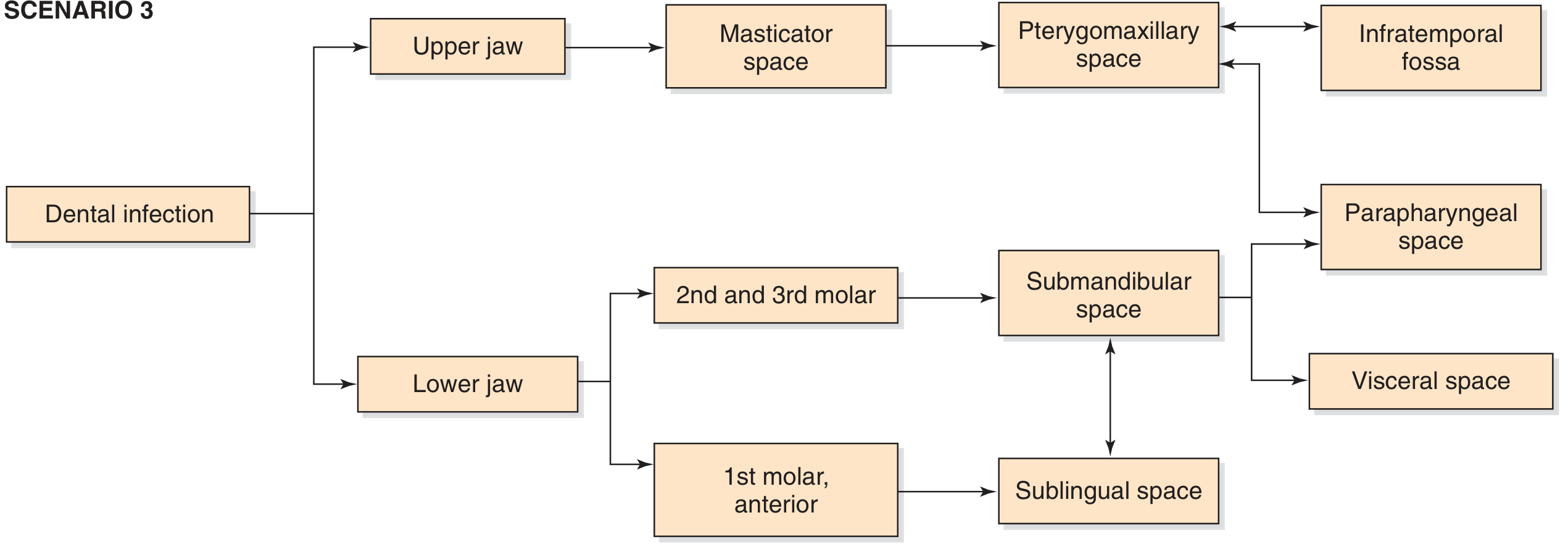
Step 3 - Primary space involvement → Submandibular space is the primary target.
Step 4 - Secondary spread:
- Submandibular space → Sublingual space (via free posterior border of mylohyoid) → tongue elevation
- Submandibular space → Submental space → bilateral spread
- Submandibular space → Parapharyngeal space → airway compromise
- Parapharyngeal space → Retropharyngeal space → posterior spread
- Retropharyngeal space → Danger space → Mediastinum (descending necrotizing mediastinitis)
- Masticator space → Temporal space → infratemporal fossa → parapharyngeal space
- Parapharyngeal space → Carotid sheath → mediastinum
IV. Ludwig's Angina - The Classic Complication
Definition: Rapidly spreading, bilateral gangrenous cellulitis involving all three primary mandibular spaces (bilateral sublingual + bilateral submandibular + submental spaces) simultaneously.
Origin: Described by Wilhelm Friedrich von Ludwig in 1836. Most commonly caused by odontogenic infection - particularly from lower second and third molars.
Bacteriology: Polymicrobial - mixed aerobic and anaerobic organisms (Streptococcus viridans, Staphylococcus aureus, Bacteroides, Fusobacterium, Peptostreptococcus). Anaerobes dominate.
Pathophysiology
- Bilateral space involvement leads to massive swelling of the floor of mouth.
- Tongue becomes grossly elevated, swollen, protuberant, and immobile.
- Brawny, woody, non-pitting induration of the submandibular region (above the hyoid).
- Minimal or no fluctuance (this is cellulitis, not a frank abscess).
- Rapid extension posteriorly into the parapharyngeal space threatens the airway.
Clinical Features
- Trismus (masticator space involvement)
- Elevated tongue - floor of mouth swelling (sublingual space)
- Brawny submandibular induration (submandibular space)
- Dysphonia ("hot potato voice")
- Dysphagia and drooling
- Systemic sepsis (fever, tachycardia, leukocytosis)
- Stridor and dyspnea - impending airway obstruction (a surgical emergency)
Management Priorities
- Airway first: Awake fiberoptic intubation preferred if >50% obstruction; elective tracheostomy if intubation not anticipated to be achievable safely; always have a tracheostomy set in the room.
- IV antibiotics: Broad-spectrum covering aerobic/anaerobic organisms (penicillin + metronidazole, or clindamycin, or piperacillin-tazobactam in severe cases).
- Surgical drainage: Multiple extraoral incisions to decompress all involved spaces.
- Source control: Extraction of the offending tooth.
V. Other Complications and Pathways
| Complication | Route | Clinical Result |
|---|---|---|
| Descending necrotizing mediastinitis | Parapharyngeal → Retropharyngeal → Danger space → Posterior mediastinum | Life-threatening; mortality 40-50% |
| Cavernous sinus thrombosis | Pterygoid plexus → emissary veins | Septic thrombosis, meningitis |
| Carotid artery erosion | Posterior PPS abscess | Mycotic aneurysm, rupture |
| Internal jugular vein thrombosis (Lemierre's syndrome) | Parapharyngeal space | Septic emboli to lungs |
| Airway obstruction | Ludwig's angina, parapharyngeal abscess | Emergency tracheostomy |
| Osteomyelitis of mandible | Direct extension | Chronic non-healing ulcer |
VI. Imaging
- CT with contrast (neck + chest): Gold standard. Defines space involvement, presence of gas (necrotizing infection), abscess vs. phlegmon, airway calibre, and mediastinal extension. Fluid with peripheral enhancement = abscess; homogeneous enhancement = phlegmon/cellulitis.
- MRI: Better soft tissue resolution; useful when CT inconclusive.
- Panoramic X-ray (OPG): Initial dental assessment; shows periapical pathology, bone loss.
- Ultrasound: Useful for superficial spaces; limited for retropharyngeal and parapharyngeal spaces.
VII. Summary Table - Key Spaces
| Space | Primary/Secondary | Key Boundaries | Contents | Source from Lower 3rd Molar |
|---|---|---|---|---|
| Submandibular | Primary | Below mylohyoid, above investing fascia | Submandibular gland, lymph nodes, facial vessels | Direct (apex below mylohyoid line) |
| Sublingual | Primary | Above mylohyoid, below oral mucosa | Sublingual gland, Wharton's duct, lingual & hypoglossal nerves | Indirect (via posterior border of mylohyoid from submandibular) |
| Submental | Primary | Between digastric bellies, above investing fascia | Submental lymph nodes | Indirect (via submandibular) |
| Masticator | Secondary | Between masseter laterally, medial pterygoid medially | Masseter, pterygoids, ramus, inferior alveolar nerve | Direct (buccal/lingual perforation + pericoronitis) |
| Parapharyngeal | Secondary | Superior constrictor medially, pterygoid/mandible laterally | Pre-styloid: fat, vessels; Post-styloid: carotid sheath | Via submandibular or masticator space |
| Retropharyngeal | Secondary | Posterior pharyngeal wall to alar fascia | Retropharyngeal lymph nodes | Via parapharyngeal space |
| Danger space | Secondary | Alar to prevertebral fascia | Loose areolar tissue only | Via retropharyngeal → mediastinum |
Conclusion
Infections from the lower third molar spread in a predictable but potentially lethal pattern dictated by fascial anatomy. The mylohyoid line is the single most important anatomical landmark - it determines whether the sublingual or submandibular space is primarily involved, with the lower 3rd molar almost always directing infection below this line into the submandibular space. From there, spread can involve multiple communicating fascial spaces, culminating in Ludwig's angina, parapharyngeal abscess, or the catastrophic danger space pathway to the mediastinum. Early recognition, aggressive airway management, appropriate antibiotics, and timely surgical drainage are the cornerstones of management.
Sources: Cummings Otolaryngology Head and Neck Surgery, Chapter 9 (Deep Neck Infections) | Scott-Brown's Otorhinolaryngology Head & Neck Surgery | Tintinalli's Emergency Medicine, Chapter 245 (Facial Space Infections)
This is a shared conversation. Sign in to Orris to start your own chat.