
Testicular torsion
testicular torsion anatomy bell clapper deformity
"testicular torsion"[MeSH Terms] AND management
testicular torsion ultrasound Doppler scrotal
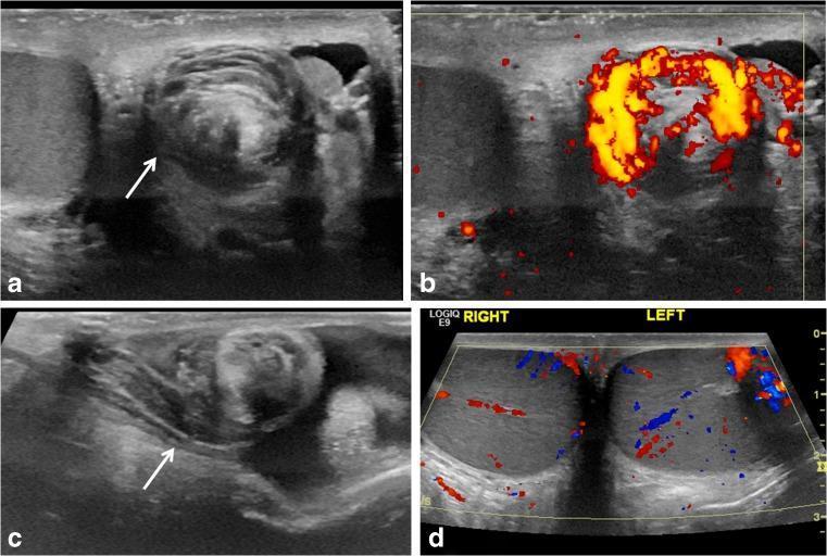
This composite of scrotal ultrasound images demonstrates the 'whirlpool sign,' a pathognomonic finding for testicular torsion. (a) Grayscale transverse ultrasound shows an eddy-like swirling of the spermatic cord (white arrow), indicating a spiral twist within the scrotal sac. (b) Power Doppler ultrasound of the same region reveals a concentric pattern of preserved blood flow within the vessels of the twisted cord, often seen in early or incomplete torsion. (c) Longitudinal grayscale ultrasound shows an abrupt spiral twisting of the spermatic cord at the level of the external inguinal ring (white arrow). (d) Transverse color Doppler ultrasound provides a bilateral comparison of the right and left testes; in this case of intermittent torsion, intratesticular vascular flow appears preserved and symmetrical bilaterally post-detorsion. These images illustrate key diagnostic markers for pediatric testicular torsion, emphasizing that the whirlpool sign is a direct sonographic indicator of cord rotation, even when intratesticular flow remains visible on Doppler interrogation.
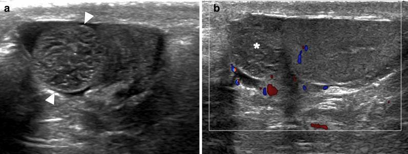
This dual-panel diagnostic ultrasound image illustrates a case of torsion of the appendix testis in a 9-year-old patient. Panel (a) is a grayscale ultrasound showing a well-defined, round, solid-appearing extratesticular lesion (arrowheads) located adjacent to the upper pole of the left testis. The lesion exhibits a mildly heterogeneous echotexture compared to the adjacent testicular parenchyma. Panel (b) shows a color flow Doppler ultrasound of the same region. The lesion (asterisk) demonstrates a complete absence of internal vascularity (avascularity), which is a key diagnostic feature of torsion. In contrast, normal blood flow signals (red and blue) are visible in the surrounding scrotal tissues and the adjacent testis. Additional secondary signs visible include minimal reactive hydrocele and mild scrotal wall thickening. This imaging presentation is characteristic of a torsed testicular appendage, a common cause of acute scrotum in the pediatric population, requiring differentiation from testicular torsion or epididymo-orchitis.
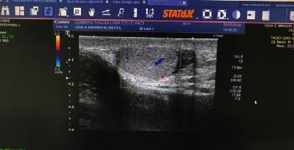
This diagnostic image is a scrotal ultrasound with Color Doppler of the left testicle. The grayscale (B-mode) imaging shows an oval-shaped testis with relatively homogeneous, isoechoic parenchymal echogenicity. The testicular capsule is clearly defined against the surrounding scrotal soft tissues. A Color Doppler sampling box is centered over the testicular parenchyma, demonstrating internal vascular flow represented by sparse red and blue signals, which indicate blood flow direction relative to the transducer. The user interface includes a vertical depth scale in centimeters on the left side of the scan window and a velocity color bar (set to 2.2 cm/s) for the Doppler settings. Textual annotations on the interface identify the anatomical region as 'TESTICULO ESQUERDO' (left testicle). This imaging modality is essential for evaluating testicular viability and perfusion in clinical scenarios such as suspected testicular torsion, orchitis, or scrotal trauma.
Testicular Torsion
Definition and Pathophysiology
- Intravaginal torsion - twisting of the cord within the tunica vaginalis; the common form in adolescents and adults. Almost always associated with the bell-clapper deformity - a congenitally abnormal fixation of the tunica vaginalis to the testicle that allows excessive testicular mobility.
- Extravaginal torsion - twisting of the entire cord including the tunica vaginalis; occurs in neonates (70% prenatal, 30% postnatal).
- Rosen's Emergency Medicine, p. 1401
- Bailey and Love's Short Practice of Surgery 28th Ed, p. 947
Epidemiology
- Incidence: approximately 1:4,000 males under 25 years of age
- Bimodal age distribution: neonatal period and puberty (peak 12-18 years)
- Can occur at any age
Clinical Features
| Feature | Finding |
|---|---|
| Pain onset | Sudden, severe - often awakens from sleep |
| Pain location | Scrotal, lower abdominal, or inguinal |
| Associated symptoms | Nausea and vomiting (common) |
| Testis position | High-riding, transversely oriented ("transverse lie") |
| Cremasteric reflex | Absent (most important sign, though not 100% specific) |
| Epididymis position | Displaced anteriorly from its normal posterior location |
| Skin changes | Erythema and oedema appear later |
-
Prior similar episodes suggest intermittent torsion in a predisposed testis
-
Because torsion can present with abdominal pain alone, the scrotum should be examined in ALL patients with abdominal pain
-
No single finding reliably rules in or rules out torsion
-
Rosen's Emergency Medicine, p. 1401
Differential Diagnosis of the Acute Scrotum
| Parameter | Testicular Torsion | Appendix Torsion | Epididymitis |
|---|---|---|---|
| Age | <1 yr, puberty | 7-14 years | Adult |
| Onset | Hours | 1-2 days | Days to weeks |
| Pain location | Entire testicle | Upper pole | Epididymis |
| Testis position | High-riding, transverse | Normal, vertical | Normal, vertical |
| Systemic symptoms | Nausea, vomiting | None | Possibly fever |
| Cremasteric reflex | Absent | Intact | Intact |
| Pyuria | Rare | No | Yes |
| Ultrasound | Hypoechoic, decreased/absent flow, cord twist | Focal lesion, normal flow | Hyperemic epididymis, increased flow |
| Treatment | Surgery (emergency) | Supportive/analgesia | Antibiotics |
- Rosen's Emergency Medicine, Table 85.9
Diagnostic Imaging
- Sensitivity 96-100%, specificity 84-95%
- Torsed testis: hypoechoic, enlarged, absent or decreased intratesticular flow
- Whirlpool sign - spiraling of the spermatic cord on ultrasound - is pathognomonic
- False negatives occur early in the course (when flow may still be present) and with intermittent torsion
- Imaging of the spermatic cord for twisting reduces false-negative rates
- MRI and radionuclide scanning are more time-consuming and have been largely replaced by ultrasound
Time-to-Salvage Relationship
| Time from onset | Testicular salvage rate |
|---|---|
| < 6 hours | >90% |
| 6-24 hours | Rapidly declining |
| > 24 hours | <10% (poor viability) |
Management
1. Immediate steps
- Emergent urology consultation - this is the first and most important step
- Provide analgesia: systemic or spermatic cord block
- Do not delay urological evaluation for imaging or any other maneuver
2. Manual detorsion (if urologist not immediately available)
- Rotate the affected testicle away from the midline (like "opening a book outward")
- If pain worsens or no relief: try the opposite direction (>1/3 of torsions are laterally oriented)
- If only partial relief: attempt further untwisting past 360 degrees (higher degrees of torsion may be present)
- Success = immediate symptom relief
- This is a temporizing measure only - surgical evaluation still mandatory afterward
3. Definitive surgical management
- Emergent scrotal exploration and detorsion
- After detorsion, testicular viability is assessed
- Only frankly gangrenous testes should be removed - some severely compromised testes survive; those that later atrophy are not harmful
- If viable: three-point fixation (orchiopexy) with non-absorbable sutures, or dartos pouch technique
- Because the anatomical anomaly (bell-clapper deformity) is bilateral, the contralateral testis must also be fixed at the same operation
- Always consent the patient preoperatively for possible orchiectomy
Neonatal torsion (special considerations)
-
Prenatal torsion: salvage rate nearly nonexistent; contralateral orchiopexy on urgent basis to prevent asynchronous contralateral torsion
-
Postnatal torsion (acute): salvage rate up to 44%; treat as a surgical emergency
-
The risk of extravaginal torsion is present only in the first few weeks of life
-
Rosen's Emergency Medicine, p. 1584-1590
-
Bailey and Love's Short Practice of Surgery 28th Ed, p. 947-962
-
Campbell-Walsh-Wein Urology, p. 1699-1704