
Scalp notes according to exam point of view for 5 marks
Reading File
Now let me search the medical library for scalp anatomy content.
Finding Sources
Searching the Web
scalp anatomy layers exam notes 5 marks surgery
Reading File
Finding Sources
Reading File
Reading File
Reading File
Reading File
Reading File
Loading Image
I now have all the information needed for comprehensive exam notes on the scalp. Here is the complete 5-mark answer:
SCALP - Exam Notes (5 Marks)
Definition & Extent
The scalp is the soft tissue covering the cranial vault. It extends:
- Anteriorly: Superciliary arches (supraorbital margins)
- Posteriorly: External occipital protuberance and superior nuchal lines
- Laterally: Zygomatic arch and external acoustic meatus
Layers of the Scalp - Mnemonic "SCALP"
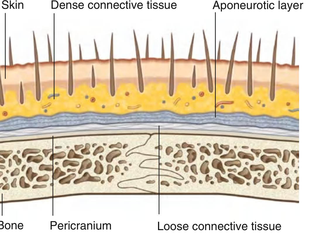
| Layer | Name | Key Features |
|---|---|---|
| S | Skin | Thickest skin of the body; contains hair follicles, sebaceous glands, sweat glands |
| C | Connective tissue (dense) | Fibro-fatty layer; contains arteries, veins, nerves; vessels held open by fibrous tissue - causes profuse bleeding when cut |
| A | Aponeurotic layer (Galea aponeurotica) | Tendon of occipitofrontalis muscle; connects frontal and occipital bellies |
| L | Loose areolar tissue | Allows movement of scalp; "danger space" - infections and blood spread freely here; can track to eyelids |
| P | Pericranium | Periosteum of outer skull; attached to bone but removable except at suture lines |
Key point: The first three layers (S, C, A) are tightly bound together and move as one unit - this is the "scalp proper." It is torn away in scalping injuries.
Arterial Supply
Five arteries supply the scalp - arise from periphery and anastomose freely at the vertex:
From Internal Carotid (via Ophthalmic artery):
- Supratrochlear artery - anterior forehead, near midline
- Supra-orbital artery - forehead and anterior scalp
From External Carotid artery:
- Superficial temporal artery - lateral scalp (largest supply)
- Posterior auricular artery - area posterior to ear
- Occipital artery - posterior scalp
Clinical point: Because arteries run in the dense connective tissue (layer 2) and are held open by fibrous tissue, scalp wounds bleed profusely and do not self-seal. Bleeding is predominantly arterial.
Venous Drainage
Veins accompany arteries and bear similar names:
- Supratrochlear + Supra-orbital veins → angular vein → facial vein → internal jugular vein
- Superficial temporal vein → retromandibular vein
- Posterior auricular vein → retromandibular vein
- Occipital vein → suboccipital venous plexus
Important: Scalp veins communicate with diploic veins of skull and dural venous sinuses via emissary veins - this is why scalp infections can spread intracranially (danger of erysipelas and osteomyelitis).
Nerve Supply (Sensory)
Anterior to ears and vertex - Branches of Trigeminal nerve (CN V):
- Supratrochlear nerve (V1) - medial forehead
- Supra-orbital nerve (V1) - lateral forehead
- Zygomaticotemporal nerve (V2) - small anterior temporal area
- Auriculotemporal nerve (V3) - temporal region and anterior to ear
Posterior to ears and vertex - Cervical nerves (C2, C3):
- Greater occipital nerve (C2 posterior ramus) - large area of posterior scalp up to vertex
- Lesser occipital nerve (C2 anterior ramus) - posterior to ear
- Great auricular nerve (C2, C3) - just posterior to ear
- Third occipital nerve (C3 posterior ramus) - small lower posterior area
Motor: Occipitofrontalis muscle is supplied by facial nerve (CN VII) - frontal belly by temporal branch, occipital belly by posterior auricular branch.
Lymphatic Drainage
| Region | Primary nodes | Secondary nodes |
|---|---|---|
| Posterior scalp | Occipital nodes | Upper deep cervical |
| Upper scalp (posterior to vertex) | Mastoid (retro-auricular) nodes | Upper deep cervical |
| Upper scalp (anterior to vertex) | Pre-auricular and parotid nodes | Deep cervical |
| Forehead | Submandibular nodes | - |
Clinical Significance (High-Yield)
| Feature | Explanation |
|---|---|
| Profuse bleeding | Dense fibrous tissue holds cut vessels open; prevents retraction |
| "Dangerous area" (Layer 4) | Loose areolar tissue - pus/blood spreads freely; can track to eyelids anteriorly |
| Scalping injury | First three layers (SCA) torn off as one unit |
| Subgaleal hematoma | Blood in loose areolar space (layer 4); crosses suture lines - limited only by attachments of galea |
| Cephalohematoma (neonates) | Blood between pericranium and skull; does NOT cross suture lines |
| Emissary veins | Connect scalp veins to dural sinuses; scalp infections can cause cavernous sinus thrombosis |
| Subaponeurotic abscess | Spreads widely under galea; treat urgently to prevent intracranial spread |
Sources: Gray's Anatomy for Students; P C Dikshit Textbook of Forensic Medicine and Toxicology
This is a shared conversation. Sign in to Orris to start your own chat.