
Meningitis signs in MRI
meningitis MRI signs findings radiology leptomeningeal enhancement
MRI Signs of Meningitis
1. Direct Signs
Leptomeningeal Enhancement (post-contrast T1 / CE-FLAIR)
- Pyogenic/viral meningitis: thin, smooth, diffuse enhancement
- TB/fungal/carcinomatous meningitis: thick, nodular, predominantly basal enhancement
- Best detected on post-contrast FLAIR rather than standard post-contrast T1, because CE-FLAIR suppresses normal vascular enhancement and makes pathological leptomeningeal enhancement more conspicuous.
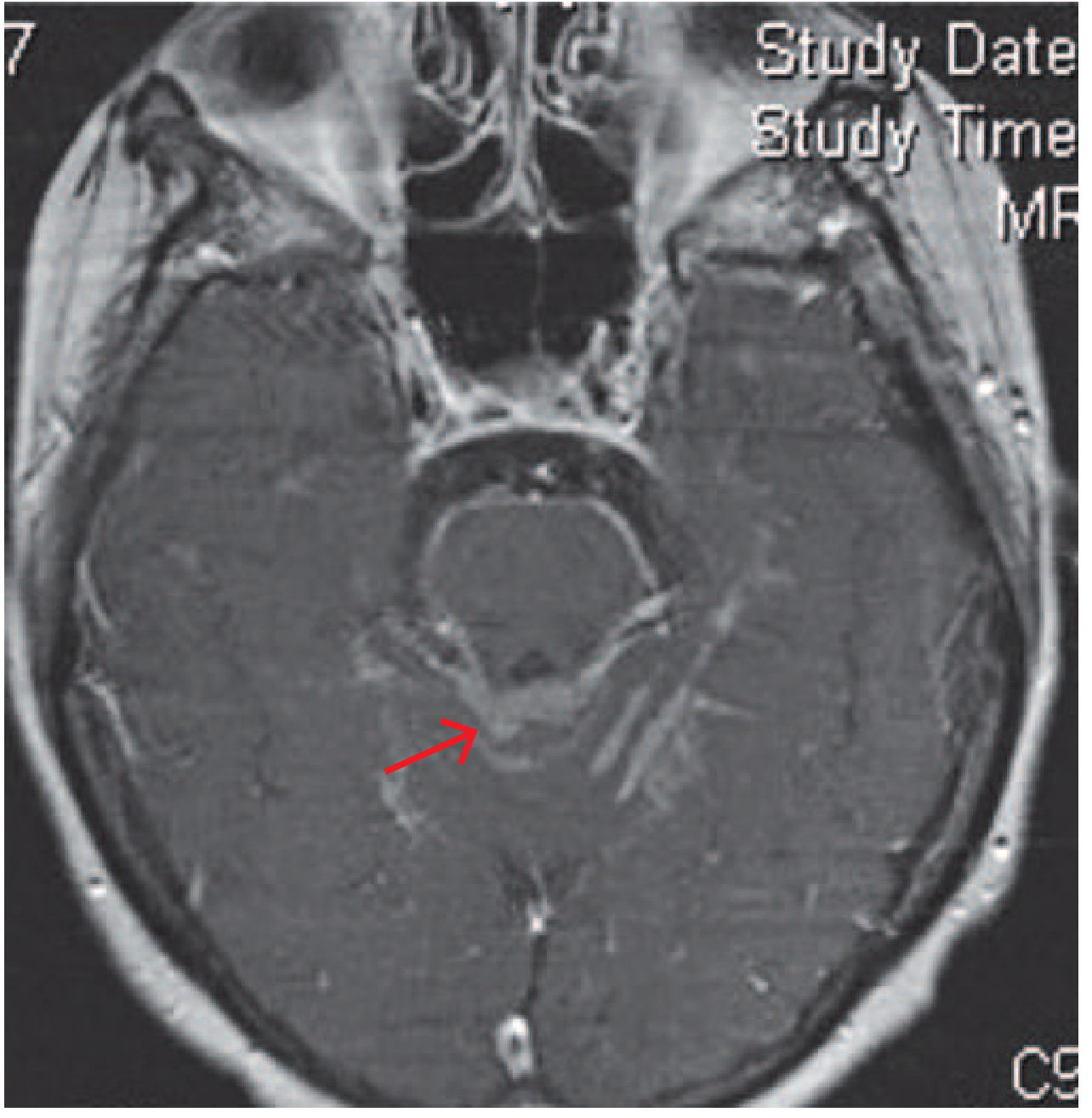
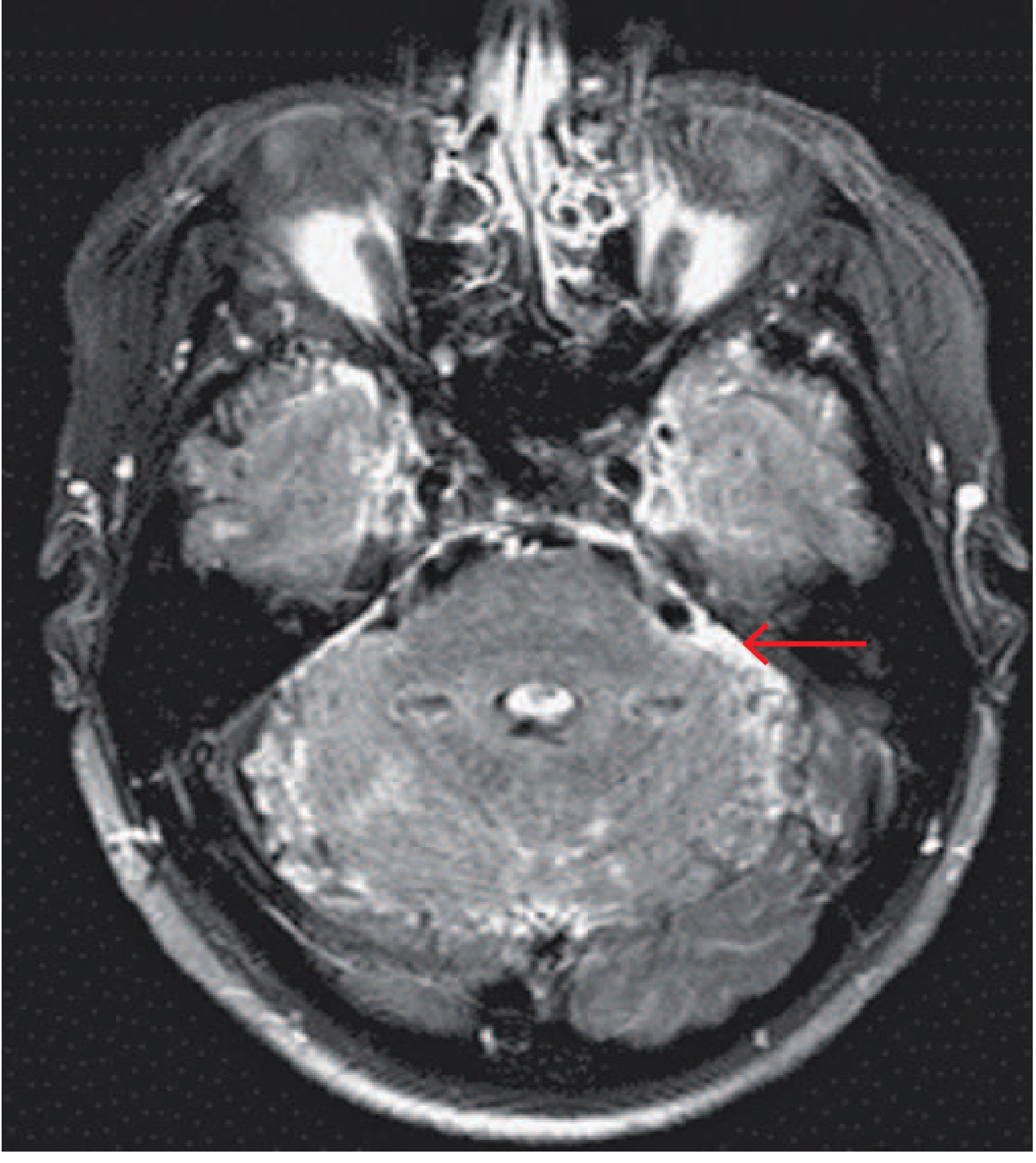
FLAIR Sulcal Hyperintensity (pre-contrast)
DWI Subarachnoid High Signal
2. MRI Sequences Summary
| Sequence | Key finding |
|---|---|
| Post-contrast T1 | Leptomeningeal enhancement |
| Post-contrast FLAIR | Most sensitive for leptomeningeal enhancement (suppresses vessels) |
| Pre-contrast FLAIR | Sulcal hyperintensity from raised CSF protein |
| DWI | Subarachnoid restricted diffusion (poor prognosis marker); also shows empyema/abscess |
| T2/FLAIR parenchyma | Cerebritis, edema, infarction |
| MRV | Venous sinus thrombosis |
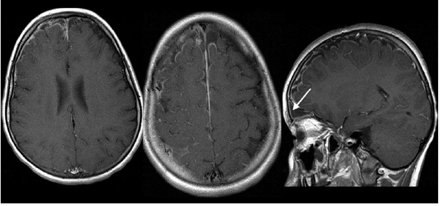
3. Complications - MRI Signs
| Complication | MRI Sign |
|---|---|
| Cerebritis | T2/FLAIR hyperintensity in cortex + white matter; gyral swelling; ill-defined enhancement |
| Brain abscess | Peripheral rim enhancement with central necrotic cavity (T1 dark, T2 bright); restricted diffusion in the core |
| Subdural effusion (sterile) | CSF-signal subdural collection, no enhancement |
| Subdural empyema | Intermediate T1 signal (protein-rich), restricted diffusion, pachymeningeal (dural) + leptomeningeal enhancement |
| Ventriculitis | Debris layered posteriorly in ventricles; ependymal T2 hyperintensity and contrast enhancement of the ependyma; can see intraventricular purulent material with DWI restriction |
| Hydrocephalus | Dilatation of ventricles - communicating type (blocked CSF absorption) most common; non-communicating if outlet foramina blocked |
| Infarction (arterial) | DWI restriction in large vessel territories OR perforator territories (basal ganglia, internal capsule) due to vasculitis of vessels traversing exudate |
| Venous sinus thrombosis | Loss of T2 flow void in sinus; expanded sinus; MRV filling defect; hemorrhagic venous infarct |
| Labyrinthitis ossificans (late) | T2 hypointensity in cochlear lumen post-meningitis |
4. Etiology-Specific Patterns
Bacterial (Pyogenic)
- Usually normal in uncomplicated cases
- When abnormal: thin smooth leptomeningeal enhancement, sulcal FLAIR signal
- Complications (empyema, abscess, ventriculitis) show restricted diffusion on DWI
- Infarcts in 30% of neonates - Grainger & Allison's Diagnostic Radiology
Tuberculous Meningitis
- Basal meningeal enhancement (interpeduncular cistern, middle cerebral artery cisterns) - hallmark
- Communicating hydrocephalus in 50% of adults, 85% of children
- Perforator territory infarcts (basal ganglia, internal capsule, brainstem)
- Tuberculomas: T1 low, T2 low (caseated) with rim or target-sign enhancement
- MRI superior to CT for basal enhancement, hydrocephalus, and infarcts
Fungal (Cryptococcus)
- Often unremarkable on MRI
- Gelatinous pseudocysts along Virchow-Robin spaces (basal ganglia, midbrain) - isointense to CSF, bilateral, symmetric
- Basal leptomeningeal enhancement when present
- Cryptococcomas = granulomas in basal ganglia, similar to tuberculomas
Carcinomatous / Chronic Meningitis
- Thick nodular leptomeningeal enhancement, often in basal cisterns
- More irregular vs. the thin smooth enhancement of acute infection
- Cranial nerve enhancement (follow nerves for thickening/signal)
Key Practical Points
- CE-FLAIR > CE-T1 for detecting leptomeningeal enhancement (suppresses background vascular signal)
- Normal MRI does not exclude meningitis - LP is required
- MRI before LP is indicated when: focal neurological signs, papilloedema, GCS <12, uncontrolled seizures
- DWI restriction in subarachnoid spaces = poor prognosis marker
- Basal enhancement + hydrocephalus + perforator infarcts = TB until proven otherwise
Cerebellar signs in tonsillar ectopia
Chiari I malformation tonsillar ectopia cerebellar signs clinical features
Cerebellar Signs in Tonsillar Ectopia (Chiari I Malformation)
Overview
- Compression of the cerebellar tonsils, caudal brainstem, and upper cervical cord at the foramen magnum
- Disruption of CSF flow dynamics at the craniocervical junction
- Syringomyelia from pressure-wave transmission into the central canal of the cord (in ~50-70% of symptomatic cases)
Age-Adjusted Normal Limits for Tonsillar Position
| Decade of Life | Upper Limit of Normal (below foramen magnum) |
|---|---|
| 1st (children) | 6 mm |
| 2nd - 3rd | 5 mm |
| 4th - 8th | 4 mm |
| 9th | 3 mm |
Cerebellar and Posterior Fossa Signs
1. Headache - "Chiari Headache"
- Suboccipital, pressing, continuous (waxing and waning, not episodic)
- May radiate behind the eyes or to the vertex
- Almost never hemicranial
- Triggered or worsened by Valsalva maneuvers - coughing, sneezing, laughing, straining, bearing down
- Exacerbations can be explosive rather than throbbing
2. Cerebellar Ataxia
- Gait ataxia with difficulty in tandem standing and walking
- Most typical finding on neurological exam: a vestibular-generated dysequilibrium
- Limb ataxia may be present but is less prominent than gait ataxia
- Favored by Chiari over syrinx as the cause: when ataxia is present alongside nystagmus - Adams & Victor's Principles of Neurology, 12th Ed.
3. Nystagmus - The Most Specific Ocular Sign
| Type | Notes |
|---|---|
| Downbeat nystagmus (DBN) | Hallmark - both spontaneous and positional; may have torsional component; worse on lateral gaze |
| Positional nystagmus | |
| Primary-position unidirectional horizontal nystagmus | |
| Periodic alternating nystagmus (PAN) | |
| Rebound nystagmus (including torsional rebound) | |
| Convergence nystagmus | |
| Divergence nystagmus / divergence paralysis |
Key caveat: Despite DBN being the "classic" finding, it is rarely seen even with tonsillar herniation as striking as 20 mm. Nystagmus is difficult to appreciate even with Fresnel lens examination - Bradley & Daroff's Neurology, 12th Ed.
4. Other Ocular Motor Abnormalities
- Impaired smooth pursuit (especially downward gaze)
- Impaired VOR cancellation
- Saccadic dysmetria (hypermetric or hypometric saccades)
- Skew deviation - accentuated or alternating on lateral gaze
- Internuclear ophthalmoplegia (INO)
- Square-wave jerks
5. Oscillopsia and Vestibular Symptoms
- Oscillopsia brought on or accentuated by head movements
- Dizziness and vertigo (75.8% prevalence in symptomatic CM-I vs 17-18% in tonsillar ectopia without CM-I criteria)
- Cervical pain with head movement
Long Tract and Brainstem Signs (When Medulla Descends)
| System | Signs |
|---|---|
| Corticospinal | Spastic quadriparesis/paraparesis; prominent in lower limbs |
| Sensory | Sensory tract involvement in lower extremities |
| Cranial nerves (lower) | Dysphagia, hoarseness, palatal weakness (IX, X involvement) |
| Respiratory | Sleep-related breathing disorders, central apnea |
| Lower cranial nerve | Dysarthria, tongue wasting (if syringobulbia co-exists) |
| Sphincter | Bladder and bowel dysfunction (usually from syrinx) |
Syringomyelia Overlay (CM-I Associated)
- Cape-like dissociated sensory loss (pain and temperature lost, light touch preserved) over shoulders and arms
- Wasting and weakness of the hands/upper limbs
- Painless burns or injuries to the hands (classic presentation)
- Segmental areflexia in upper limbs with hyperreflexia below
Special Points
- Children <3 years: vomiting and gastric reflux may be the sole manifestations
- Pediatric presentation: oropharyngeal dysfunction, scoliosis, failure to thrive
- Neurological exam is often completely normal - even with significant herniation
- Symptoms are typically exacerbated by any action that increases intrathoracic or intraabdominal pressure (Valsalva)
- At least a quarter of symptomatic patients first develop symptoms following minor head or neck injury
MRI Evaluation
- Best sequence: T2-weighted sagittal MRI of brain + cervical spine
- Assesses: tonsil position, posterior fossa volume, syrinx, brainstem compression
- Phase-contrast (cine) MRI: evaluates CSF flow at foramen magnum - pulsatile systolic tonsillar descent and obstruction of CSF flow are positive findings predictive of surgical response
- Dynamic MRI useful when clinical presentation is equivocal
Tests for cerebellar signs
clinical examination tests cerebellar signs ataxia dysmetria assessment neurology
Clinical Tests for Cerebellar Signs
Mnemonic: DANISH
| Letter | Sign |
|---|---|
| D | Dysdiadochokinesia |
| A | Ataxia (gait/truncal) |
| N | Nystagmus |
| I | Intention tremor |
| S | Slurred (scanning) speech |
| H | Hypotonia / past-pointing |
Domain 1: Gait and Stance
Normal Gait Observation
- Variable stride length
- Veering to one side (toward the side of the lesion in unilateral disease)
- Wide-based gait - compensatory in moderate-severe ataxia
- Staggering, inability to maintain a straight line
Tandem (Heel-to-Toe) Walking
Single-leg Stance / Hopping
Detecting subtle difficulty: observe patients running or walking up/down stairs - these stress the system more than level walking - Bradley & Daroff's Neurology in Clinical Practice
Domain 2: Stance and Truncal Stability
Romberg's Test
- Stand with feet together, arms by sides
- First with eyes open, then eyes closed
- Positive Romberg = patient falls when eyes are closed but not open → implies proprioceptive or vestibular deficit (not pure cerebellum)
- Cerebellar lesion: patient is unsteady with both eyes open and closed (the cerebellum cannot integrate the remaining senses - it is a processing, not sensing, problem)
Balance requires input from proprioception, vestibular system, and vision. At least 2 are needed. Romberg removes vision. If the patient falls only with eyes closed, the cerebellar integration is intact but one of the other two systems is deficient - Shambaugh Surgery of the Ear
Sharpened (Tandem) Romberg
Sitting Without Back Support
Domain 3: Upper Limb (Hand) Tests
1. Finger-Nose-Finger Test (FNF)
- Their own nose
- The examiner's outstretched index finger
- Intention tremor - oscillation that increases as the target is approached (pathognomonic of cerebellar disease)
- Past-pointing (dysmetria) - over- or undershooting the target
- Decomposition of movement - jerky, broken up action
- Perform with eyes open and then eyes closed (closed removes visual correction and exaggerates cerebellar dysmetria; worsening = cerebellar; remaining the same = proprioceptive problem)
2. Finger Chase Test
3. Dysdiadochokinesia Test (Rapid Alternating Movements)
- Rate - normal is rapid and regular
- Rhythm - should be uniform
- Amplitude - should be consistent
4. Rebound Test (Stewart-Holmes Sign)
5. Hyperdysmetria / Overshoot on Resistance
Domain 4: Lower Limb Tests
Heel-Shin Test
- Patient lies supine
- Lifts one leg, places the heel on the opposite knee, then slides it down the shin to the ankle
- Repeat several times, then with the other leg
Domain 5: Speech
Scanning/Staccato Speech
- Slow with irregular force and rhythm
- Unnecessary hesitations between words
- Words broken into separate syllables (scanning or syllabic speech)
- May sound explosive or slurred
- Distinct from dysarthria of upper/lower motor neuron origin
Domain 6: Eye Movement Examination
| Test | What to Look For | Implication |
|---|---|---|
| Fixation | Square-wave jerks (small involuntary saccades breaking fixation) | Common in Friedreich ataxia |
| Smooth pursuit | Saccadic (cogwheel) pursuit instead of smooth tracking | Cerebellar hemisphere dysfunction |
| Saccades | Dysmetric (hypo- or hypermetric) saccades | Multiple ataxia types |
| Gaze-evoked nystagmus | Nystagmus in eccentric gaze (end-gaze) | Many cerebellar disorders |
| Downbeat nystagmus (DBN) | Fast phase beating downward, worse on lateral gaze | Cervicomedullary junction / flocculus |
| Rebound nystagmus | Nystagmus on returning to primary gaze after sustained lateral gaze | Cerebellar |
| VOR cancellation | Ask patient to fix on their own thumb while rotating in a chair - inability to suppress VOR | Flocculonodular lobe |
SARA Scale (Formal Clinical Rating)
| Domain | Test |
|---|---|
| 1 | Gait |
| 2 | Stance |
| 3 | Sitting |
| 4 | Speech disturbance |
| 5 | Finger chase |
| 6 | Nose-finger test |
| 7 | Fast alternating hand movements |
| 8 | Heel-shin slide |
Summary: Localizing Value
| Sign | Localization |
|---|---|
| Gait ataxia + truncal sway | Vermis / midline (e.g., alcohol, medulloblastoma) |
| Limb dysmetria (ipsilateral) | Cerebellar hemisphere (same side as lesion) |
| Intention tremor | Cerebellar outflow (dentate nucleus / superior cerebellar peduncle) |
| Downbeat nystagmus | Flocculus / cervicomedullary junction |
| Dysdiadochokinesia | Cerebellar hemisphere |
| Scanning speech | Vermis / bilateral hemispheres |
A key principle: cerebellar signs are ipsilateral to the lesion (unlike upper motor neuron signs which cross). The patient veers toward the side of the lesion. - Bradley & Daroff's Neurology in Clinical Practice
Chiari malformation
Chiari malformation types classification pathophysiology management 2024
Chiari Malformation
Historical Background
Classification
| Type | Definition | Key Associations |
|---|---|---|
| Type I (CM-I) | Caudal displacement of cerebellar tonsils ≥5 mm below foramen magnum; conical "peg-like" shape | Syringomyelia (50-70%); most common type; presents in adults |
| Type II (CM-II) | Displacement of cerebellar vermis, tonsils, fourth ventricle, and medulla below foramen magnum; "beaking" of midbrain | Near 100% association with myelomeningocele; hydrocephalus |
| Type III | Herniation of cerebellum into a high cervical myelomeningocele | Extremely rare |
| Type IV | Cerebellar hypoplasia/agenesis alone | Probably unrelated to other types |
| Chiari 0 | Syringomyelia with CSF flow obstruction but no tonsillar descent | Controversial subtype |
| Chiari 1.5 | Features of CM-I + brainstem herniation, but without myelomeningocele | Uncommon |
Chiari I Malformation - Deep Dive
Epidemiology
- Prevalence ~0.1-1% of the general population (Goldman-Cecil: ~1% on cranial MRI)
- Women affected 3x more often than men
- Hereditary tendency - transmissibility rate approaching 12%
- Majority of patients are asymptomatic; symptoms usually emerge in adolescence or early adulthood
Pathophysiology - Five Mechanisms of Tonsillar Herniation
| Mechanism | Examples |
|---|---|
| "Squeeze down" - Cranial constriction | Classic CM-I, craniosynostosis, achondroplasia, Paget disease |
| "Pull down" - Spinal cord tethering | Tethered cord syndrome, CM-II |
| "Shake down" - Cranial settling | Ehlers-Danlos syndrome, connective tissue disorders, craniocervical instability |
| "Push down" - Intracranial hypertension | Hydrocephalus, posterior fossa tumors/cysts, subdural hematoma |
| "Suck down" - Intraspinal hypotension | CSF leaks, prolonged lumboperitoneal shunting, dural ectasia |
MRI Findings
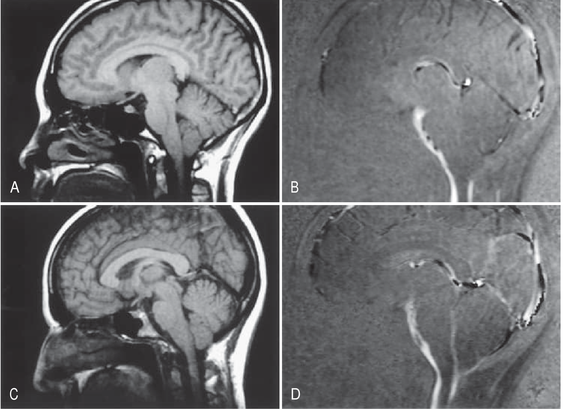
Chiari I - T2 sagittal MRI showing low-lying tonsils (8 mm below foramen magnum) and syringomyelia
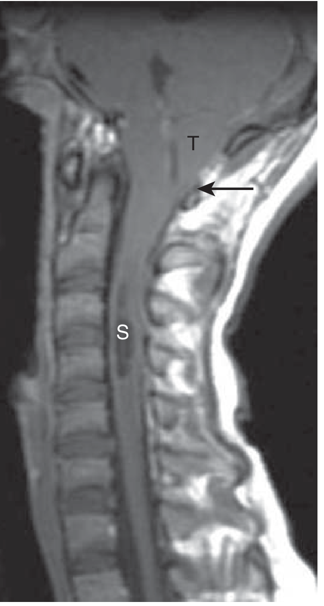
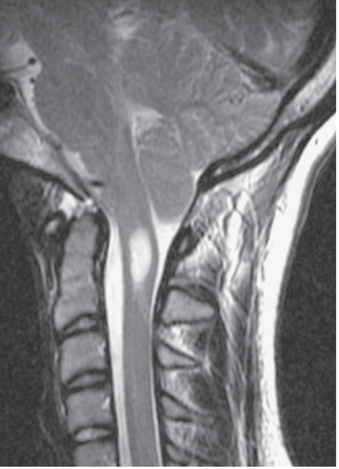
Key MRI Features
- Best sequence: T2-weighted sagittal brain + cervical spine
- Tonsil descent measured from McRae line (basion to opisthion)
- Tonsils appear peg-shaped (pointed, not rounded) in CM-I
- Crowded foramen magnum; obliteration of cisterna magna
- Cervical syrinx (central T2-bright cavity in cord)
- Phase-contrast (cine) MRI: obstruction of CSF flow at foramen magnum is a positive finding predicting surgical response
Age-Adjusted Normal Limits
| Decade | Max normal descent |
|---|---|
| 1st | 6 mm |
| 2nd-3rd | 5 mm |
| 4th-8th | 4 mm |
| 9th | 3 mm |
Clinical Features (CM-I)
Symptom Frequency (n=364 patients, Milhorat et al. 1999)
| Symptom | % of Patients |
|---|---|
| Suboccipital headache (often retro-orbital; exertional/postural) | 81% |
| Ocular disturbances (floaters, blurring, photophobia, diplopia) | 78% |
| Acoustic/vestibular (dizziness, disequilibrium, tinnitus) | 74% |
| Dysesthesias (numbness, tingling, burning) | 59% |
| Chronic fatigue | 58% |
| Bulbar and coordinative problems (dysphagia, dysarthria, sleep apnea, tremor) | 52% |
| Segmental pain | 44% |
| Impaired memory/concentration | 39% |
| Cervical pain | 34% |
| Low back pain | 24% |
| Urinary incontinence | 17% |
The "Chiari Headache"
- Suboccipital, pressing, continuous (not episodic, not hemicranial)
- Radiates behind eyes or to vertex
- Exacerbated by Valsalva: coughing, sneezing, laughing, straining, bearing down
- Exacerbations can be explosive - not throbbing
- May respond to carbonic anhydrase inhibitors (topiramate, acetazolamide)
- Does NOT respond to triptans or beta-blockers
Neurological Syndromes in CM-I (Adams & Victor)
- Raised ICP - headache
- Progressive cerebellar ataxia
- Progressive spastic quadriparesis (long tract compression)
- Downbeating nystagmus
- Cervical syringomyelia syndrome - segmental amyotrophy + dissociated sensory loss (pain/temperature lost, light touch preserved) in hands/arms
Important mimics: multiple sclerosis, foramen magnum tumor. Onset may be acute after neck hyperextension (dental work, hairdressing, chiropractic manipulation).
Children
- <3 years: vomiting and gastric reflux may be the sole symptoms
- Older children: oropharyngeal dysfunction, scoliosis, failure to thrive
Chiari II Malformation
- Nearly always associated with myelomeningocele (lumbosacral)
- Involves herniation of vermis, tonsils, fourth ventricle, and medulla (brainstem descent)
- Midbrain "beaking" on sagittal MRI (posterior midbrain elongation)
- Foramen magnum is enlarged (unlike CM-I where it is constricted)
- Hydrocephalus is invariably associated
CM-II Clinical Features
- Lower cranial nerve abnormalities: laryngeal stridor, tongue fasciculations, sternomastoid paralysis (head lag on pull-to-sit), facial weakness, deafness, bilateral abducens palsies
- Progressive hydrocephalus
- Cerebellar signs emerge later in childhood/adolescence
- Seizures, cognitive delay, paraplegia, sphincter dysfunction (from associated myelomeningocele)
Posterior Cranial Fossa Morphology Comparison
| Condition | Occipital Bone | Posterior Fossa Volume | Foramen Magnum |
|---|---|---|---|
| CM-I classic | Small | Small | Small |
| CM-II (with myelodysplasia) | Small | Small | Large |
| CM-I with tethered cord | Normal | Normal | Large |
| CM-I with cranial settling | Normal | Normal | Normal |
Management
CM-I
| Approach | Indication |
|---|---|
| Observation | Asymptomatic incidental finding |
| Medical | Headache: carbonic anhydrase inhibitors, NSAIDs, head elevation, avoidance of Valsalva activities |
| Surgical - Posterior fossa decompression (PFD) | Significant functional impairment from persistent headache despite medical therapy OR progressive syringomyelia |
| Surgery NOT indicated for | Chronic fatigue alone, musculoskeletal pain, vertigo alone, prophylaxis |
- Headache and neck pain respond best to surgery
- Ataxia and nystagmus tend to persist after decompression
- Syrinx usually resolves or stabilizes after PFD
CM-II
- Surgical repair of myelomeningocele
- VP or ventriculoperitoneal shunting for hydrocephalus
- Bladder management (intermittent catheterization)
- Prognosis depends on severity of brain abnormalities
Meningitis vs encephalitis clinical signs and symptoms
meningitis vs encephalitis clinical signs symptoms differences diagnosis
Meningitis vs Encephalitis: Clinical Signs and Symptoms
Conceptual Distinction
| Meningitis | Encephalitis | |
|---|---|---|
| Definition | Inflammation of the meninges (leptomeninges - arachnoid + pia mater) | Inflammation of the brain parenchyma itself |
| Key difference | Meningeal irritation signs dominate; brain function relatively preserved early | Brain dysfunction is the cardinal feature - altered consciousness, seizures, focal deficits |
| Overlap | When both coexist = meningoencephalitis |
"Encephalitis is inflammation of the brain parenchyma, characterized by neurologic dysfunction early in the course of illness." - Frameworks for Internal Medicine
Meningitis - Clinical Features
Core Triad (present together in only ~44% of adults)
- Fever
- Headache - severe, generalized (87% of bacterial cases)
- Neck stiffness (nuchal rigidity) (83% of bacterial cases)
Symptoms (Bacterial Meningitis - 696 cases, van de Beek et al.)
| Symptom | Frequency |
|---|---|
| Headache | 87% |
| Neck stiffness | 83% |
| Nausea | 74% |
| Full fever + neck stiffness + AMS triad | 44% |
| Focal neurological deficits | 33% |
| Aphasia | 23% |
| Hemiparesis | 7% |
Classic Meningeal Signs (Signs of Meningeal Irritation)
1. Nuchal Rigidity
- Resistance to passive neck flexion (forward bending)
- Specific to meningitis - resistance is predominantly on forward flexion only
- Rotation from side to side is preserved (helps distinguish from cervical spondylosis, which resists movement in all directions)
- Disappears in deep coma
- Stiffness in the first few degrees of flexion is more specific; stiffness in the latter part is more sensitive but less specific - Adams & Victor
2. Kernig's Sign
- Hip flexed to 90°, then attempt to extend the knee
- Positive: inability/pain on extending the leg (hamstring spasm due to nerve root irritation)
- Sensitivity: ~30% for bacterial meningitis
3. Brudzinski's Sign
- Passive forward flexion of the neck causes involuntary flexion of the hips and knees
- Part of a "flexor protective reflex"
- Sensitivity: ~30% for bacterial meningitis
Both Kernig and Brudzinski signs have low sensitivity (~30%) individually but high specificity. Their absence does not exclude meningitis - Goldman-Cecil Medicine
4. Jolt Accentuation
- Horizontal rotation of the head at 2-3 Hz worsens the headache
- Sensitivity ~97%, specificity lower - useful screening sign
Other Signs by Etiology
| Finding | Suggests |
|---|---|
| Petechial/purpuric rash (lower body) | Meningococcemia - treat immediately |
| Waterhouse-Friderichsen syndrome (adrenal hemorrhage, shock, DIC) | Fulminant meningococcal septicemia |
| Herpes labialis | Pneumococcal meningitis |
| Preceding ear/sinus/lung infection | Pneumococcal or H. influenzae |
| Battle's sign (bruising behind ear), CSF rhinorrhea, periorbital ecchymoses | Basilar skull fracture → meningitis |
| Hyponatremia (SIADH) | H. influenzae meningitis |
Complications Producing Additional Signs
- Cranial nerve palsies (III, IV, VI, VII) - 5-10% of adults
- Seizures (focal or generalized) - 20-30%
- Papilledema - raised ICP
- Sensorineural deafness (CN VIII, cochlear spread)
- Altered mental status progressing to stupor and coma as disease advances
Encephalitis - Clinical Features
Diagnostic Criteria (IDSA/International Encephalitis Consortium)
- Fever (>38°C) within 72 hours
- New-onset seizures
- New focal neurological findings
- CSF pleocytosis (>5 WBC/µL)
- Abnormal neuroimaging (new parenchymal changes)
- Abnormal EEG consistent with encephalitis
Core Clinical Features
| Feature | Notes |
|---|---|
| Altered consciousness | The cardinal sign - confusion, disorientation, personality change, stupor, coma; present in 70-90% of HSV encephalitis |
| Fever | Present in ~75-80% of HSV encephalitis |
| Headache | 60-70% |
| Seizures (focal or generalized) | 55% of HSV encephalitis; focal seizures are particularly suggestive |
| Personality/behavioral change | 60% - disinhibition, aggression, bizarre behavior; especially frontal/temporal lobe involvement |
| Memory disturbance | 35% - anterograde or retrograde amnesia |
| Aphasia | 40-60% (HSV - temporal lobe) |
| Motor deficits | 40% - hemiparesis, focal weakness |
| Focal neurological signs | Reflect area of brain involved |
| Photophobia | Present (shared with meningitis) |
| Neck stiffness | May be present if meninges also involved |
Side-by-Side Comparison
| Feature | Meningitis | Encephalitis |
|---|---|---|
| Primary pathology | Meningeal inflammation | Brain parenchymal inflammation |
| Fever | ✓ Very common | ✓ Very common |
| Headache | ✓ Very common (87%) | ✓ Common (60-70%) |
| Neck stiffness | ✓✓ Cardinal sign (83%) | +/- (only if meninges involved) |
| Kernig/Brudzinski | ✓ (low sensitivity ~30%) | Absent (unless meningoencephalitis) |
| Photophobia/phonophobia | ✓ Common | +/- |
| Altered consciousness | Late sign (as disease progresses) | Early, cardinal feature |
| Seizures | 20-30% (complication) | 55% (prominent early feature) |
| Focal neurological signs | 33% (complication) | Common, often early |
| Personality/behavioral change | Absent | 60% - characteristic |
| Aphasia | 23% (complication) | 40-60% (temporal involvement) |
| Memory disturbance | Absent | 35% |
| Level of consciousness | Relatively preserved initially | Impaired early |
| EEG | Normal or diffuse slowing | Focal slowing / periodic lateralized discharges (HSV) |
| MRI parenchyma | Usually normal (meningeal enhancement) | Parenchymal T2 changes (temporal lobes in HSV) |
| Rash (petechiae) | Meningococcemia | Absent |
CSF Comparison
| Parameter | Bacterial Meningitis | Viral Meningitis | Viral Encephalitis |
|---|---|---|---|
| Opening pressure | ↑↑ (>250 mm H₂O) | Normal/slight ↑ | Normal/slight ↑ |
| Appearance | Turbid/cloudy | Clear | Clear |
| WBC | >1000/µL (neutrophils) | 10-500 (lymphocytes) | 10-500 (lymphocytes) |
| Protein | ↑↑ (>1 g/L) | Slight ↑ | Slight ↑ |
| Glucose | ↓ (<2.2 mmol/L; CSF:serum <0.4) | Normal | Normal (or slight ↓) |
| Gram stain | + in 60-90% | Negative | Negative |
Key Distinguishing Principle
Thyroid eye signs
thyroid eye disease Graves ophthalmopathy named clinical signs lid retraction proptosis
https://eyewiki.org/Thyroid_Eye_Disease
NOSPECS classification thyroid eye disease clinical activity score CAS staging
Thyroid Eye Signs (Thyroid Eye Disease / Graves' Ophthalmopathy)
Background
Pathophysiology in Brief
| Tissue Target | Effect |
|---|---|
| Orbital fibroblasts / adipocytes | Expansion of orbital fat → proptosis |
| Extraocular muscles | Inflammation → fibrosis → restrictive myopathy |
| Eyelid tarsal muscles (Müller) | Sympathetic overactivation → lid retraction |
| Optic nerve at orbital apex | Compressed by enlarged muscles → optic neuropathy |
The Complete List of Named (Eponymous) Signs
Eyelid Signs (Upper Lid)
| Eponymous Sign | Description | Mechanism |
|---|---|---|
| Dalrymple sign | Upper eyelid retraction - sclera visible above the limbus (white scleral show at the top) | Most common sign in TED (~74-90%); Müller muscle sympathetic overactivity + levator fibrosis |
| Von Graefe sign | Lid lag on downgaze - upper lid lags behind the globe as the patient looks downward | Levator fibrosis / tethering; failure of smooth lid descent |
| Boston sign | Jerky, irregular movements of the upper eyelid on downgaze | Fibrotic lid tethering causing irregular descent |
| Stellwag sign | Incomplete and infrequent blinking + staring appearance | Lid retraction + sympathetic overactivity |
| Grove sign | Resistance to pulling the retracted upper lid downward | Fibrosis of the levator palpebrae |
| Gifford sign | Difficulty in everting the upper eyelid | Lid stiffness from infiltration |
| Gellineck sign | Abnormal pigmentation of the upper eyelid | Melanin deposition |
Eyelid Signs (Lower Lid)
| Sign | Description |
|---|---|
| Enroth sign | Lower eyelid edema - puffiness of the lower lid |
| Griffith sign | Lower lid lag on upgaze - lower lid does not follow the globe upward |
Conjunctival Signs
| Sign | Description |
|---|---|
| Goldzeiher sign | Conjunctival injection (redness), especially over the insertions of rectus muscles |
| Chemosis | Conjunctival edema - boggy, gelatinous appearance |
Extraocular Muscle Signs
| Sign | Description |
|---|---|
| Möbius sign | Poor or absent convergence |
| Ballet sign | Restriction/paralysis of one or more extraocular muscles |
| Suker sign | Poor fixation in abduction |
| Jendrassik sign | Paralysis of all extraocular muscles (severe) |
Pupillary Signs
| Sign | Description |
|---|---|
| Knies sign | Uneven pupillary dilation in dim light |
| Cowen sign | Jerky pupillary light reaction |
Generalized / Other Signs
| Sign | Description |
|---|---|
| Vigouroux sign | Eyelid fullness (periorbital soft tissue swelling) |
| Joffroy sign | Absent forehead creases on superior gaze (frontalis fails to contract because the eye can already see upward without frontalis help, due to lid retraction) |
| Rosenbach sign | Fine tremor of the closed eyelids |
The Four Clinical Domains (Critical Sequelae)
1. Eyelid Disorders
- Lid retraction (Dalrymple - #1 most common sign)
- Lid lag (Von Graefe)
- Lagophthalmos - inability to fully close the eyes (exposure risk)
- Staring / wide-eyed appearance
- Reduced blink rate (Stellwag)
- Periorbital edema
2. Ocular Surface Disorders (Active Phase)
- Conjunctival injection (Goldzeiher)
- Chemosis (conjunctival edema)
- Exposure keratopathy - from lagophthalmos and incomplete blinking
- Gritty sensation, photophobia, tearing, dry eye
- Corneal ulceration (sight-threatening)
3. Motility Disorders / Extraocular Muscle Disease
- Restrictive myopathy - "tight" not "weak" muscle (fibrosis)
- Muscle involvement order - mnemonic: "I'M SO" (in decreasing frequency):
- I - Inferior rectus (most common → hypotropia, elevation deficit)
- M - Medial rectus (→ esotropia, abduction deficit)
- S - Superior rectus
- O - Obliques (least common)
- Lateral rectus is rarely affected; XT in TED should raise suspicion for co-existing myasthenia gravis
- Vertical diplopia (asymmetric IR involvement) is the most common presentation
- Diplopia often worse in the morning and improves through the day - Localization in Clinical Neurology
4. Optic Neuropathy (Sight-Threatening)
- Caused by compression of optic nerve at orbital apex by enlarged extraocular muscles
- Signs: ↓ visual acuity, ↓ colour vision, RAPD (relative afferent pupillary defect), visual field defect
- Proptosis alone can stretch the optic nerve
- Rare but serious - present in 3-5% of TED cases
Proptosis (Exophthalmos)
- Present in up to one-third of thyroid patients
- Purely axial (straight forward) - distinguishes from other orbital masses
- Measured with Hertel exophthalmometer (normal <21 mm; asymmetry >2 mm significant)
- Increased resistance to retropulsion on pressing the globe back
- Most common cause of bilateral proptosis in adults
Disease Phases (Rundle Curve)
| Phase | Duration | Features |
|---|---|---|
| Active/inflammatory | 6-18 months | Proptosis, retraction, chemosis, injection, pain |
| Static | Variable | Inflammation decreases, minimal improvement |
| Quiescent/fibrotic | Chronic | Fixed restrictive changes; appearance may not fully normalise |
Clinical Activity Score (CAS) - 7-Point Scale
- Spontaneous orbital pain
- Gaze-evoked orbital pain
- Eyelid swelling (due to active TED)
- Eyelid erythema
- Conjunctival redness
- Chemosis
- Inflammation of caruncle or plica
NOSPECS Classification
| Class | Findings |
|---|---|
| N | No symptoms or signs |
| O | Only signs (lid retraction, stare, lid lag) |
| S | Soft tissue involvement (edema, injection, chemosis) |
| P | Proptosis (>3 mm above normal) |
| E | Extraocular muscle involvement (diplopia) |
| C | Corneal involvement |
| S | Sight loss (optic neuropathy) |
Urgent Referral Criteria
- Rapid deterioration in visual acuity
- Visual acuity <6/18
- Decreased colour vision
- RAPD
- Corneal ulceration
- Ocular nerve palsies
- Unacceptable cosmetic progression
Thalassemia
thalassemia alpha beta types pathophysiology clinical features treatment 2024
Thalassemia
Overview
- Decreased red cell production (ineffective erythropoiesis)
- Decreased red cell lifespan (hemolysis from unpaired globin chain precipitation)
Genetics and Molecular Basis
| Feature | α-Thalassemia | β-Thalassemia |
|---|---|---|
| Gene | HBA1 & HBA2 on chromosome 16 | HBB on chromosome 11 |
| Gene copies | 4 α-globin genes (2 per chromosome) | 2 β-globin genes (1 per chromosome) |
| Mutation type | Mainly deletions | Mainly point mutations (>100 known) |
| Mutation classes | - | β⁰ (absent synthesis) / β⁺ (reduced synthesis) |
β-Thalassemia Mutations (3 classes)
- Splicing mutations - most common cause of β⁺; create ectopic splice sites in introns
- Promoter region mutations - reduce transcription 75-80%; cause β⁺
- Chain terminator mutations (nonsense / frameshift) - most common cause of β⁰; no functional β-globin produced - Robbins & Cotran Pathologic Basis of Disease
Pathogenesis of β-Thalassemia
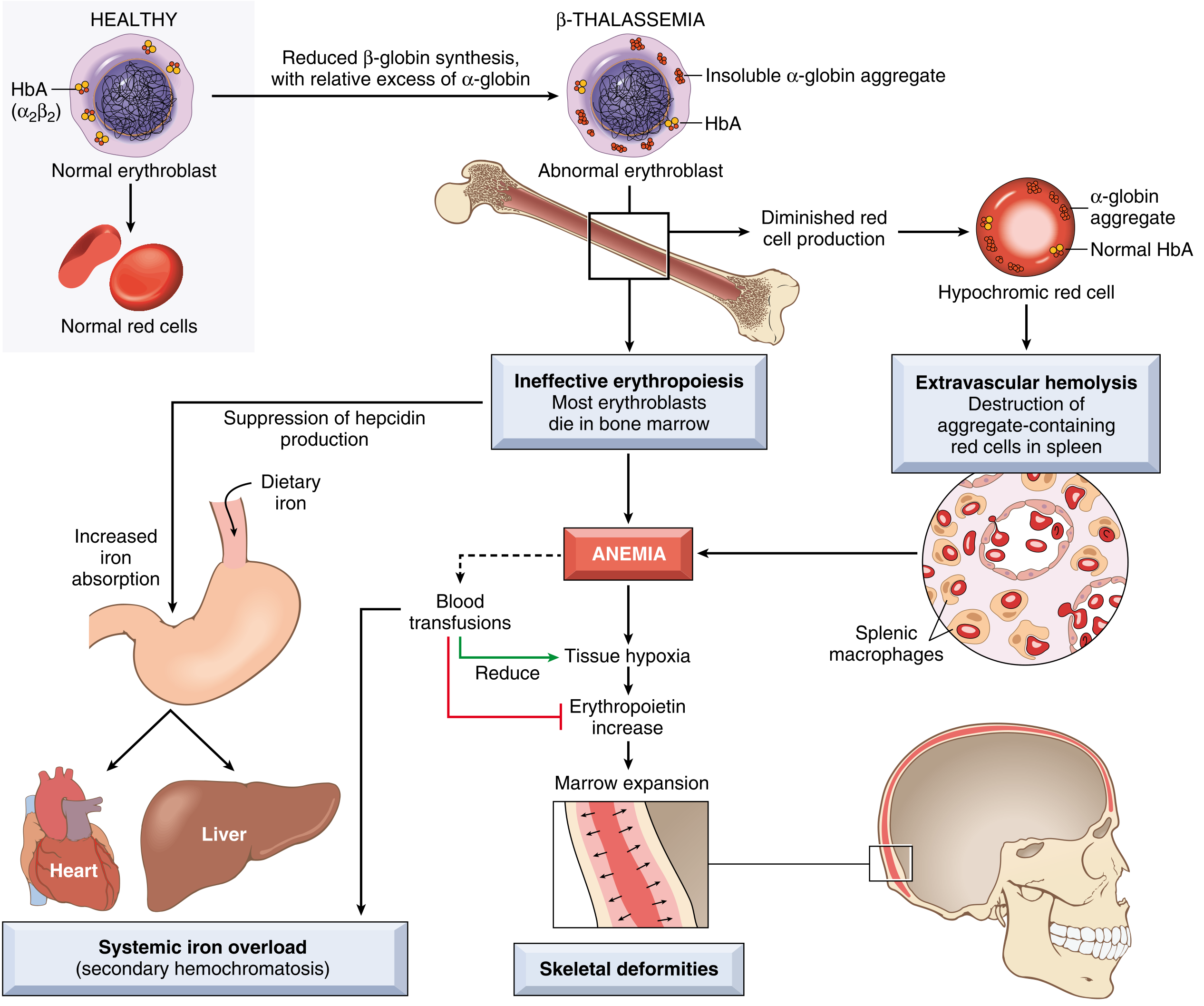
- Reduced β-globin → relative excess of α-globin
- Unpaired α-chains are insoluble - precipitate within erythroblasts
- → Ineffective erythropoiesis (most erythroblasts die in marrow)
- → Extravascular hemolysis (aggregate-containing cells destroyed in spleen)
- → Severe anemia → tissue hypoxia → EPO ↑ → marrow expansion → skeletal deformities
- Erythroferrone (from expanding erythroid mass) suppresses hepcidin → ↑ gut iron absorption → iron overload
- Plus transfusions add further iron → secondary hemochromatosis
β-Thalassemia - Clinical Syndromes
β-Thalassemia Minor (Trait)
- Genotype: β⁺/β or β⁰/β (heterozygous)
- Mild microcytic, hypochromic anemia - usually asymptomatic
- Splenomegaly may be present
- Blood smear: microcytosis, hypochromia, basophilic stippling
- Elevated HbA₂ (4-6%) - diagnostic hallmark
- Often detected incidentally on CBC evaluation
β-Thalassemia Intermedia
- Genotype: β⁺/β⁺, β⁺/β⁰, or unusual heterozygous forms
- Moderate anemia (Hb usually >7 g/dL), does not always require regular transfusions
- Splenomegaly, bone marrow hyperplasia, elevated HbF
- Iron overload develops but is less extreme than major
- Variable transfusion requirements
β-Thalassemia Major (Cooley's Anemia)
- Genotype: β⁰/β⁰, β⁺/β⁰, or β⁺/β⁺ (homozygous/compound heterozygous)
- Presents at 6-9 months of age when HbF → HbA switch occurs
- Hb 3-6 g/dL if untransfused
Clinical Features
| System | Features |
|---|---|
| Hematologic | Severe microcytic hypochromic anemia; anisocytosis, poikilocytosis, target cells, basophilic stippling, nucleated RBCs, fragmented cells |
| Growth | Growth retardation, failure to thrive, severe cachexia (erythroid precursors steal nutrients) |
| Hepatosplenomegaly | Massive - from extramedullary hematopoiesis (spleen up to 1500 g); liver and lymph nodes also enlarged |
| Skeletal ("crew-cut" skull) | Marrow erodes cortical bone → frontal bossing, prominent cheekbones, maxillary hyperplasia; "hair-on-end" on skull X-ray; osteoporosis |
| Iron overload | Secondary hemochromatosis from transfusions + increased gut absorption |
| Cardiac | Dilated cardiomyopathy from iron deposition → leading cause of death in transfused patients |
| Hepatic | Cirrhosis, hepatic failure from iron deposition |
| Endocrine | Diabetes mellitus, hypogonadism, hypothyroidism, hypoparathyroidism (from iron in endocrine glands) |
| Infection | Increased susceptibility (functional asplenia from hypersplenism) |
| Jaundice | From ongoing hemolysis |
| Gallstones | Pigment stones from chronic hemolysis |
α-Thalassemia - Clinical Syndromes
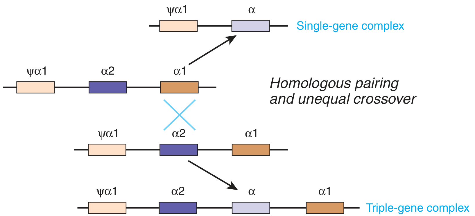
| Clinical State | Functional α Genes | Genotype | α-Chain Production | Features |
|---|---|---|---|---|
| Normal | 4 | αα/αα | 100% | Normal |
| Silent carrier | 3 | αα/α- | 75% | No anemia, no signs; diagnosed by molecular testing |
| α-Thalassemia trait (minor) | 2 | α-/α- or αα/-- | 50% | Mild microcytic anemia; asymptomatic; normal HbA₂ |
| HbH disease | 1 | α-/-- | 25% | Moderate-severe hemolytic anemia; β₄ tetramers (HbH); splenomegaly; worsens with oxidant stress |
| Hb Bart's / Hydrops fetalis | 0 | --/-- | 0% | Lethal - γ₄ tetramers (Hb Bart's); severe intrauterine hypoxia; stillbirth or neonatal death |
Key Note on Genotypes
- Southeast Asian carriers often have both deletions in cis (--/αα) → offspring can inherit --/-- → Hb Bart's is possible
- Other populations: deletions in trans (α-/α-) → --/-- is not possible in offspring
HbH Disease
- β₄ (HbH) tetramers are unstable → Heinz bodies → splenic removal
- Chronic hypochromic microcytic anemia with hemolytic crises on oxidant stress
- Avoid oxidant drugs (primaquine, sulfonamides, nitrofurantoin, dapsone, naphthalene)
- Unlike β-thalassemia: hemolysis > ineffective erythropoiesis, but bone marrow expansion can still cause deformities
Blood Film Findings
| Finding | Significance |
|---|---|
| Microcytosis + hypochromia | Reduced HbA synthesis |
| Target cells (codocytes) | Excess membrane relative to Hb content |
| Basophilic stippling | Ribosomal RNA aggregates |
| Anisocytosis / poikilocytosis | Marked variation in size and shape |
| Nucleated RBCs (normoblasts) | Stress erythropoiesis, extramedullary release |
| Heinz bodies (with supravital stain) | Precipitated HbH (in α-thalassemia) |
| Elevated reticulocyte count | But lower than expected due to ineffective erythropoiesis |
Diagnosis
| Test | Finding |
|---|---|
| CBC | Microcytic (low MCV), hypochromic (low MCH), low Hb |
| Peripheral blood smear | Target cells, basophilic stippling, nucleated RBCs |
| Hb electrophoresis / HPLC | ↑ HbA₂ (β-thalassemia minor); ↑ HbF (major/intermedia); HbH (α-thalassemia); Hb Bart's (hydrops) |
| HbA₂ level | >3.5% = β-thalassemia trait (4-6% is classic) |
| Molecular genetic testing | Definitive - identifies specific mutations |
| Serum iron / ferritin / TIBC | Normal or elevated (distinguish from iron deficiency where ferritin is low) |
| Serum bilirubin | Elevated (hemolysis) |
Key distinction: Iron deficiency anemia vs β-thalassemia trait - both cause microcytic hypochromic anemia. Iron deficiency: ↓ ferritin, ↑ TIBC, normal HbA₂. β-thalassemia trait: normal iron stores, elevated HbA₂.
Management
β-Thalassemia Major
| Intervention | Details |
|---|---|
| Regular blood transfusions | Every 2-4 weeks; maintain Hb >9-10 g/dL; suppresses ineffective erythropoiesis |
| Iron chelation | Mandatory with transfusions; options: Desferrioxamine (parenteral), Deferiprone (oral), Deferasirox (oral) |
| Folic acid supplementation | For increased erythropoietic demand |
| Splenectomy | When hypersplenism increases transfusion requirements |
| Hematopoietic stem cell transplantation (HSCT) | Curative; increasing use with matched donor |
| Gene therapy | Lentiviral delivery of functional β-globin gene into patient HSCs - now approved (betibeglogene gene therapy) |
| CRISPR-based therapy | Reactivation of HbF production (e.g., Casgevy - approved for transfusion-dependent β-thalassemia) |
β-Thalassemia Minor
- No treatment needed
- Genetic counseling for at-risk couples
- Avoid unnecessary iron supplementation
α-Thalassemia (HbH disease)
- Supportive - avoid oxidant drugs and infections
- Folic acid supplementation
- Transfusions during hemolytic crises
Complications of Iron Overload (Secondary Hemochromatosis)
- Heart → dilated cardiomyopathy, arrhythmias (leading cause of death)
- Liver → cirrhosis, hepatocellular carcinoma
- Pancreas → diabetes mellitus ("bronze diabetes")
- Pituitary/gonads → growth retardation, delayed puberty, infertility
- Thyroid/parathyroid → hypothyroidism, hypoparathyroidism
- Skin → bronze discoloration
Prognosis
- Untransfused β-thalassemia major: death in early childhood
- With transfusion + chelation: survival into 3rd decade common; overall outlook remains guarded
- With HSCT or gene therapy: potentially curative
- β-thalassemia minor: normal life expectancy; no treatment needed
Sick sinus syndrome
sick sinus syndrome pathophysiology clinical features ECG management pacemaker
Sick Sinus Syndrome (Sinus Node Dysfunction)
Definition
Arrhythmias Encompassed by SSS
| Arrhythmia | Description |
|---|---|
| Inappropriate sinus bradycardia | HR <60 bpm not explained by physiology (drugs, fitness, sleep) |
| Sinus arrest | SA node fails to fire; atrial asystole (no P waves); pauses >3 sec during daytime are pathological |
| Sinoatrial (SA) exit block | SA node fires but impulse fails to traverse perinodal tissue; PP interval is a multiple of the basic PP interval |
| Tachycardia-bradycardia (tachy-brady) syndrome | Alternating paroxysmal atrial tachyarrhythmias (AF, flutter, SVT) and bradycardia; most dangerous - sudden pause after tachycardia termination |
| Chronotropic incompetence | Inability to increase heart rate appropriately with exercise; max HR <100 bpm on exercise |
| Atrial fibrillation with slow ventricular response | Indicates combined SA and AV nodal disease |
ECG Images
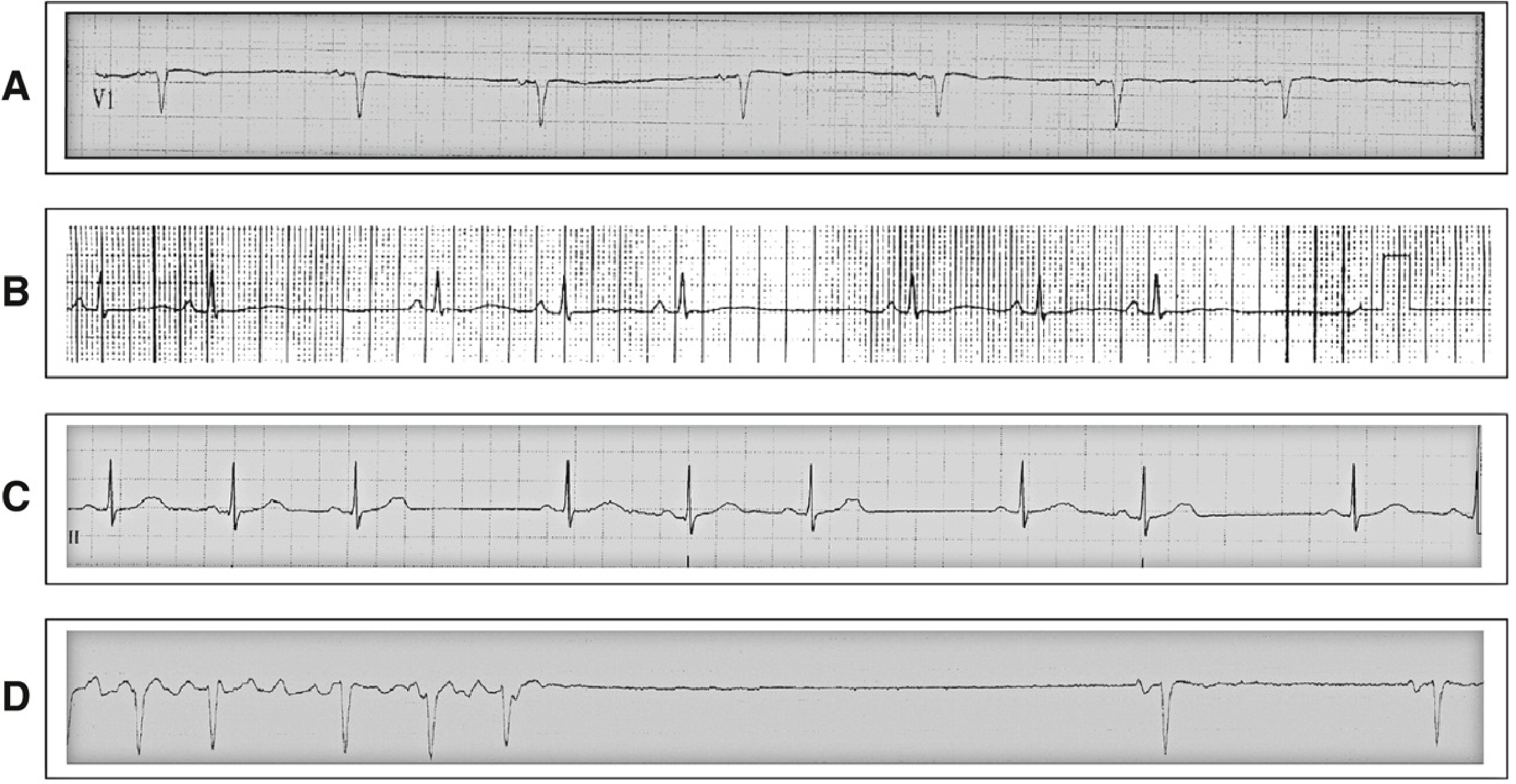
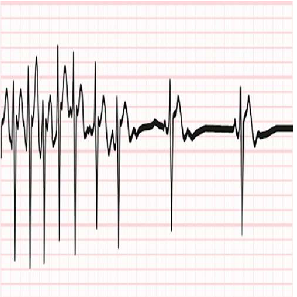
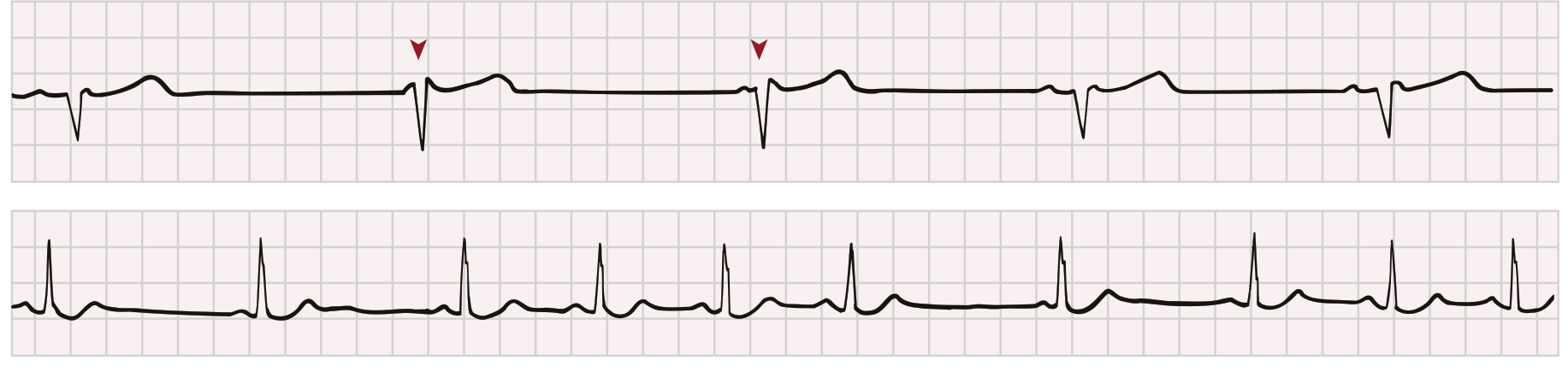
SA Exit Block - Types
| Type | ECG Pattern |
|---|---|
| 1st degree | Cannot be seen on surface ECG (delayed but not blocked) |
| 2nd degree Type I (Wenckebach) | Progressive PP shortening → pause; pause <2x shortest PP |
| 2nd degree Type II | Pause = exact multiple of basic PP interval (constant PP before and after) |
| 3rd degree (complete) | Complete absence of sinus P waves |
Epidemiology
- Predominately affects the elderly (1 in 600 cardiac patients >65 years)
- Incidence increases with age; median age ~68 years at diagnosis
- No significant sex predilection
- Familial forms exist (autosomal dominant with variable penetrance, or recessive)
Causes
Intrinsic Causes (Direct SA Node Disease)
| Category | Examples |
|---|---|
| Degenerative/fibrotic | Age-related fibrosis of the SA node (most common) - sclerodegenerative process involving SA node, AV node, bundle of His |
| Ischemic | RCA occlusion (SA node artery is a branch of RCA in ~60%), acute MI |
| Cardiomyopathy | Dilated, hypertrophic |
| Inflammatory | Myocarditis, pericarditis |
| Infiltrative | Amyloidosis, sarcoidosis, hemochromatosis |
| Surgical | Post-cardiac surgery (especially Mustard/Fontan procedures for congenital heart disease) |
| Congenital | SA node structural abnormalities |
Extrinsic Causes (Reversible - Must Exclude First)
| Category | Examples |
|---|---|
| Autonomic | Excessive vagal tone (athletes, vasovagal, carotid sinus hypersensitivity) |
| Drugs | Beta-blockers, calcium channel blockers (diltiazem, verapamil), digoxin, amiodarone, ivabradine, antiarrhythmics (class I) |
| Metabolic | Hypothyroidism, hypothermia, hypoxia, hypo/hyperkalemia |
| Raised ICP | Cushing reflex |
| Sleep apnea | Nocturnal pauses (physiologic at night) |
Genetic Basis (Familial SSS)
| Gene | Product | Mechanism |
|---|---|---|
| SCN5A | Nav1.5 (cardiac sodium channel) | Loss-of-function → reduced automaticity; can co-present with Brugada, CCD, LQT3 |
| HCN4 | I_f "funny" pacemaker current | Loss-of-function → impaired automaticity; can cause severe bradycardia + TdP |
| ANK2 | Ankyrin-B (membrane adaptor) | Disrupts ion channel localization |
| MYH6 | Alpha-myosin heavy chain | Structural SA node disease |
Pathology
- Fibrosis and fatty infiltration of the SA node
- Nodal-atrial discontinuity
- Inflammatory/degenerative changes in peri-nodal nerves and ganglia
- Occlusion of the SA nodal artery
- Process often extends to the AV node and His-Purkinje system (hence associated AV block)
Clinical Features
Symptoms (from end-organ hypoperfusion)
| Symptom | Notes |
|---|---|
| Fatigue, exercise intolerance | Most common; from chronotropic incompetence |
| Dizziness, lightheadedness | Transient cerebral hypoperfusion |
| Pre-syncope / syncope | Prolonged pauses (tachy-brady) - most dangerous presentation |
| Palpitations | From tachyarrhythmia component |
| Worsening heart failure | From low output |
| Stroke / TIA | From AF component (thromboembolic risk) |
Important: Symptoms must correlate with ECG findings. ECG abnormalities without symptoms are not sufficient for SSS diagnosis. Bradycardia during sleep or in athletes may be physiologic.
Physical Examination
- Bradycardia (may be irregular if with AF)
- Irregular pulse during tachy-brady episodes
- Signs of heart failure if severe
- Often normal between episodes
Diagnosis
| Test | Use |
|---|---|
| 12-lead ECG | Sinus bradycardia, pauses, SA exit block; often normal between episodes |
| 24-48 hour Holter monitoring | First-line for frequent symptoms |
| Extended ambulatory ECG (2-4 weeks) | For infrequent symptoms |
| Implantable loop recorder (ILR) | Gold standard for very infrequent syncope; records up to 3 years |
| Exercise stress test | Reveals chronotropic incompetence |
| Electrophysiology (EP) study | Sinus node recovery time (SNRT); corrected SNRT >550 ms = abnormal; SA conduction time; less sensitive than monitoring |
| Carotid sinus massage | Assess carotid sinus hypersensitivity |
| Thyroid function, electrolytes | Exclude reversible causes |
| Drug review | Identify causative medications |
Management
1. Treat Reversible Causes First
- Withdraw offending drugs (beta-blockers, CCBs, digoxin, antiarrhythmics)
- Correct electrolyte abnormalities, hypothyroidism
- Treat sleep apnea
2. Permanent Pacemaker - Definitive Treatment
- Symptomatic sinus bradycardia
- Symptomatic sinus pauses
- Symptomatic chronotropic incompetence
- Tachy-brady syndrome (pacemaker allows safe use of rate-controlling drugs for tachyarrhythmia)
| Mode | Use |
|---|---|
| AAIR (atrial pacing, rate-adaptive) | SSS without AV block; preserves normal AV conduction |
| DDDR (dual-chamber, rate-adaptive) | Preferred - SSS with risk of AV block (common in SND); reduces AF risk vs ventricular-only pacing |
| Minimize RV pacing | Dual-chamber devices that minimize unnecessary RV pacing reduce AF incidence and HF risk |
Key principle: Atrial pacing is preferred over ventricular pacing in SSS - reduces AF risk and preserves AV synchrony. Dual-chamber pacing is recommended because of the ~15-20% rate of associated AV block in SND - Braunwald's Heart Disease
3. Anticoagulation
- Patients with AF component are at increased stroke risk → anticoagulate per CHA₂DS₂-VASc score
4. Rate Control in Tachy-Brady
- Pacemaker must be implanted before starting rate-control drugs (beta-blockers, CCBs) to prevent dangerous pauses
Prognosis
- SSS is slowly progressive over time
- Underlying cardiac disease, not SSS itself, drives mortality
- Risk of sudden cardiac death is low with appropriate pacing
- AF develops in up to 50% over time - significant stroke risk
- Pacemaker implantation controls symptoms but does not alter the natural history of the underlying disease
Levosulpride effect on blood pressure
levosulpiride effect on blood pressure hypertension hypotension mechanism
Find recent clinical guidelines on thyroid disorders