In top
It looks like your message got cut off! You started with "In top" - could you finish your thought? What would you like to know or do?
Anatomy of ear
Reading File
Finding Sources
Finding Sources
Reading File
Reading File
Reading File
Reading File
Reading File
Finding Sources
Reading File
Finding Sources
Reading File
Reading File
Reading File
Loading Image
Reading File
Finding Sources
Reading File
Loading Image
Reading File
Reading File
Now I have comprehensive information from authoritative otolaryngology textbooks to give a thorough answer. Let me compile the complete anatomy.
Anatomy of the Ear
The ear is divided into three parts: the external ear, middle ear, and inner ear. Each has distinct anatomy and function.
1. External Ear
Auricle (Pinna)
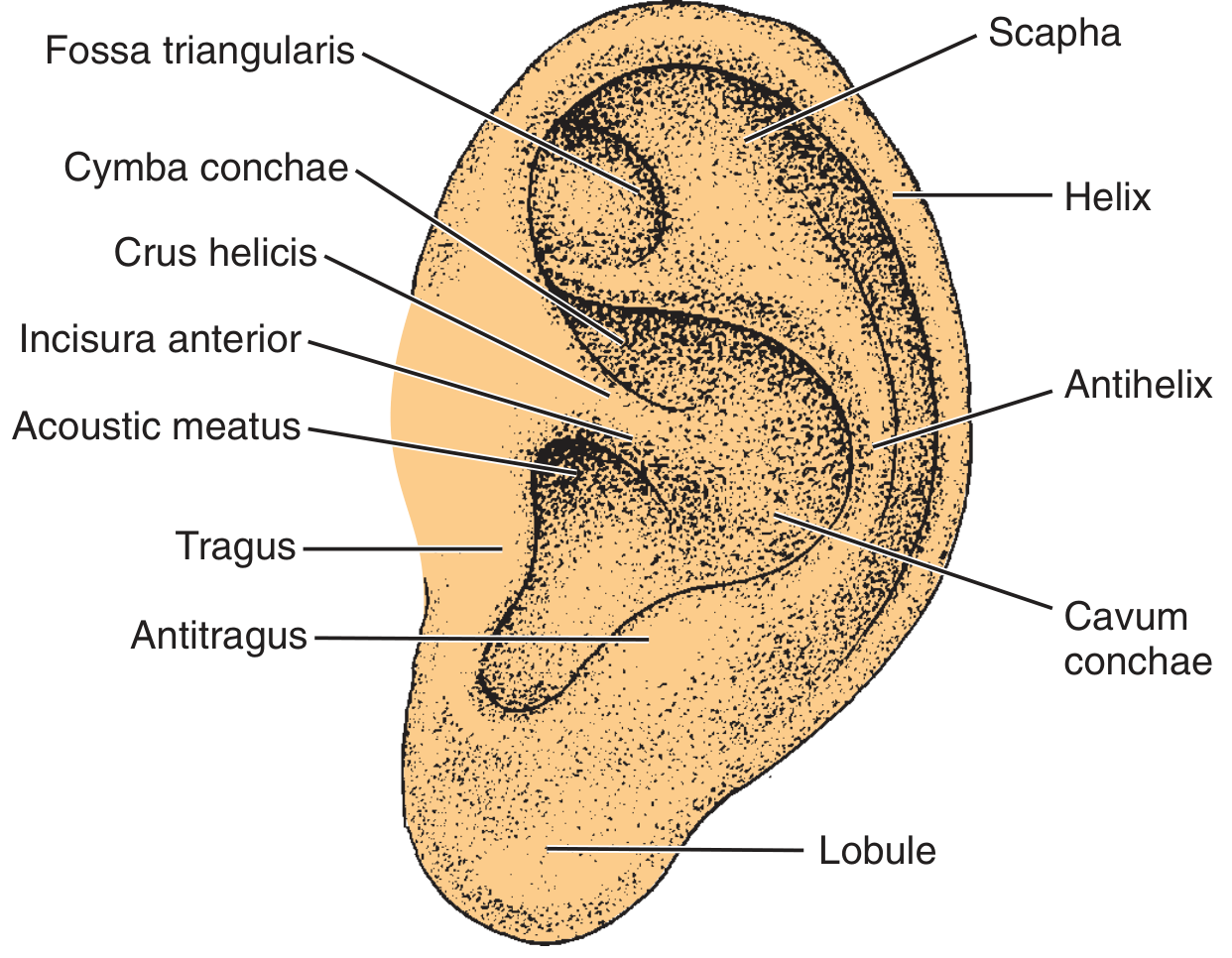
The auricle is a funnel-shaped cartilaginous structure covered by keratinizing squamous epithelium over a framework of elastic cartilage. Its surface landmarks include:
| Structure | Description |
|---|---|
| Helix | Outer curved rim of the auricle |
| Antihelix | Inner curved ridge parallel to the helix |
| Scapha | Groove between helix and antihelix |
| Fossa triangularis | Depression above where the antihelix divides |
| Cymba conchae | Upper part of the concave bowl |
| Cavum conchae | Lower, deeper part of the concave bowl |
| Tragus | Small projection anterior to the canal opening |
| Antitragus | Small projection opposite the tragus |
| Crus helicis | Root of the helix crossing the concha |
| Incisura anterior | Notch between tragus and crus helicis |
| Lobule | Soft, cartilage-free inferior part |
The blood supply arises from the external carotid artery via the posterior auricular and superficial temporal vessels.
External Auditory Canal (EAC)
- Total length: ~2.5 cm in adults
- Lateral one-third: cartilaginous (membranous), lined with thick skin containing sebaceous glands, apocrine (ceruminous) glands, and hair follicles; this is where cerumen is produced
- Medial two-thirds: bony, formed by the tympanic part of the temporal bone; lined by thin skin adherent to bone, with no glands or hair follicles
- The isthmus (bony-cartilaginous junction) is the narrowest point
- Fissures of Santorini: transverse slits in the cartilaginous portion that can allow spread of infection to surrounding soft tissue
- Foramen of Huschke: developmental defect in the anterior bony canal that may allow disease to spread to the parotid gland
Cerumen is slightly acidic (pH 6.0-6.5) and hydrophobic. The EAC has a self-cleansing mechanism: sloughed epithelium migrates centrifugally from the TM to the outer canal.
2. Middle Ear
Tympanic Membrane (TM)
The TM has four layers:
- Squamous epithelium (outer)
- Radiating fibrous layer
- Circular fibrous layer
- Mucosal layer (inner)
- Total area: 70-80 mm²
- Functional vibrating surface: ~55 mm²
- Divided into pars tensa (the larger, taut portion) and pars flaccida (Shrapnell membrane, the small superior portion above the lateral process of the malleus)
Middle Ear Cavity
The middle ear (tympanic cavity) is divided into regions:
- Epitympanum (attic): superior compartment, contains the head of the malleus and body of the incus; bounded superiorly by the tegmen tympani (bone forming the floor of the middle cranial fossa)
- Mesotympanum: main cavity at the level of the TM
- Hypotympanum: below the TM
- Prussak space: just medial to the pars flaccida and lateral to the head and neck of the malleus - a common site for cholesteatoma formation
Ossicles
The three ossicles form a mechanical chain from the TM to the oval window:
Malleus
- Head, Neck, Manubrium (handle), Anterior process, Lateral process
- The manubrium is embedded in the TM
Incus
- Body, Short process (posteriorly to incudal fossa), Long process with lenticular process (articulates with stapes)
- Most vulnerable ossicle: its long process has a single nutrient vessel with no collateral circulation
Stapes
- Anterior crus, Posterior crus, Capitulum (head), Footplate
- Footplate average dimensions: 1.41 mm × 2.99 mm
- Footplate sits in the oval window, secured by the annular ligament
Ossicular joints:
- Malleo-incudal joint: diarthrodial
- Incudo-stapedial joint: diarthrodial
- Stapedial-labyrinthine joint: syndesmotic
Middle Ear Muscles:
- Tensor tympani: attaches to the medial surface of the upper manubrium; innervated by the medial pterygoid nerve (V3)
- Stapedius: smallest skeletal muscle; attaches to the posterior neck of the stapes from the pyramidal eminence; innervated by the facial nerve (CN VII)
Eustachian Tube
- Connects the middle ear to the nasopharynx
- Maintains equalization of middle ear pressure
- The superior/lateral semicanal contains the tensor tympani; the deeper canal contains the Eustachian tube
Key Middle Ear Landmarks
- Cochleariform process: landmark for the midportion of the tympanic facial nerve segment
- Pyramidal eminence: landmark for the second genu of the facial nerve
- Oval window: where the stapes footplate articulates
- Round window: covered by the secondary tympanic membrane; allows hydraulic compensation of cochlear pressure waves
- Aditus ad antrum: opening connecting epitympanum to the mastoid antrum
3. Inner Ear
The inner ear sits within the petrous part of the temporal bone (otic capsule) and has two components: the cochlea (hearing) and the vestibular labyrinth (balance).
Cochlea
- Bony spiral making 2.5 turns
- Divided into three fluid-filled compartments:
- Scala vestibuli: filled with perilymph, above the basilar membrane
- Scala media (cochlear duct): filled with endolymph, contains the organ of Corti
- Scala tympani: filled with perilymph, below the basilar membrane; ends at the round window
Organ of Corti (sensory organ of hearing):
- Sits on the basilar membrane
- Contains inner hair cells (IHCs) and outer hair cells (OHCs)
- IHCs: responsible for 95% of all afferent nerve responses
- OHCs: motile, act as a "cochlear amplifier" by adjusting basilar membrane stiffness
- Covered by the tectorial membrane
Tonotopic organization: High frequencies are processed at the base of the cochlea; low frequencies at the apex.
Fluid compartments:
- Endolymph (in scala media): high K+, low Na+ (similar to intracellular fluid); produced by the stria vascularis
- Perilymph (in scala vestibuli and tympani): high Na+, low K+ (similar to extracellular fluid)
Vestibular Labyrinth
Semicircular canals (detect angular/rotational acceleration):
- Superior (anterior) SCC
- Lateral (horizontal) SCC
- Posterior SCC
- Each has an ampulla containing a crista ampullaris with hair cells embedded in the cupula
Otolith organs (detect linear acceleration and gravity):
- Utricle: macula in horizontal plane; detects horizontal linear acceleration
- Saccule: macula in vertical plane; detects vertical linear acceleration
- Maculae contain hair cells embedded in the otolithic membrane (contains calcium carbonate crystals called otoliths/otoconia)
Vestibular Nerve Innervation

- Superior vestibular nerve: innervates the utricle, superior SCC, lateral SCC, and (via the Voit nerve) part of the saccule
- Inferior vestibular nerve: innervates the saccule and posterior SCC
Cochlear (Auditory) Pathway
- Hair cells → Spiral ganglion cells → Cochlear nerve
- Cochlear nucleus (ipsilateral, at pontomedullary junction)
- Superior olivary complex (bilateral)
- Lateral lemniscus → Inferior colliculus
- Medial geniculate body (thalamus)
- Primary auditory cortex (Heschl's gyri, temporal lobe)
Temporal Bone Overview
The temporal bone houses the entire ear and has four embryologically distinct parts:
| Part | Key features |
|---|---|
| Squamous | Lateral wall of middle fossa; zygomatic process |
| Mastoid | Contains mastoid air cells and antrum; mastoid process formed by SCM and digastric pull |
| Petrous | Houses inner ear; arcuate eminence = superior SCC prominence |
| Tympanic | Forms walls of EAC and middle ear floor |
The mastoid antrum is the central mastoid air cell, communicating with the epitympanum via the aditus ad antrum. Pneumatization of the mastoid occurs after birth and is complete by puberty.
Sources:
- Cummings Otolaryngology Head and Neck Surgery - Chapter 126: Anatomy of the Temporal Bone, External Ear, and Middle Ear
- KJ Lee's Essential Otolaryngology - Chapter 13-14: Ear Anatomy and Physiology
- Miller's Anesthesia (10e) - Chapter 66: Synopsis of ENT Anatomy
This is a shared conversation. Sign in to Orris to start your own chat.