
Peditric case of infective endocarditis
"infective endocarditis"[MeSH Terms] AND "child"[MeSH Terms]
infective endocarditis vegetation echocardiogram
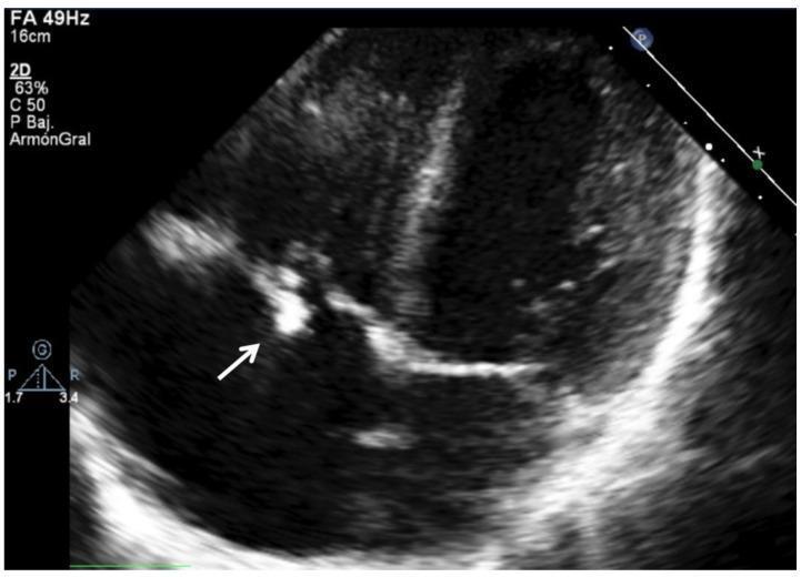
This diagnostic image is a 2D transthoracic echocardiogram (TTE) in a pediatric patient, focusing on the right heart chambers. The image demonstrates a prominent, hyperechoic (bright) irregular mass attached to the tricuspid valve leaflet, as indicated by the white arrow. This echogenic structure is characteristic of a vegetation, a hallmark of infective endocarditis. The valve leaflets themselves appear thickened and irregular, which disrupts normal coaptation. The clinical context involves a 16-year-old male with a history of congenital heart disease (restrictive perimembranous VSD) and tricuspid regurgitation, now presenting with Viridans Streptococcus endocarditis. The image illustrates the typical ultrasonographic presentation of right-sided infective endocarditis, showing how the vegetation appears as a distinct, reflective cluster against the darker, less echogenic blood-filled chambers and myocardium. This finding is a major Duke criterion for the diagnosis of infective endocarditis.
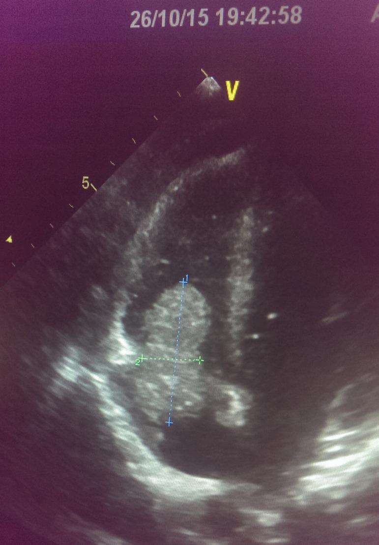
A diagnostic ultrasound (echocardiogram) image depicting a prominent vegetation associated with infective endocarditis. The image shows a large, roughly oval-shaped, echogenic mass situated within a right-sided cardiac chamber. This mass exhibits a heterogeneous internal texture and irregular, well-defined borders. Electronic calipers (marked with '+' and '1' and '2') are seen measuring the longitudinal and transverse dimensions of the structure. The vegetation appears to be attached to the tricuspid valve, where it potentially obstructs normal leaflet excursion or causes valvular regurgitation. This clinical finding is particularly relevant in patients with permanent indwelling vascular access, such as dialysis catheters, which are known risk factors for right-sided infective endocarditis. The image illustrates the typical appearance of valvular vegetation as a mobile, echogenic intracardiac mass, essential for the diagnosis of endocarditis based on the Modified Duke Criteria.
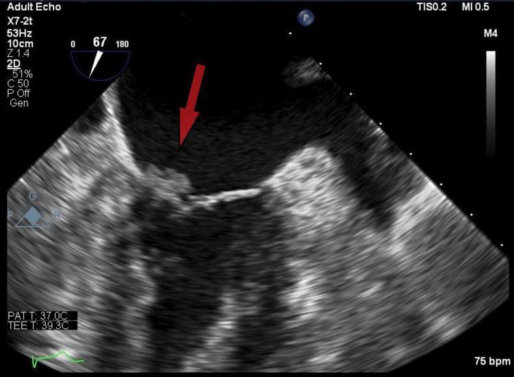
Diagnostic ultrasound image, specifically a transesophageal echocardiogram (TEE) in a mid-esophageal view, focusing on the mitral valve apparatus. A red arrow identifies a distinct, irregular, and heterogeneously echogenic mass attached to the atrial surface of the anterior mitral valve leaflet. This mobile density is characteristic of a vegetation. The imaging demonstrates the vegetation protruding towards the left ventricular outflow tract (LVOT). Clinical context suggests this finding is consistent with non-bacterial thrombotic endocarditis (marantic endocarditis), particularly given the underlying malignancy noted in the patient's history. This visual is intended for medical education regarding the sonographic identification of valvular vegetations, the differentiation between infective and non-infective endocarditis, and the evaluation of embolic risk in cardiac patients.
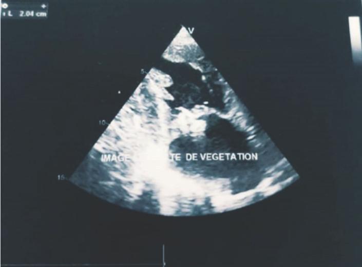
This diagnostic image is a transthoracic echocardiogram in a parasternal long-axis view showing the cardiac chambers. The primary finding is a large, irregularly shaped, echogenic mass attached to the anterior leaflet of the mitral valve, consistent with infective endocarditis vegetation. A digital measurement caliper at the top left indicates a size of approximately 2.04 cm (20 mm). The vegetation appears mobile and occupies a significant portion of the mitral valve apparatus, which is clinically associated with severe mitral regurgitation and a high risk of embolic events. The image demonstrates the classic sonographic appearance of valvular vegetation used for diagnosing infective endocarditis according to the Modified Duke Criteria. Key anatomical landmarks visible include the left atrium, left ventricle, and the mitral valve leaflets.
Pediatric Infective Endocarditis - Comprehensive Clinical Review
Overview & Epidemiology
- Annual incidence: 0.05 to 0.12 cases per 1,000 pediatric admissions (low, but significant morbidity)
- Mortality: 10-15% despite declining trends
- Major complications: septic emboli, valvular dysfunction, congestive heart failure
Risk Factors in Children
| Clinical Condition | Key Notes |
|---|---|
| Congenital heart disease | Unrepaired cyanotic CHD (including palliative shunts and conduits) |
| Completely repaired defects with prosthetic material within 6 months | |
| Repaired CHD with residual defects adjacent to prosthetic patch/device | |
| Prosthetic cardiac valves | High risk; add rifampin to empiric regimen |
| Cardiac transplantation | Especially with subsequent valvulopathy |
| Previous IE | Significant predictor |
| Central venous catheters | Higher risk for S. aureus; especially neonates |
| Rheumatic heart disease | Less common in developed countries now |
Pathogenesis
- Damaged endocardium - from turbulent flow, jet lesions, prosthetic material, or pacemaker wires
- Circulating bacteria - from bacteremia (dental procedures, GI/GU manipulation, skin infections, IV drug use, or trivial daily activities like tooth brushing)
Microbiology
| Organism | Context |
|---|---|
| Streptococcus viridans | Most common in children with underlying heart disease; subacute presentation |
| Staphylococcus aureus | Most common in children without heart disease; also healthcare-acquired; acute, destructive presentation |
| Gram-negatives (Klebsiella, Enterobacter) | Neonates - rare |
| HACEK group | Haemophilus, Actinobacillus, Cardiobacterium, Eikenella, Kingella - normal oral flora; slow-growing gram-negatives |
| Candida spp. | Neonates and children with indwelling catheters |
| Culture-negative | ~10% of cases (prior antibiotics, fastidious organisms) |
Classification
| Acute IE | Subacute IE | |
|---|---|---|
| Causative organism | Virulent (e.g., S. aureus) | Less virulent (e.g., S. viridans) |
| Valve | Previously normal or diseased | Usually diseased |
| Onset | Rapid, fulminant | Insidious, weeks to months |
| Fever | High spiking fevers, clinical sepsis | Low-grade or absent |
| Prognosis | High morbidity/mortality | Recovers with antibiotics |
Clinical Features
- Fever (most common) - unexplained or persistent fever is the hallmark
- Fatigue, weakness, arthralgias, myalgias, weight loss, rigors, diaphoresis (older children)
- Subtler presentations in infants
- In cyanotic CHD patients: pneumonia, new neurologic deficits, hematuria, or skin rashes should trigger IE suspicion
- Fever + tachycardia + new or changing cardiac murmur (present in 90% of left-sided lesions)
- Splinter hemorrhages - linear subungual hemorrhages in nail beds
- Janeway lesions - painless erythematous macules on palms/soles (septic emboli)
- Osler nodes - tender subcutaneous nodules on fingers (immune complex deposition)
- Roth spots - retinal hemorrhages with pale centers
- Splenomegaly (subacute disease)
- CNS focal neurologic deficits (cerebral emboli)
- Rales/decreased breath sounds (septic pulmonary emboli - right-sided IE)
Echocardiographic Findings
Diagnosis
Modified Duke Criteria
Major Criteria:
- Positive blood cultures:
- 2 positive cultures for a typical IE organism, OR
- Persistently positive cultures (same organism over 12 h, or 3+ cultures with ≥1 h between first and last), OR
- Single positive culture for Coxiella burnetii
- Endocardial involvement on echocardiography:
- Vegetation, abscess, new valvular regurgitation, or prosthetic valve dehiscence
Minor Criteria:
- Fever ≥38°C (100.4°F)
- Predisposing heart condition or injection drug use
- Vascular phenomena (emboli, mycotic aneurysm, intracranial hemorrhage)
- Immunologic phenomena (elevated RF, immune complex GN, Osler nodes, Roth spots)
- Positive blood culture not meeting major criterion
Classification:
- Definite IE: 2 major criteria, OR 1 major + 3 minor criteria, OR 5 minor criteria
- Possible IE: 1 major + 1 minor criterion, OR 3 minor criteria
Investigations:
- Blood cultures: 3 separate samples in first 24 hours; 1-3 mL (infants), 3-5 mL (toddlers/preschool), 10 mL (school-age/adolescents). Repeat at 48 h if negative.
- CBC: Anemia of chronic disease; leukocytosis may be absent
- ESR/CRP: Usually elevated (mean ESR ~55 mm/h)
- Rheumatoid factor: Positive in subacute/chronic disease
- Urinalysis: Proteinuria and microscopic hematuria in ~50% (immune complex GN)
- TTE: More sensitive in children than adults (better acoustic windows); reveals vegetations, abscess, valvular regurgitation
- TEE: Better sensitivity than TTE for prosthetic valves and perivalvular extension
Treatment
Empiric Antibiotic Therapy (ED initiation after cultures):
- Vancomycin 15 mg/kg IV q6h (max 2 g per dose) - for MRSA coverage
- + Gentamicin 5-7.5 mg/kg IV q24h - synergistic bactericidal effect
- Penicillin G 50,000-100,000 units/kg IV q6h, OR
- Ceftriaxone 100 mg/kg IV daily
- Prolonged parenteral antibiotic course (4-6 weeks typically required for complete eradication)
- Hospitalization mandatory
- Pediatric cardiology consultation indicated
- Pediatric cardiac surgery consultation for hemodynamic instability
Surgical Indications:
- Uncontrolled infection despite adequate antibiotics
- Heart failure from valvular dysfunction
- Large vegetations at high embolic risk
- Perivalvular abscess/extension
Prophylaxis
- Prosthetic cardiac valve or prosthetic material used for valve repair
- Previous IE
- Unrepaired cyanotic CHD (including palliative shunts/conduits)
- Completely repaired CHD with prosthetic material within the first 6 months after procedure
- Repaired CHD with residual defect at/adjacent to prosthetic patch or device
- Cardiac transplantation with subsequent valvulopathy
- Oral: Amoxicillin 50 mg/kg PO
- Parenteral: Ampicillin 50 mg/kg IM/IV
- Penicillin-allergic:
- Cephalexin 50 mg/kg PO, OR
- Clindamycin 20 mg/kg PO/IV, OR
- Azithromycin 15 mg/kg PO
Complications
| Complication | Mechanism |
|---|---|
| Septic emboli | Friable vegetations shed embolic material |
| Valvular dysfunction/destruction | Direct tissue invasion; ring abscess |
| Heart failure | Valvular regurgitation or obstruction |
| Cerebral emboli/stroke | Left-sided IE; mycotic aneurysm |
| Septic pulmonary emboli | Right-sided IE (tricuspid valve) |
| Immune complex glomerulonephritis | Hematuria, proteinuria, renal failure |
| Mycotic aneurysms | Septic emboli infect arterial walls |
| Metastatic abscesses | Septic infarcts at embolization sites |
Recent Evidence Update
Key Pediatric-Specific Pearls
- 90% have structural heart disease - always evaluate for underlying CHD
- Neonates are special - gram-negatives and Candida are important pathogens; often linked to central venous catheters
- Subacute presentations mimic common illness - maintain suspicion for persistent unexplained fever in any child with CHD
- TTE is preferred initially in children (better acoustic windows than adults)
- HACEK organisms are characteristic of IE in children and adolescents with normal dental hygiene and CHD
- Right-sided IE (tricuspid) is associated with central venous catheter use and presents with septic pulmonary emboli
- Outcome depends heavily on the infecting organism and speed of diagnosis and treatment