
make 3 min detailed disscusion on glaucoma to make video. give description in pointers
Reading File
Now let me search the medical library for comprehensive glaucoma content.
Finding Sources
Searching the Web
glaucoma pathophysiology types diagnosis treatment overview 2024
Now let me pull content from the best ophthalmology and pathology sources simultaneously.
Reading File
Reading File
Reading File
Excellent - I have rich textbook content. Let me fetch the Kanski ophthalmology (detailed clinical) and Katzung pharmacology sections for treatment details, and get the diagnostic image.
Reading File
Reading File
Loading Image
I now have comprehensive content from Robbins Pathology, Goodman & Gilman's Pharmacology, and Kanski's Ophthalmology. Here is the full 3-minute video script in detailed bullet-point format:
🎬 Glaucoma - 3-Minute Video Script (Detailed Pointer Discussion)
🔷 SEGMENT 1: INTRODUCTION (0:00 - 0:25)
- Glaucoma is not a single disease - it is a group of eye diseases that share one common endpoint: progressive optic nerve damage leading to irreversible vision loss and blindness
- It is the leading cause of irreversible blindness worldwide, with prevalence growing as populations age
- The optic nerve transmits visual signals from the retina to the brain - when it is damaged, vision is permanently lost
- Key concept: Intraocular pressure (IOP) is the only known modifiable risk factor, and all current treatments target IOP reduction
- Normal IOP range: 10-21 mmHg; glaucomatous damage often occurs above this, but some patients develop damage even at normal pressures ("normal-tension glaucoma")
🔷 SEGMENT 2: ANATOMY & PATHOPHYSIOLOGY (0:25 - 1:00)
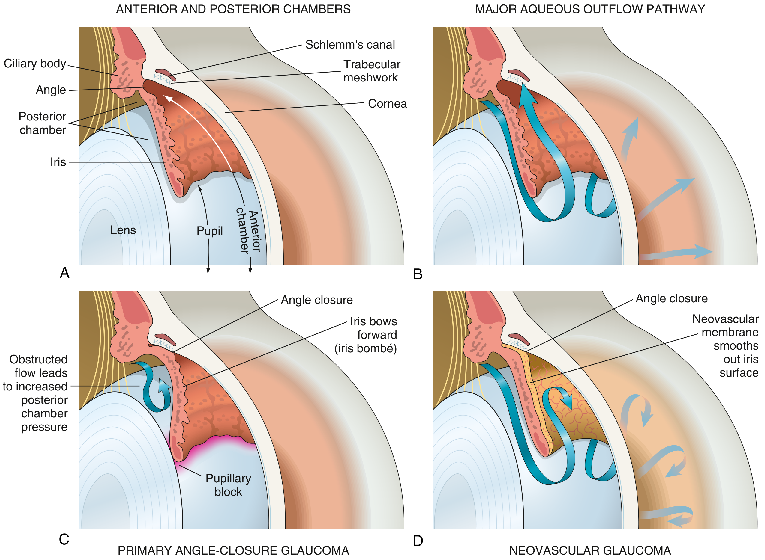
(Use the diagram image above - Robbins Pathology Fig. 21.38)
- The eye continuously produces aqueous humor from the ciliary body in the posterior chamber
- Aqueous flows through the pupil into the anterior chamber, then drains out via the trabecular meshwork into Schlemm's canal
- This drainage system maintains stable IOP
- When drainage is impaired - fluid accumulates - IOP rises - pressure is transmitted to the optic nerve head - retinal ganglion cell axons are crushed and die
- The result is characteristic "cupping" of the optic disc: the neuroretinal rim is lost, and the central cup enlarges (increased cup-to-disc ratio)
- Retinal nerve fiber layer (RNFL) progressively thins, causing visual field defects
- Most glaucomatous defects occur within 30° of the fixation point (central field) - threatening reading and fine detail vision in advanced disease
🔷 SEGMENT 3: TYPES OF GLAUCOMA (1:00 - 1:40)
1. Primary Open-Angle Glaucoma (POAG) - Most Common
- Drainage angle between cornea and iris is open and normal-appearing
- Problem is increased resistance within the trabecular meshwork itself
- Develops slowly and silently - no pain, no redness - often undetected until late
- Peripheral vision lost first; patients may not notice until >40% of optic nerve fibers are gone
2. Angle-Closure Glaucoma (ACG)
- The anterior chamber angle is physically narrowed or closed
- In acute ACG: transient iris apposition to the lens blocks aqueous flow from posterior to anterior chamber → pressure builds → iris bows forward ("iris bombé") → obstructs the trabecular meshwork completely
- Presents as a medical emergency: sudden severe eye pain, headache, nausea/vomiting, halos around lights, mid-dilated non-reactive pupil, red eye
- More common in hypermetropic (far-sighted) eyes with shallow anterior chambers
3. Secondary Glaucomas
- Neovascular glaucoma: new blood vessels (from diabetic retinopathy, central retinal vein occlusion) grow a membrane over the iris and trabecular meshwork → blocks outflow
- Pigmentary glaucoma: pigment granules from iris float into and clog trabecular meshwork
- Exfoliative glaucoma: flaky protein material deposits on lens and trabecular meshwork
- Traumatic glaucoma: red blood cells or inflammatory debris block drainage after injury
4. Normal-Tension Glaucoma
- IOP is within normal range, yet the optic nerve shows classic glaucomatous damage
- Mechanism may involve vascular insufficiency to the optic nerve head
5. Congenital Glaucoma
- Due to maldevelopment of the trabecular meshwork (trabeculodysgenesis)
- Presents with buphthalmos (enlarged globe/"ox eye"), tearing, photophobia in infants
🔷 SEGMENT 4: RISK FACTORS (1:40 - 1:55)
- Elevated IOP - the single most important modifiable risk factor
- Age - prevalence rises sharply after age 40
- Family history / genetics - first-degree relatives have 4-9x higher risk
- African descent - higher prevalence, earlier onset, more severe disease in POAG
- Myopia (near-sightedness) - associated with POAG
- Hypermetropia (far-sightedness) - predisposes to angle-closure
- Diabetes mellitus and systemic hypertension - possible additional risk
- Corticosteroid use (topical or systemic) - can raise IOP significantly ("steroid glaucoma")
- Thin central cornea - structural risk factor
🔷 SEGMENT 5: DIAGNOSIS (1:55 - 2:20)
A full glaucoma workup includes:
- Tonometry - measures IOP (Goldmann applanation is gold standard); IOP >21 mmHg is suspicious
- Ophthalmoscopy / Slit-lamp exam - examines optic disc for cupping, notching, hemorrhages
- Cup-to-disc ratio - normal is <0.5; glaucomatous cupping often >0.6-0.7
- Gonioscopy - uses a special lens to directly examine the drainage angle (open vs. closed)
- Perimetry (visual field testing) - automated static perimetry (e.g., Humphrey 24-2 or 30-2 pattern) detects characteristic visual field defects:
- Nasal step
- Arcuate (Bjerrum) scotoma
- Paracentral scotoma
- Advanced: tunnel vision
- Optical Coherence Tomography (OCT) - measures RNFL thickness objectively; detects nerve fiber loss before visual field changes
- Corneal pachymetry - measures corneal thickness (thin cornea = underestimated true IOP)
🔷 SEGMENT 6: TREATMENT (2:20 - 2:55)
A. Medical (First-Line)
1. Prostaglandin Analogues (PGAs) - First-line
- Latanoprost, travoprost, bimatoprost, tafluprost
- Once-daily dosing, potent IOP reduction (~25-35%)
- Mechanism: increase uveoscleral outflow by relaxing ciliary muscle; also some trabecular outflow effect
- Side effects: iris pigmentation darkening, eyelash growth (hypertrichosis), periorbital fat atrophy
- Replaced beta-blockers as first-line due to fewer systemic effects
2. Beta-Adrenergic Blockers
- Timolol (non-selective), betaxolol (beta-1 selective)
- Mechanism: reduce aqueous humor production by blocking beta-2 receptors in ciliary body epithelium
- Contraindicated in asthma, COPD, heart block, bradycardia
3. Alpha-2 Adrenergic Agonists
- Brimonidine, apraclonidine
- Dual mechanism: reduce aqueous production + enhance uveoscleral outflow
- Useful when PGAs or beta-blockers are contraindicated
4. Carbonic Anhydrase Inhibitors (CAIs)
- Topical: dorzolamide, brinzolamide
- Oral: acetazolamide (for acute ACG crisis)
- Mechanism: block carbonic anhydrase in ciliary body → reduce aqueous secretion
5. Rho-Kinase (ROCK) Inhibitors
- Netarsudil - newest class; increases trabecular outflow by relaxing the meshwork cells
B. Laser Treatment
- Selective Laser Trabeculoplasty (SLT) - targets trabecular meshwork cells, improves drainage; used in open-angle glaucoma; can replace or supplement drops; repeatable
- Laser Peripheral Iridotomy (LPI) - creates a small hole in the iris, bypassing pupillary block; treatment of choice for angle-closure glaucoma; also used prophylactically in the fellow eye
C. Surgical Treatment
- Trabeculectomy - surgical creation of a new drainage channel (fistula) from anterior chamber to subconjunctival space, forming a "bleb"; gold standard surgical procedure; often augmented with mitomycin C to prevent scarring
- Glaucoma Drainage Devices (GDDs) - tube-shunt implants (Ahmed, Baerveldt, Molteno) to divert aqueous to a plate posteriorly; preferred in complex/refractory cases
- Minimally Invasive Glaucoma Surgery (MIGS) - newer options (iStent, Hydrus, OMNI) with lower risk profile; used in mild-moderate disease often combined with cataract surgery
- Cyclodestruction - diode laser ciliary body ablation to reduce aqueous production; used as last resort when other options fail
🔷 SEGMENT 7: KEY TAKEAWAYS & CLOSING (2:55 - 3:00)
- Glaucoma = silent thief of sight - damage is irreversible and often asymptomatic until late
- Early detection through regular eye exams is the most effective prevention strategy, especially after age 40 or with risk factors
- All treatment (medical, laser, surgical) aims at lowering IOP to slow or halt progression
- No cure exists - but with early diagnosis and consistent treatment, most patients preserve useful vision throughout their lifetime
Sources consulted:
- Kanski's Clinical Ophthalmology, 10th Ed.
- Robbins & Kumar Basic Pathology (9780323790185)
- Goodman & Gilman's Pharmacological Basis of Therapeutics
- Goldman-Cecil Medicine, Harriet Lane Handbook
its too much wordy give it in few words no need to give too long explanation
🎬 Glaucoma – 3-Min Video Script (Short Pointers)
1. WHAT IS GLAUCOMA? (0:00–0:20)
- Group of eye diseases → optic nerve damage → vision loss
- Leading cause of irreversible blindness worldwide
- Key culprit: raised intraocular pressure (IOP)
- Normal IOP: 10–21 mmHg
2. HOW DOES IT HAPPEN? (0:20–0:45)
- Eye produces aqueous humor (ciliary body)
- Flows: posterior chamber → pupil → anterior chamber → trabecular meshwork → Schlemm's canal
- Blockage in drainage → fluid builds up → IOP rises → optic nerve compressed → nerve fibers die
- Result: optic disc cupping + visual field loss
3. TYPES (0:45–1:20)
| Type | Key Feature |
|---|---|
| Primary Open-Angle | Most common; silent; slow onset |
| Angle-Closure | Emergency; sudden pain, red eye, halos |
| Normal-Tension | Nerve damage despite normal IOP |
| Secondary | Due to diabetes, trauma, steroids |
| Congenital | Birth defect; enlarged globe (buphthalmos) |
4. RISK FACTORS (1:20–1:35)
- High IOP
- Age >40
- Family history
- African descent
- Diabetes, hypertension
- Steroid use
- Thin cornea
5. SYMPTOMS (1:35–1:45)
- Open-angle: no symptoms until late (silent thief of sight)
- Angle-closure: sudden eye pain, headache, nausea, blurred vision, halos
6. DIAGNOSIS (1:45–2:05)
- Tonometry – measures IOP
- Ophthalmoscopy – checks optic disc cupping (C:D ratio >0.6 suspicious)
- Gonioscopy – views drainage angle
- Visual field test (Perimetry) – detects field defects (arcuate scotoma, tunnel vision)
- OCT – measures retinal nerve fiber thickness
7. TREATMENT (2:05–2:50)
💊 Medical (Drops)
- Prostaglandin analogues (latanoprost) – 1st line; increase outflow
- Beta-blockers (timolol) – reduce aqueous production
- Alpha-2 agonists (brimonidine) – reduce production + increase outflow
- Carbonic anhydrase inhibitors (dorzolamide, acetazolamide) – reduce secretion
🔦 Laser
- SLT – opens trabecular meshwork (open-angle)
- Laser iridotomy – bypasses pupil block (angle-closure)
🔪 Surgery
- Trabeculectomy – new drainage channel; gold standard
- Tube shunts – for complex cases
- MIGS – minimally invasive; combined with cataract surgery
8. KEY TAKEAWAYS (2:50–3:00)
- Glaucoma = silent, irreversible, but manageable
- Regular eye exams after age 40 = best prevention
- Goal of all treatment = lower IOP, preserve vision
- No cure, but early treatment saves sight ✅
This is a shared conversation. Sign in to Orris to start your own chat.