
Digestive system for vocational course telangana
Reading File
The digestive system covers the organs and processes involved in breaking down food, absorbing nutrients, and eliminating waste. Let me search the medical library for comprehensive content.
Searching sources...
Finding Sources
Finding Sources
Searching the Web
digestive system vocational course Telangana class 11 12 biology notes
Reading File
Finding Sources
Reading File
Reading File
Reading File
Reading File
Respiratory system with images for vocational course telangana
Finding Sources
Finding Sources
Reading File
Finding Sources
Reading File
Reading File
Finding Sources
Reading File
Finding Sources
Finding Sources
Reading File
Reading File
Now let me fetch some key images to include in the answer:
Loading Image
Loading Image
Reading File
Loading Image
Finding Sources
Reading File
Loading Image
Now I have excellent content and images. Let me compile the comprehensive answer.
🫁 Respiratory System - Vocational Course (Telangana)
1. Introduction
The respiratory system is responsible for external respiration - extracting oxygen (O₂) from air and releasing carbon dioxide (CO₂) from the blood. It also helps in vocalization (speech), olfaction (smell), and filtering/warming inspired air.
The system has three principal functions:
- Air conduction - moving air to and from the lungs
- Air filtration - warming, moistening, and cleaning inhaled air
- Gas exchange (respiration) - O₂ and CO₂ exchange at the alveoli
2. Divisions of the Respiratory System
| Division | Structures |
|---|---|
| Upper Respiratory Tract | Nose, nasal cavity, paranasal sinuses, pharynx |
| Lower Respiratory Tract | Larynx, trachea, bronchi, bronchioles, lungs |
Clinically:
- Upper airways = all structures above the larynx (nasal cavities, sinuses, pharynx)
- Lower airways = larynx, trachea, bronchial tree down to alveoli
(Color Atlas of Human Anatomy, Vol 2)
3. Organs of the Respiratory System
A. Nose and Nasal Cavity
- The nose filters, warms, and humidifies incoming air
- Divided into vestibule, respiratory region, and olfactory region
- Lined by respiratory epithelium - ciliated pseudostratified columnar cells, goblet cells, basal cells
- The olfactory region (roof of nasal cavity) contains bipolar olfactory receptor neurons with G-protein-coupled receptors for smell
- Rich vascular network warms air; mucus traps particles
B. Pharynx
- Posterior continuation of oral and nasal cavities
- Directs food into the esophagus and air into the larynx
- The two passages intersect here - hence why we can choke
C. Larynx
- Connects pharynx to trachea
- Contains vocal folds (vocal cords) - vibrate to produce sound
- Lined by respiratory mucosa except vocal cord surfaces (stratified squamous epithelium)
- The epiglottis covers the airway during swallowing
D. Trachea
- Extends from larynx into the mediastinum, then divides into right and left main (primary) bronchi
- Wall has 4 layers:
- Mucosa - ciliated pseudostratified epithelium on thick basement membrane
- Submucosa - dense irregular connective tissue
- Cartilaginous layer - C-shaped hyaline cartilage rings (keep airway open)
- Adventitia - binds trachea to adjacent structures
(Histology: A Text and Atlas)
4. The Bronchial Tree - Airways
The airway system branches 23 times (generations) from trachea to alveolar sacs:
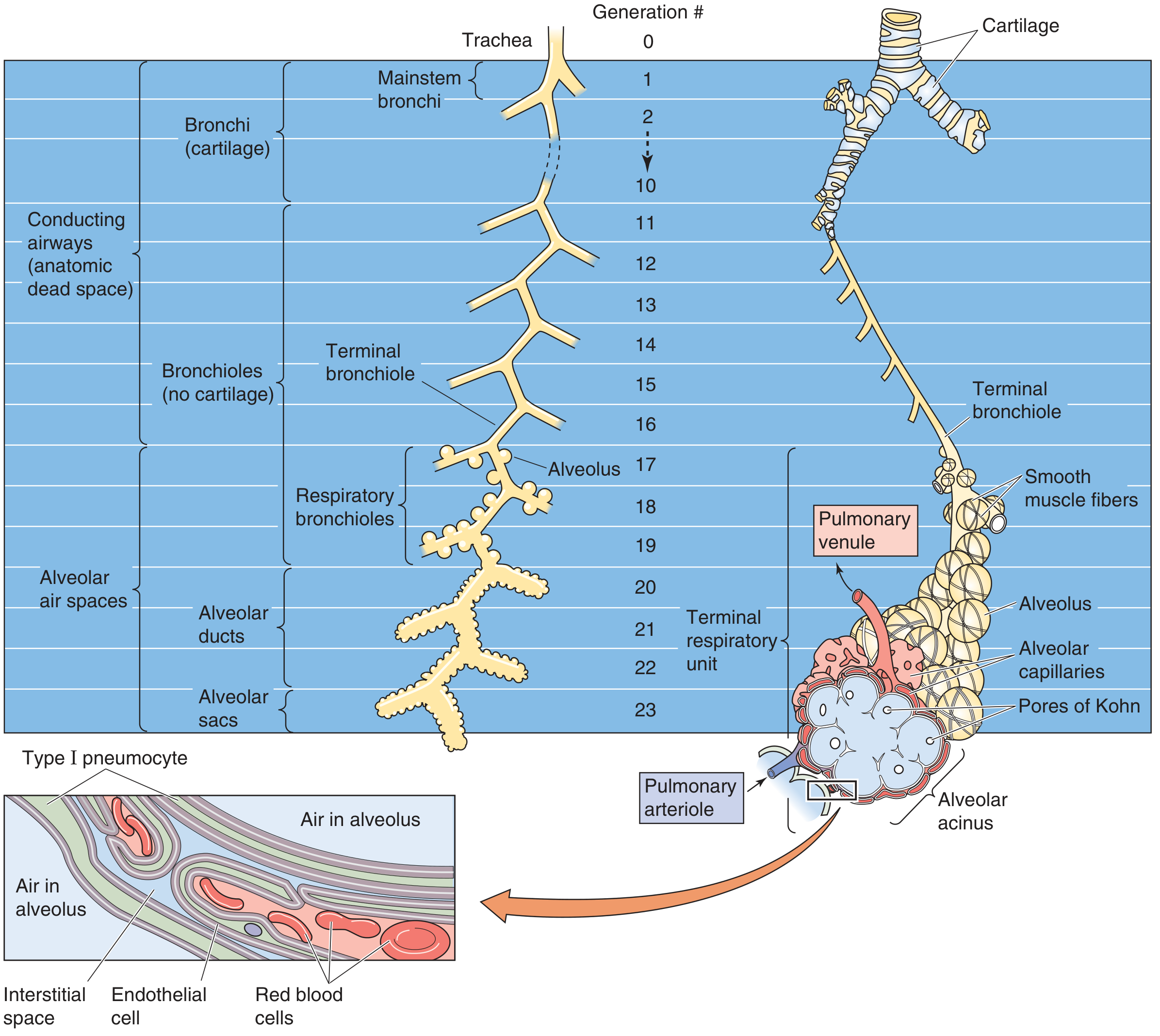
Conducting Zone (Generations 0-16)
- Moves air by convection - NO gas exchange occurs here
- Forms the anatomical dead space (~150 mL)
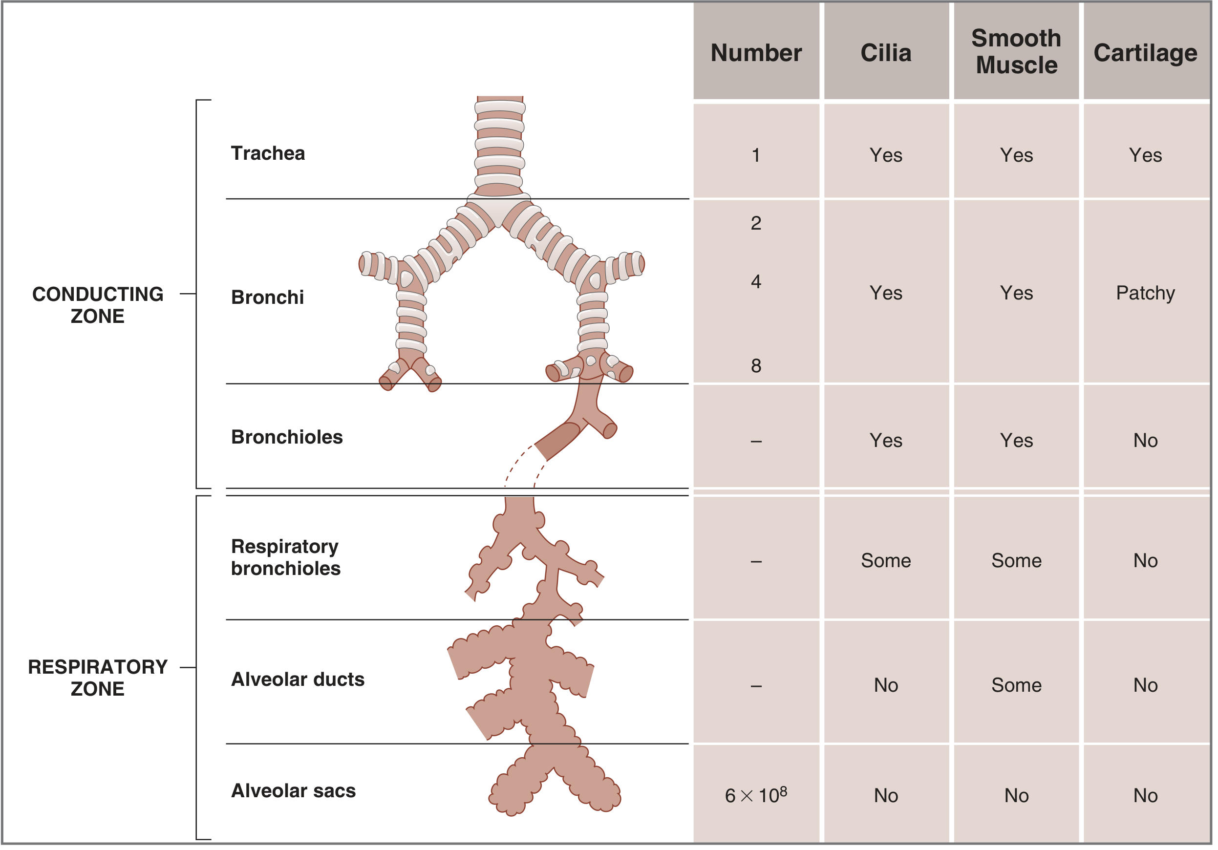
- Airways progressively lose: cilia → mucous glands → cartilage
| Structure | Key Feature |
|---|---|
| Trachea (gen 0) | C-shaped cartilage rings, cilia |
| Mainstem bronchi (gen 1) | Cartilage, cilia, smooth muscle |
| Bronchi (gen 2-10) | Cartilage plates, cilia |
| Bronchioles (gen 11-16) | No cartilage, cilia, smooth muscle |
| Terminal bronchioles (gen 16) | Last conducting airway, club cells |
Respiratory Zone (Generations 17-23)
- Site of actual gas exchange
- Alveoli line the walls
| Structure | Feature |
|---|---|
| Respiratory bronchioles | First site of gas exchange, some alveoli |
| Alveolar ducts | Completely lined with alveoli |
| Alveolar sacs (gen 23) | Terminal clusters of alveoli |
5. The Lungs
- Two lungs occupying the thoracic cavity, each enclosed in a pleural cavity lined by serous membrane (pleura)
- Right lung: 3 lobes (upper, middle, lower)
- Left lung: 2 lobes (upper, lower) - smaller due to cardiac notch
- Each lung has ~300 million alveoli, total surface area ~200 m² for gas exchange
- Pulmonary circulation: Right ventricle → pulmonary artery → lung capillaries → pulmonary vein → left atrium
- Blood flow is highest at the base and lowest at the apex when standing (gravity)
6. The Alveoli - Site of Gas Exchange
- Alveoli are tiny air sacs (~200 µm diameter), pouchlike evaginations of respiratory bronchioles, ducts, and sacs
- Alveolar wall (septum) = the air-blood barrier, very thin for rapid diffusion
Cell Types in Alveoli
| Cell | Function |
|---|---|
| Type I pneumocytes | Extremely thin squamous cells, cover 95% of alveolar surface, form the diffusion barrier |
| Type II pneumocytes | Secrete surfactant (reduces surface tension, prevents alveolar collapse), contain lamellar bodies |
| Alveolar macrophages | Phagocytose dust, debris, and pathogens; migrate to bronchioles for clearance |
Surfactant
- Produced by Type II cells
- Reduces surface tension in alveoli
- Without surfactant - alveoli collapse (atelectasis); seen in premature babies as Respiratory Distress Syndrome
Air-Blood Barrier (from alveolar air to blood)
- Thin layer of surfactant
- Type I alveolar cell + basal lamina
- Capillary endothelial cell + basal lamina (often fused with above)
Gas exchange occurs by simple diffusion down concentration gradients:
- O₂ moves from alveolus (high) → blood (low)
- CO₂ moves from blood (high) → alveolus (low)
7. Mechanics of Breathing
Inspiration (Active process)
- Diaphragm contracts and moves downward
- External intercostal muscles contract, ribs move up and out
- Thoracic volume increases → lung pressure drops below atmospheric → air flows in
- Innervated by the phrenic nerve (C3, C4, C5)
Expiration (Passive at rest)
- Respiratory muscles relax
- Elastic recoil of lungs and chest wall pushes air out
- During exercise, internal intercostal muscles and abdominal muscles actively assist
Lung Volumes (Spirometry)
| Volume/Capacity | Value | Description |
|---|---|---|
| Tidal Volume (TV) | ~500 mL | Normal quiet breathing |
| Inspiratory Reserve Volume (IRV) | ~3000 mL | Extra air after normal inspiration |
| Expiratory Reserve Volume (ERV) | ~1200 mL | Extra air pushed out after normal expiration |
| Residual Volume (RV) | ~1200 mL | Air remaining after maximal expiration (cannot be measured by spirometer) |
| Total Lung Capacity (TLC) | ~5-6 L | All volumes combined |
| Functional Residual Capacity (FRC) | ~2400 mL | ERV + RV |
(Costanzo Physiology 7th Edition)
8. Control of Breathing
Breathing is automatic and rhythmic, controlled by the brainstem:
- Medulla oblongata - contains the Central Pattern Generator (CPG), sets the basic rhythm of inspiration/expiration
- Pons - fine-tunes the rhythm
- Respiratory-related neurons (RRNs) fire during specific phases of the respiratory cycle
Chemoreceptors (sensors that adjust breathing)
| Receptor | Location | Senses |
|---|---|---|
| Central chemoreceptors | Medulla | Rise in CO₂ / fall in pH of CSF |
| Peripheral chemoreceptors | Carotid body, aortic body | Fall in O₂, rise in CO₂, fall in pH |
When CO₂ rises or O₂ falls → breathing rate and depth increase automatically.
Autonomic Control of Airway Diameter
| System | Effect on Airways |
|---|---|
| Sympathetic (β₂ adrenergic) | Bronchodilation - widens airways (e.g., salbutamol/albuterol in asthma) |
| Parasympathetic (muscarinic) | Bronchoconstriction - narrows airways |
9. Pulmonary Blood Supply
The lung has two circulations:
- Pulmonary circulation - for gas exchange; right ventricle → pulmonary artery → capillaries around alveoli → pulmonary vein → left atrium
- Bronchial circulation - for nutrition of airway walls; branches of aorta supply bronchi and bronchioles
10. Summary: Path of Air (Inspiration)
Nostrils/Mouth
    ↓
Nasal Cavity / Oral Cavity (filters, warms, humidifies)
    ↓
Pharynx
    ↓
Larynx (past vocal cords, epiglottis protects)
    ↓
Trachea (supported by C-shaped cartilage rings)
    ↓
Right & Left Main Bronchi
    ↓
Secondary (lobar) Bronchi → Segmental Bronchi
    ↓
Bronchioles → Terminal Bronchioles (gen 16)
[End of conducting zone - dead space]
    ↓
Respiratory Bronchioles (gen 17-19)
    ↓
Alveolar Ducts (gen 20-22)
    ↓
Alveolar Sacs → Alveoli (gen 23)
[GAS EXCHANGE: O₂ in, CO₂ out]
11. Common Disorders (Vocational Relevance)
| Disorder | Cause | Key Feature |
|---|---|---|
| Asthma | Airway inflammation, bronchoconstriction | Wheezing, treated with bronchodilators |
| Pneumonia | Infection (bacterial/viral) | Alveoli fill with fluid/pus |
| Tuberculosis (TB) | Mycobacterium tuberculosis | Lung granulomas, cough, fever |
| COPD | Smoking, emphysema | Destruction of alveolar walls, loss of surface area |
| Neonatal RDS | Surfactant deficiency in premature babies | Alveolar collapse |
Sources:
- Histology: A Text and Atlas with Correlated Cell and Molecular Biology (Ross & Pawlina)
- Costanzo Physiology, 7th Edition
- Medical Physiology (Boron & Boulpaep)
- Color Atlas of Human Anatomy, Vol. 2: Internal Organs
Sterilization with images
Finding Sources
Finding Sources
Your credits are exhausted. You can purchase a top-up or subscribe to a plan to continue using Orris. If you're already subscribed, your daily credits will reset tomorrow.
Make a pdfvwith images
Your credits are exhausted. You can purchase a top-up or subscribe to a plan to continue using Orris. If you're already subscribed, your daily credits will reset tomorrow.
Generate a PDF on respiratory system with diagrams
Your credits are exhausted. You can purchase a top-up or subscribe to a plan to continue using Orris. If you're already subscribed, your daily credits will reset tomorrow.
This is a shared conversation. Sign in to Orris to start your own chat.