
Structure of heart
Reading File
Reading File
Finding Sources
Finding Sources
Searching PubMed
"Heart"[MeSH Terms] AND anatomy
Searching the Web
heart anatomy chambers valves coronary circulation authoritative
Searching Images
labeled diagram external and internal anatomy of human heart chambers valves great vessels
Reading File
Reading File
Reading File
query_images_db
Structure of the heart
The heart is a hollow muscular organ in the middle mediastinum, between the lungs. It is enclosed in the pericardium and functions as a double pump: the right side sends blood to the lungs, while the left side sends blood to the body.
1. External features
- Apex: pointed lower end, directed downwards, forwards, and to the left. It is formed mainly by the left ventricle.
- Base: broad posterior surface, formed mainly by the left atrium.
- Surfaces:
- Sternocostal/anterior surface
- Diaphragmatic/inferior surface
- Right and left pulmonary surfaces
- Grooves (sulci): contain coronary vessels and fat.
- Coronary sulcus separates atria from ventricles.
- Anterior and posterior interventricular sulci separate the ventricles.
2. Coverings and wall
The heart is enclosed by a double-walled pericardial sac:
- Fibrous pericardium
- Serous pericardium: parietal and visceral layers
The visceral serous layer is also called the epicardium.
The heart wall has three layers:
- Epicardium: outer layer
- Myocardium: thick muscular middle layer responsible for contraction
- Endocardium: smooth inner lining of the chambers and valves
The myocardium is thickest in the left ventricle, because it pumps blood through the high-pressure systemic circulation.
3. Chambers
The heart has four chambers:
| Right side | Left side |
|---|---|
| Right atrium receives deoxygenated blood from the superior vena cava, inferior vena cava, and coronary sinus. | Left atrium receives oxygenated blood from four pulmonary veins. |
| Right ventricle pumps blood to the lungs through the pulmonary trunk. | Left ventricle pumps blood to the body through the aorta. |
The right and left sides are separated by the interatrial septum and interventricular septum.
4. Valves
Four valves keep blood moving in one direction and prevent backflow:
- Tricuspid valve: between right atrium and right ventricle
- Pulmonary valve: between right ventricle and pulmonary trunk
- Mitral (bicuspid) valve: between left atrium and left ventricle
- Aortic valve: between left ventricle and aorta
The tricuspid and mitral valves are atrioventricular valves. Their cusps are supported by chordae tendineae attached to papillary muscles, which prevent valve prolapse during ventricular contraction. The aortic and pulmonary valves are semilunar valves.
Goldman-Cecil Medicine International Edition, 2 Volume Set, section “Anatomy of the Heart”
Goldman-Cecil Medicine International Edition, 2 Volume Set, section “Anatomy of the Heart”
5. Great blood vessels
- Superior vena cava: brings blood from the upper body to the right atrium.
- Inferior vena cava: brings blood from the lower body to the right atrium.
- Pulmonary trunk and pulmonary arteries: carry deoxygenated blood from the right ventricle to the lungs.
- Pulmonary veins: bring oxygenated blood from the lungs to the left atrium.
- Aorta: carries oxygenated blood from the left ventricle to the whole body.
6. Blood flow through the heart
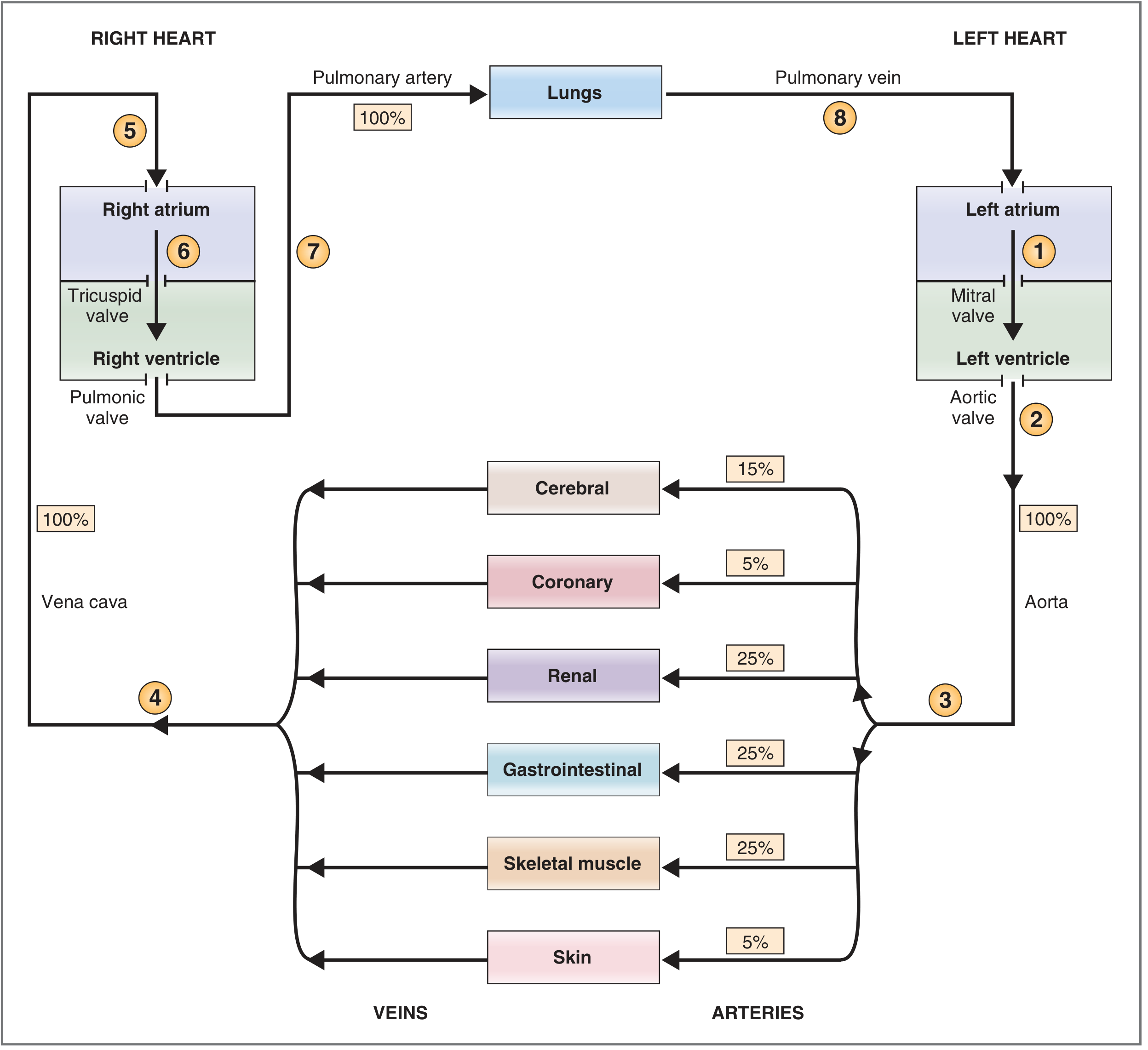
Body → venae cavae → right atrium → tricuspid valve → right ventricle → pulmonary valve → pulmonary arteries → lungs → pulmonary veins → left atrium → mitral valve → left ventricle → aortic valve → aorta → body
The right and left hearts work in series: the right ventricle supplies the pulmonary circulation and the left ventricle supplies the systemic circulation. NHLBI overview describes this one-way flow and the role of the four valves.
This is a shared conversation. Sign in to Orris to start your own chat.