
pulmonary surfactant . Synthesis ,regulation, function and clinical explain ?
I'll start by reading the medical library skill, then search for comprehensive information on pulmonary surfactant.
Reading File
Now I'll search the medical library and PubMed simultaneously for comprehensive information on pulmonary surfactant.
Finding Sources
Finding Sources
Searching PubMed
pulmonary surfactant synthesis regulation function
Excellent results. Now I'll read the key textbook sections in parallel - Fishman's chapter on pulmonary surfactant (the main dedicated chapter) plus the Murray & Nadel sections.
Reading File
Reading File
Reading File
Excellent content. Now let me read the synthesis/regulation section from Fishman's block1 and search for images, plus get more detail on the clinical disorders.
Reading File
Reading File
Searching Images
pulmonary surfactant synthesis type II pneumocyte alveolus
Let me read the key synthesis/regulation content from Fishman's and fetch the alveolar ultrastructure image already found.
Reading File
Loading Image
Excellent. Let me fetch the surfactant metabolism diagram (Fig 5-2) and also read further for the regulation and clinical RDS sections.
Loading Image
Reading File
I now have comprehensive content from the textbooks plus the two key diagrams. Let me compile the full detailed answer.
Pulmonary Surfactant: Synthesis, Regulation, Function, and Clinical Significance
Overview
Pulmonary surfactant is an emulsion of phospholipids, cholesterol, and specific apoproteins that coats the inner surface of alveoli. Its primary role is to reduce surface tension at the air-liquid interface, preventing alveolar collapse at end-expiration. Without it, the ~70 dyn/cm² surface tension generated at a simple air-water interface would cause widespread alveolar collapse and respiratory failure.
- Fishman's Pulmonary Diseases and Disorders, p. 122
1. Composition
Surfactant isolated by bronchoalveolar lavage consists of heterogeneous phospholipid-protein aggregates:
| Component | Proportion | Role |
|---|---|---|
| Phospholipids | 80-90% of mass | Surface tension reduction |
| - DPPC (dipalmitoylphosphatidylcholine) | ~50-60% of lipid | Primary surface tension reducer |
| - Phosphatidylglycerol (PG) | 5-10% of lipid | Film stability |
| - Phosphatidylcholine (PC) | 70-80% of lipid | Major lipid component |
| Cholesterol | ~10% | Film fluidity |
| Surfactant Proteins (SP-A, B, C, D) | ~10% | Film formation, immunity |
DPPC is distinctive in that both fatty acid chains are saturated, making it capable of forming a tightly packed monolayer film at the air-liquid interface. The phospholipids are amphiphilic and spontaneously organize into surface-reducing films.
Structural forms in the alveolus:
-
Tubular myelin - the most abundant form; a highly organized, square tubular array composed of large, dense aggregates (large aggregate surfactant). This represents the extracellular pool that rapidly moves to the air-liquid interface.
-
Lamellar body contents - newly secreted, unraveling to form tubular myelin
-
Small aggregate surfactant - less dense, remnant/catabolic forms with poor surface activity, destined for uptake or degradation
-
Fishman's Pulmonary Diseases, pp. 122-123
2. Surfactant Proteins (SP-A, SP-B, SP-C, SP-D)
SP-A and SP-D (Hydrophilic collectins)
- Encoded by genes on chromosome 10 (SP-A) and chromosome 19 (SP-D)
- SP-A: Calcium-dependent carbohydrate-binding protein; promotes formation of tubular myelin; modulates alveolar macrophage function; enhances bacterial and viral clearance; regulates surfactant recycling by AT2 cells
- SP-D: Modulates surfactant homeostasis and alveolar immune defense; Sftpd-knockout mice develop severe pulmonary disease with macrophage accumulation confirming its essential role in alveolar homeostasis
SP-B (Hydrophobic)
- Encoded by SFTPB (chromosome 2); 79-amino acid protein
- Stored in lamellar bodies and secreted with phospholipids
- Critical for surfactant film formation and stability - enhances lipid adsorption, aids multilayer formation
- SP-B and SP-C together improve lung inflation, compliance, and are used in exogenous surfactant therapy
- Homozygous loss-of-function mutations → neonatal respiratory failure and death
SP-C (Hydrophobic)
-
Encoded by SFTPC (chromosome 8); 35-amino acid hydrophobic peptide; expressed exclusively in AT2 cells
-
Palmitoylated on cysteine residues (the palmitoyl groups enhance hydrophobicity and stability of the alpha-helical domain anchoring it in the lipid bilayer)
-
Enhances surface-active properties, reduces surface tension during compression, and enhances adsorption rate at the air-water interface
-
Dominant mutations in SFTPC → interstitial lung disease in neonates, children, and adults (not RDS alone)
-
Fishman's Pulmonary Diseases, pp. 123-125
3. Synthesis and Intracellular Trafficking
The AT2 (alveolar type II) pneumocyte is the sole site of surfactant synthesis and secretion.
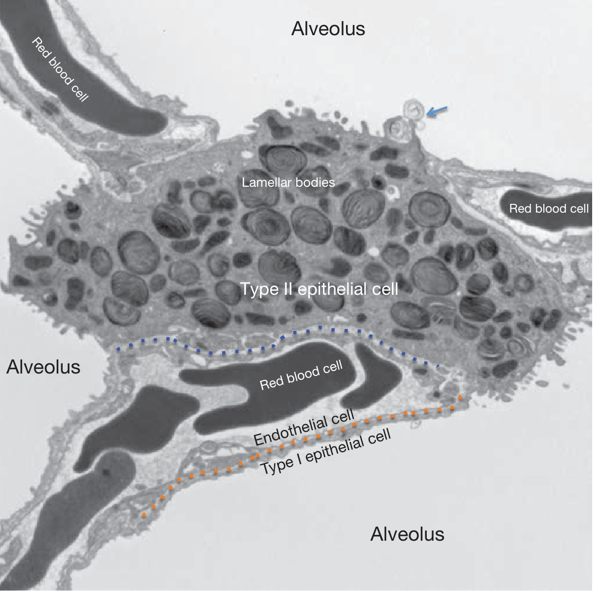
Figure: Electron micrograph of the alveolar ultrastructure. Type II cells contain prominent lamellar bodies (intracellular surfactant stores). Arrow indicates secretion into the alveolar space. - Fishman's Pulmonary Diseases, Fig 5-1
The intracellular pathway:
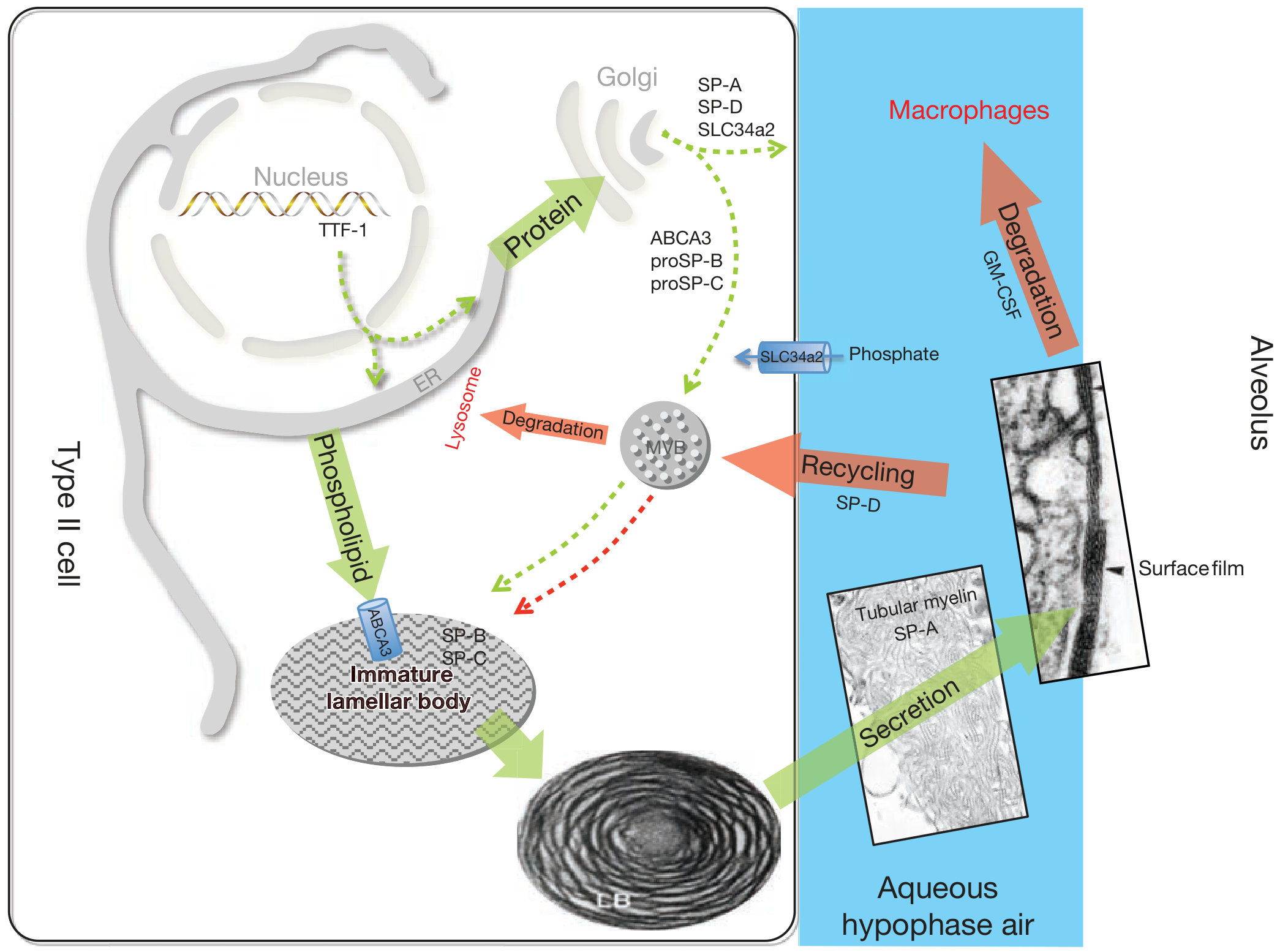
Figure: Surfactant metabolism. Surfactant proteins (proSP-B, proSP-C) and ABCA3 traffic from ER → Golgi → Multivesicular Body (MVB) → Lamellar Body (LB). Phospholipids are transported directly ER → LB via lipid transfer proteins. After secretion, SP-A promotes tubular myelin formation and the air-liquid surface film. Recycling occurs via AT2 cell uptake; degradation via macrophages and lysosomes. - Fishman's Pulmonary Diseases, Fig 5-2
Step-by-step synthesis:
- Phospholipid synthesis in the endoplasmic reticulum (ER) - DPPC, PG, PC are synthesized
- Protein synthesis - proSP-B and proSP-C are translated in the ER
- Golgi processing - proteins traffick through the Golgi apparatus; SP-A and SP-D are secreted via the conventional Golgi pathway
- Multivesicular body (MVB) - proteins (proSP-B, proSP-C) converge with phospholipids; proteolytic processing to mature peptides begins
- Lamellar body (LB) formation - the definitive intracellular storage organelle for surfactant; ABCA3 (an ABC transporter) pumps phospholipids (phosphatidylcholine, cholesterol, sphingomyelin, phosphatidylglycerol) into the LB; SP-B and SP-C are stored in the LB; the MVB serves as the integration hub for synthesis, secretion, recycling, and degradation pathways
- Secretion by exocytosis - LBs fuse with the apical plasma membrane and release contents into the alveolar space
- Extracellular assembly - in the alveolar hypophase (aqueous lining fluid), SP-A facilitates the transformation of lamellar body material into tubular myelin, which reorganizes into multilayered phospholipid surface films
TTF-1 (NKX2.1) is the master transcription factor driving AT2 cell differentiation and is critical for expression of SP-B, SP-C, and ABCA3. Haploinsufficiency causes a triad of hypothyroidism, brain abnormalities, and lung disease.
- Fishman's Pulmonary Diseases, p. 124
4. Regulation of Surfactant Synthesis
Surfactant synthesis is regulated at multiple levels:
A. Developmental/Hormonal Regulation
- Fetal lung surfactant phospholipid content increases markedly with advancing gestational age, driven by a complex signaling/transcriptional network controlling AT2 differentiation
- Glucocorticoids (corticosteroids) are the most potent inducers of lung maturation and surfactant synthesis; they upregulate SP-A, SP-B, SP-C gene expression and accelerate lamellar body development
- Thyroid hormones and beta-adrenergic agents also stimulate surfactant synthesis and secretion
- Glucocorticoid administration to mothers at risk of preterm delivery (<34 weeks) is standard practice to accelerate fetal lung maturation
B. Transcriptional Regulation
- TTF-1/NKX2.1: Master regulator of AT2 cell-specific gene expression (SP-B, SP-C, ABCA3)
- The surfactant protein genes have highly conserved promoter elements responsive to glucocorticoids, thyroid hormones, and other factors
C. Secretion Regulation
- Surfactant secretion by AT2 cells is stimulated by:
- Beta-adrenergic agonists (catecholamines) - act via cAMP
- Mechanical stretch (lung inflation/breathing movements)
- Purinergic signaling (ATP released during stretch)
- Phorbol esters (PKC activation)
- Secretion is inhibited by: SP-A (negative feedback on secretion), nitrogen mustard
D. Recycling Regulation
- ~50% of secreted surfactant is reuptaken by AT2 cells and repackaged for re-use - a highly efficient recycling system
- SP-D facilitates the reuptake and recycling of small aggregate surfactant
- Alveolar macrophages degrade the rest via lysosomal pathways; GM-CSF (granulocyte-macrophage colony-stimulating factor) is essential for macrophage clearance of surfactant - its deficiency leads to pulmonary alveolar proteinosis (PAP)
E. Fetal Maturity Tests
Because surfactant composition reflects lung maturity, amniotic fluid analysis predicts postnatal respiratory function:
-
Lecithin:Sphingomyelin (L/S) ratio ≥2.0 = lung maturity
-
Phosphatidylglycerol appearance in amniotic fluid
-
Lamellar body counts
-
Fishman's Pulmonary Diseases, p. 124; Murray & Nadel's Respiratory Medicine, p. 81
5. Functions of Pulmonary Surfactant
Primary Function - Surface Tension Reduction
- The phospholipid monolayer at the air-liquid interface lowers surface tension from ~70 dyn/cm² (at a plain air-water interface) to nearly zero at end expiration
- By Laplace's law (P = 2T/r): without surfactant, small alveoli would collapse into large ones; surfactant preferentially reduces tension more at smaller radii, stabilizing alveoli of different sizes
- This prevents atelectasis, reduces the work of breathing, and maintains FRC (functional residual capacity)
Additional Functions
| Function | Mechanism |
|---|---|
| Antioxidant protection | Scavenges free radicals, reducing oxygen toxicity |
| Anti-inflammatory | Suppresses mediator release by inflammatory cells; deactivates released mediators; suppresses lymphocyte mitogenic responses |
| Host defense | Antibacterial and antiviral properties via increased alveolar macrophage phagocytosis (opsonization by SP-A and SP-D) |
| Airway clearance | Alters physical properties of mucus; increases ciliary beat frequency |
| Airway smooth muscle relaxation | Direct relaxation of bronchial smooth muscle |
| Cytoprotection | Protects alveolar cell surfaces |
- Fishman's Pulmonary Diseases, p. 2567
6. Clinical Conditions Related to Surfactant
A. Neonatal Respiratory Distress Syndrome (RDS / Hyaline Membrane Disease)
- Cause: Deficiency of surfactant in premature infants (<34-36 weeks); AT2 cells have not yet produced adequate surfactant
- Pathophysiology: Increased alveolar surface tension → alveolar collapse → ventilation-perfusion mismatch → hypoxemia → protein-rich fluid exudate → hyaline membrane formation → respiratory failure
- Prevention: Antenatal glucocorticoids to mother (betamethasone/dexamethasone) given 24-48h before preterm delivery
- Treatment: Exogenous surfactant replacement (intratracheal instillation via endotracheal tube); newer delivery methods include nebulization and thin-catheter instillation during nasal CPAP (INSURE technique)
- Surfactant therapy reduces RDS mortality by 30-40% and reduces morbidity from pneumothorax, pulmonary interstitial emphysema, bronchopulmonary dysplasia (BPD), and intraventricular hemorrhage
- Natural surfactants (from bovine or porcine lung lavage) are superior to original synthetic surfactants; newer-generation synthetic surfactants with peptide analogues of SP-B and SP-C are in clinical trials
B. Acute Respiratory Distress Syndrome (ARDS)
- Plasma proteins that leak into alveoli during ARDS inactivate surfactant - fibrinogen, albumin, and lipids from inflammatory cells competitively displace surfactant from the air-liquid interface
- Surfactant therapy in ARDS is under investigation; recent 2025 meta-analysis (PMID 40399976) in children shows potential benefit in reducing mechanical ventilation duration
C. Meconium Aspiration Syndrome
- Meconium inactivates surfactant
- Exogenous surfactant reduces the need for mechanical ventilation and ECMO, and reduces hospital stay
D. Genetic Surfactant Dysfunction Disorders
These are caused by mutations disrupting surfactant production/function:
| Gene | Mutation Effect | Clinical Presentation |
|---|---|---|
| SFTPB | Recessive loss-of-function (121ins2 frameshift most common, ~60%) | Fatal neonatal respiratory failure; absent SP-B in BAL; alveolar proteinosis pattern |
| SFTPC | Dominant mutations (I73T most common, ~25%) | Interstitial lung disease in neonates, children, adults; variable severity |
| ABCA3 | Autosomal recessive | Fatal neonatal surfactant deficiency OR chronic respiratory insufficiency in older children; surfactant deficient in phosphatidylcholine |
| NKX2.1 (TTF-1) | Haploinsufficiency | "Brain-thyroid-lung" syndrome: hypothyroidism + brain abnormalities + lung disease |
- Murray & Nadel's Respiratory Medicine, pp. 541-555
E. Pulmonary Alveolar Proteinosis (PAP)
- Accumulation of surfactant lipids and proteins in alveoli due to defective clearance by macrophages
- Autoimmune PAP: anti-GM-CSF antibodies block macrophage maturation (most common form)
- Congenital PAP: mutations in GM-CSF receptor (CSF2RA, CSF2RB) or SP genes
- Treatment: whole-lung lavage; GM-CSF inhaled or subcutaneous (for autoimmune PAP)
F. Exogenous Surfactant Preparations
| Preparation | Source |
|---|---|
| Beractant (Survanta) | Bovine lung extract |
| Calfactant (Infasurf) | Bovine lung lavage |
| Poractant alfa (Curosurf) | Porcine lung extract |
| Lucinactant (Surfaxin) | Synthetic with SP-B peptide analogue (KL4 peptide) |
Summary
Pulmonary surfactant is synthesized exclusively by AT2 cells through a pathway involving ER → Golgi → MVB → lamellar body → exocytosis. Its composition (dominated by DPPC and surfactant proteins SP-A/B/C/D) enables it to reduce alveolar surface tension to near zero, preventing atelectasis. Regulation occurs developmentally (glucocorticoids being most important), transcriptionally (TTF-1), and dynamically (stretch, beta-adrenergic signaling). Beyond surface tension, surfactant provides immune defense, antioxidant protection, and mucociliary assistance. Deficiency - whether from prematurity, genetic mutations, or inactivation by plasma proteins - underlies several critical respiratory diseases, and exogenous surfactant replacement remains one of the most effective interventions in neonatal medicine.
Sources:
- Fishman's Pulmonary Diseases and Disorders, 2-Volume Set, Chapter 5 (Surfactant Composition, Synthesis, and Metabolism), pp. 122-125
- Murray & Nadel's Textbook of Respiratory Medicine, 2-Volume, Chapter 3 (Functional Roles of the Alveolar Unit), pp. 81-82; Chapter 23 (Surfactant Metabolic Dysfunction Disorders), pp. 541-555
- Fishman's Pulmonary Diseases, p. 2567 (Pulmonary Surfactant Clinical Use)
- Recent evidence: Surfactant + budesonide in premature infants (PMID 39787118) - 2025 meta-analysis; Surfactant therapy in pediatric ARDS (PMID 40399976) - 2025 meta-analysis
Humburger phenomenon and haldane effect explain
Reading File
Now searching the library for both phenomena simultaneously.
Finding Sources
Finding Sources
Finding Sources
Finding Sources
Good - found Haldane effect content in multiple textbooks. Now searching specifically for "chloride shift" (the biochemical name for the Hamburger phenomenon) and reading the Haldane sections in parallel.
Finding Sources
Reading File
Reading File
Excellent content. Now let me read the chloride shift (Hamburger phenomenon) section from Morgan & Mikhail and Guyton, plus fetch the Haldane effect diagram.
Reading File
Reading File
Loading Image
Loading Image
I also need the CO2 transport diagram from Guyton that shows the chloride shift mechanism. Let me fetch it.
Loading Image
I now have all the content and diagrams needed for a thorough answer. Let me compile the complete response.
Hamburger Phenomenon (Chloride Shift) and the Haldane Effect
These two phenomena are both related to CO₂ transport in the blood and are deeply interconnected. They explain how the body efficiently moves CO₂ from tissues to the lungs for elimination.
Part 1: The Hamburger Phenomenon (Chloride Shift)
Definition
The Hamburger phenomenon, also called the chloride shift, is the exchange of bicarbonate (HCO₃⁻) and chloride (Cl⁻) ions across the red blood cell (RBC) membrane that occurs as a consequence of CO₂ transport - in opposite directions at the tissues versus the lungs.
It was first described by the Dutch physiologist Hartog Jakob Hamburger in the late 19th century.
Mechanism - At the Tissue Capillaries
CO₂ produced by tissue metabolism enters the blood and follows this sequence:
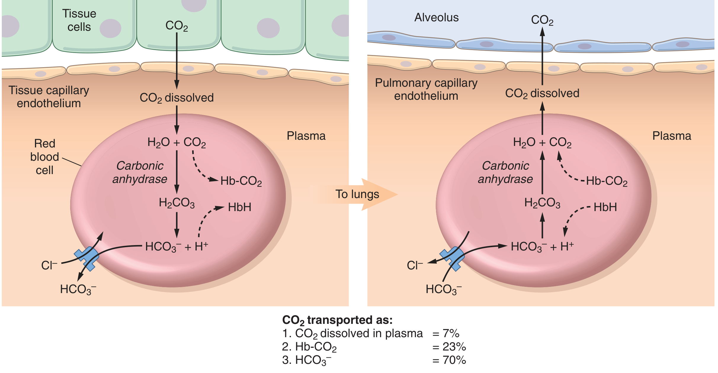
Figure: CO₂ transport in blood. Left panel - tissue capillaries (Hamburger shift: HCO₃⁻ exits RBC, Cl⁻ enters). Right panel - pulmonary capillaries (reversal: HCO₃⁻ re-enters, Cl⁻ exits). CO₂ transported as: dissolved 7%, Hb-CO₂ 23%, HCO₃⁻ 70%. - Guyton & Hall Medical Physiology, Fig 41.13
Step-by-step at tissues:
-
CO₂ diffuses from tissue cells into plasma, then into RBCs (dissolved CO₂ = ~7% of total transport)
-
Inside the RBC, CO₂ + H₂O → H₂CO₃ (catalyzed by carbonic anhydrase, ~5000× faster than in plasma - this is why the reaction is physiologically meaningful)
-
H₂CO₃ rapidly dissociates: H₂CO₃ → H⁺ + HCO₃⁻
-
The H⁺ ions are buffered by hemoglobin (histidine residues of Hb act as a buffer, absorbing H⁺): HbO₂ + H⁺ → HbH⁺ + O₂
-
HCO₃⁻ accumulates inside the RBC and diffuses out into plasma down its concentration gradient - this is the major form of CO₂ transport (~70% of total CO₂)
-
To maintain electrical neutrality (since HCO₃⁻ is leaving the cell), Cl⁻ ions move from plasma INTO the RBC via a specific bicarbonate-chloride anion exchanger (Band 3 protein / AE1) in the RBC membrane - this is the Hamburger / chloride shift
Result: Venous RBCs have a higher Cl⁻ content than arterial RBCs. Venous blood plasma has a higher HCO₃⁻ content than arterial plasma.
Reversal - At the Pulmonary Capillaries
In the lungs, the entire process reverses:
- PO₂ rises (from ~40 to ~100 mmHg) → O₂ binds hemoglobin (HbH⁺ + O₂ → HbO₂ + H⁺)
- Released H⁺ combines with HCO₃⁻: H⁺ + HCO₃⁻ → H₂CO₃ → CO₂ + H₂O
- CO₂ is released and diffuses into the alveolus
- As HCO₃⁻ is consumed, more HCO₃⁻ re-enters the RBC from plasma
- Cl⁻ exits the RBC back into plasma to maintain electrical neutrality (reverse chloride shift)
Result: Cl⁻ content of arterial RBCs is lower than venous RBCs. CO₂ is expelled from the lungs.
Key Facts Summary
| Feature | At Tissues | At Lungs |
|---|---|---|
| CO₂ movement | Enters RBC | Exits RBC |
| HCO₃⁻ movement | Exits RBC → plasma | Enters RBC from plasma |
| Cl⁻ movement | Enters RBC from plasma | Exits RBC → plasma |
| Transporter | Band 3 protein (AE1 - anion exchanger) | Band 3 protein (AE1) |
| Net effect on RBC | Cl⁻↑, HCO₃⁻↓ | Cl⁻↓, HCO₃⁻↑ |
Why it matters clinically:
-
Carbonic anhydrase inhibitors (e.g., acetazolamide) block this reaction, significantly impairing CO₂ transport - tissue PCO₂ can rise from 45 to ~80 mmHg
-
The chloride shift causes a slight increase in venous RBC volume (as Cl⁻ entry draws water in osmotically) - this slightly increases venous hematocrit (the "venous hematocrit effect")
-
Guyton & Hall Medical Physiology, p. 529; Morgan & Mikhail's Clinical Anesthesiology, p. 967
Part 2: The Haldane Effect
Definition
The Haldane effect states that deoxygenated hemoglobin (deoxyhemoglobin) carries significantly more CO₂ than oxygenated hemoglobin at the same PCO₂. Conversely, oxygenation of hemoglobin promotes release of CO₂. Named after John Scott Haldane, the Scottish physiologist.
Guyton & Hall notes: "The Haldane effect is quantitatively far more important in promoting CO₂ transport than the Bohr effect is in promoting O₂ transport."
The CO₂ Dissociation Curve - Graphic Explanation
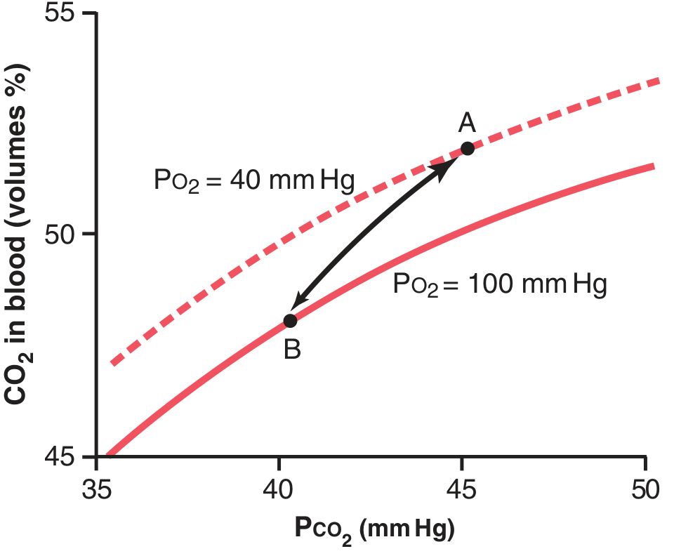
Figure: Haldane effect. At PCO₂ = 45 mmHg in tissues (Point A, PO₂ = 40 mmHg), blood carries 52 vol% CO₂. In the lungs, PCO₂ drops to 40 mmHg, but the rise in PO₂ to 100 mmHg shifts the dissociation curve downward (Point B). Blood now carries only 48 vol% CO₂ - releasing 4 vol% total (2% from PCO₂ fall + 2% additional from the Haldane effect). The Haldane effect doubles CO₂ release. - Guyton & Hall, Fig 41.15
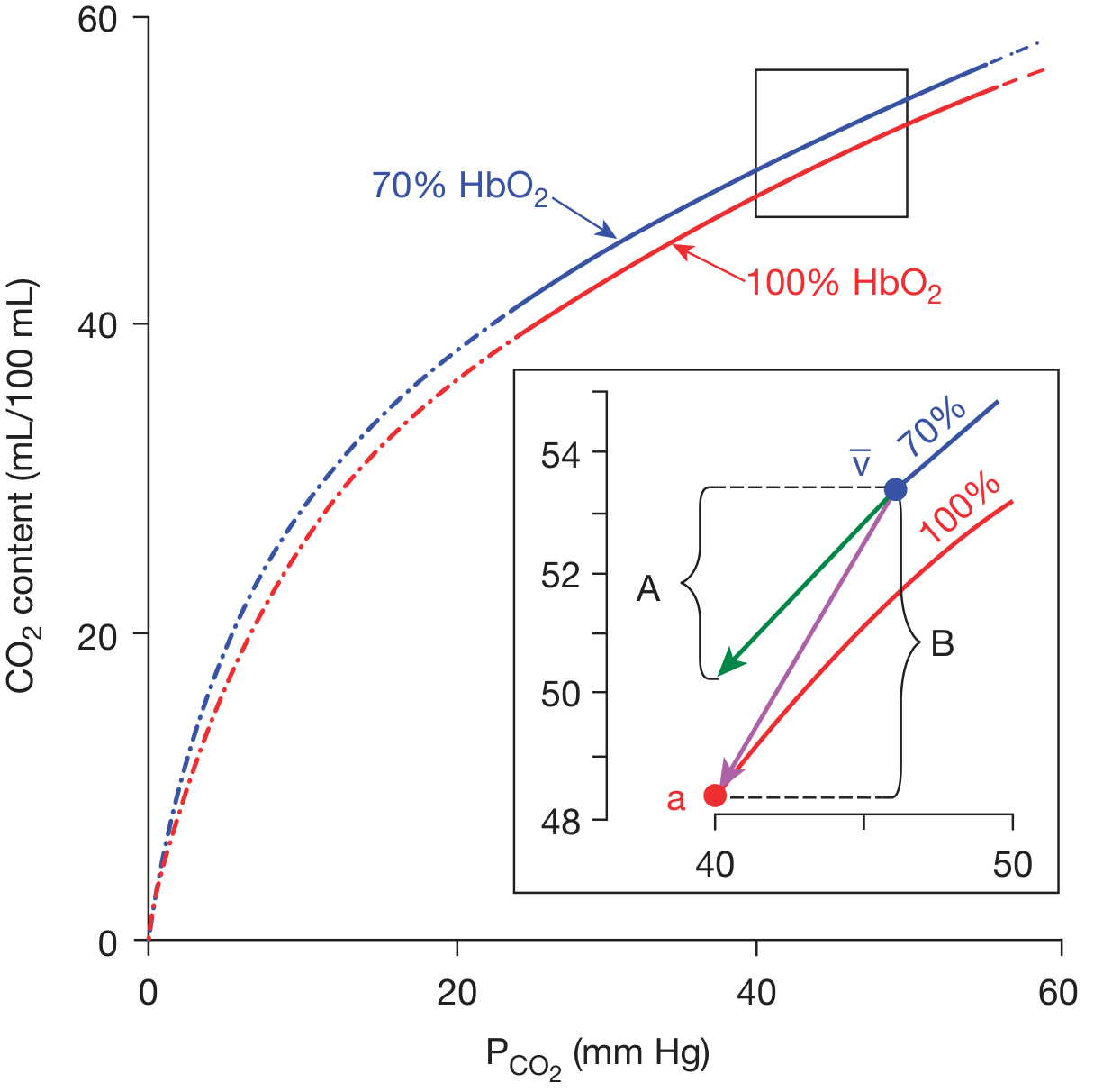
Figure: CO₂ dissociation curves at 70% (blue, venous, tissues) and 100% (red, arterial, lungs) hemoglobin oxygen saturation. Inset shows: A = CO₂ released due to falling PCO₂ alone; B = additional CO₂ released due to oxygenation (Haldane effect). - Fishman's Pulmonary Diseases, Fig 15-4
Mechanism of the Haldane Effect
The Haldane effect operates via two simultaneous mechanisms, both related to changes in hemoglobin's molecular configuration upon oxygenation:
Mechanism 1: Oxylabile Carbamate (Carbaminohemoglobin)
- CO₂ binds to N-terminal amino groups of hemoglobin to form carbaminohemoglobin (Hb-CO₂): R-NH₂ + CO₂ → R-NHCOO⁻ + H⁺
- Deoxyhemoglobin has 3.5× greater affinity for CO₂ than oxyhemoglobin (due to changes in quaternary structure upon deoxygenation that make N-terminal amino groups more available for carbamate formation)
- In tissues: O₂ is released → hemoglobin becomes deoxyhemoglobin → binds more CO₂ as carbamate
- In lungs: O₂ binds → hemoglobin becomes oxyhemoglobin → becomes a stronger acid → less tendency to form carbaminohemoglobin → CO₂ released
This carbamate mechanism accounts for approximately 1/8 of the arterio-venous CO₂ difference (its physiological importance would be twice as great, but is reduced by competition with 2,3-DPG for the same N-terminal amino groups)
Mechanism 2: Oxylabile Buffering (Bicarbonate Pathway)
- The pKa of hemoglobin changes with its oxygenation state
- Deoxyhemoglobin is a stronger base (weaker acid) than oxyhemoglobin
- In tissues: Hb deoxygenates → becomes better at buffering H⁺ → this H⁺ buffering drives the reaction CO₂ + H₂O → H₂CO₃ → H⁺ + HCO₃⁻ further to the right → more CO₂ stored as bicarbonate
- Reaction: CO₂ + H₂O + HbO₂ → HbH⁺ + HCO₃⁻ + O₂
- In lungs: O₂ binds Hb → Hb becomes oxyhemoglobin → weaker base → releases H⁺ → H⁺ + HCO₃⁻ → H₂CO₃ → CO₂ released
Both mechanisms together account for the full Haldane effect. Approximately equal contributions come from oxylabile carbamate and oxylabile buffering (bicarbonate formation).
Quantitative Significance
| Scenario | CO₂ content |
|---|---|
| Tissues (PCO₂ 45 mmHg, PO₂ 40 mmHg) | 52 vol% |
| Lungs - PCO₂ alone drops to 40 mmHg | Would only fall to 50 vol% (2 vol% released) |
| Lungs - PCO₂ drop + Haldane effect (PO₂ rises to 100 mmHg) | Falls to 48 vol% (4 vol% released) |
The Haldane effect approximately doubles the amount of CO₂ released at the lungs and approximately doubles the amount of CO₂ picked up at the tissues. Under normal conditions, the Haldane effect accounts for 40-50% of total CO₂ exchange.
- Fishman's Pulmonary Diseases, pp. 258-259; Guyton & Hall, p. 530
Comparing the Two Phenomena
| Feature | Hamburger Phenomenon | Haldane Effect |
|---|---|---|
| What it describes | Ion exchange (Cl⁻/HCO₃⁻) across RBC membrane | CO₂ carrying capacity of Hb changes with O₂ binding |
| Primary molecule involved | Band 3 anion exchanger; carbonic anhydrase | Hemoglobin (deoxy vs. oxy) |
| Effect at tissues | Cl⁻ enters RBC; HCO₃⁻ exits to plasma | Deoxy-Hb picks up more CO₂ (carbamate + bicarbonate) |
| Effect at lungs | Cl⁻ exits RBC; HCO₃⁻ re-enters | Oxy-Hb releases CO₂ |
| Reversal? | Fully reverses at lungs | Fully reverses at lungs |
| Relationship | The chloride shift is the mechanism that moves the bicarbonate produced by the Haldane effect's buffering component out of the RBC | The Haldane effect's buffering drives bicarbonate production, which then triggers the chloride shift |
Relationship to the Bohr Effect
The Bohr effect (CO₂/H⁺ reduces Hb's O₂ affinity at tissues, promoting O₂ delivery) and the Haldane effect (O₂ reduces Hb's CO₂ affinity at lungs, promoting CO₂ release) are mirror-image phenomena - the two effects synergize to simultaneously unload O₂ at tissues and load CO₂, then load O₂ at lungs and unload CO₂.
Clinical Relevance
| Condition | Relevance |
|---|---|
| Acetazolamide (carbonic anhydrase inhibitor) | Blocks the chloride shift mechanism → CO₂ transport severely impaired; used clinically for altitude sickness, glaucoma, metabolic alkalosis |
| Respiratory acidosis | Acute rise in plasma HCO₃⁻ largely due to the chloride shift - as CO₂ rises, more HCO₃⁻ is generated inside RBCs and exits in exchange for Cl⁻ |
| High-flow O₂ therapy | Oxygenation of Hb (Haldane effect) reduces CO₂ carrying capacity - can worsen hypercapnia in CO₂ retainers (COPD) by the "Haldane effect" mechanism (along with Bohr effect reversal) |
| Carbon monoxide poisoning | CO-Hb cannot participate in normal O₂/CO₂ exchange - both chloride shift and Haldane effect are impaired |
Sources:
- Guyton & Hall Textbook of Medical Physiology, pp. 529-530
- Fishman's Pulmonary Diseases and Disorders, pp. 258-259 (Haldane Effect)
- Morgan & Mikhail's Clinical Anesthesiology, 7e, pp. 967-968 (Chloride/Hamburger shift; Haldane effect)
- Murray & Nadel's Textbook of Respiratory Medicine, p. 82 (CO₂ transport and Haldane effect)
caission disease
Your credits are exhausted. You can purchase a top-up or subscribe to a plan to continue using Orris. If you're already subscribed, your daily credits will reset tomorrow.
This is a shared conversation. Sign in to Orris to start your own chat.