
Druj subluxation left
DRUJ distal radioulnar joint subluxation wrist
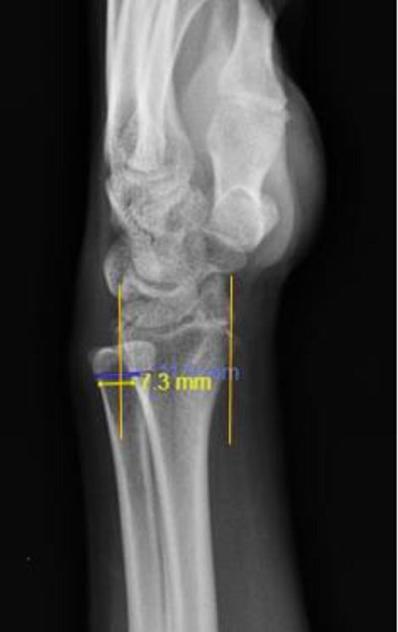
This diagnostic image is a lateral radiograph of the wrist and distal forearm demonstrating the radiographic assessment of the distal radioulnar joint (DRUJ) using the Mino method. The image shows the distal radius, distal ulna, and carpal bones in a lateral profile. Two vertical reference lines are drawn parallel to the longitudinal axis of the radius, originating from the volar and dorsal margins of the distal radial articular surface. Overlaid measurements in yellow indicate a quantitative analysis of ulnar head displacement. A specific measurement of 7.3 mm highlights the distance of the dorsal subluxation of the ulnar head relative to the dorsal reference line. According to the Mino criteria, instability or subluxation is diagnosed when more than 25% of the ulnar head width lies outside these boundaries. This visual demonstrates dorsal DRUJ subluxation, a significant finding in cases of wrist trauma or ligamentous instability, typically used by orthopedic and radiology professionals to evaluate joint congruity.
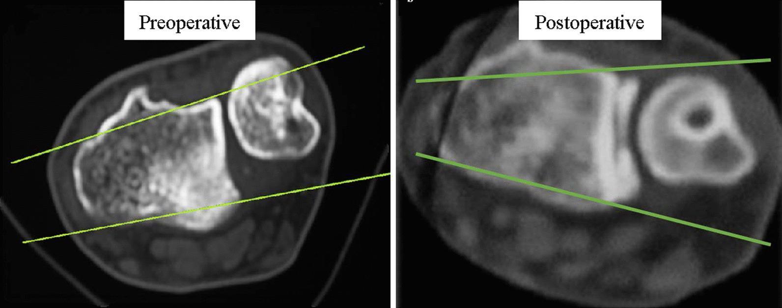
This diagnostic comparison contains two cross-sectional axial CT scans of the wrist, demonstrating the distal radioulnar joint (DRUJ) in neutral position. Both images utilize Mino's criteria, with two parallel reference lines drawn along the dorsal and palmar cortical borders of the distal radius to assess ulnar alignment. The 'Preoperative' image shows the ulnar head displaced dorsally, lying outside the space defined by the tracing lines, which indicates dorsal subluxation of the DRUJ. The 'Postoperative' image displays the ulnar head successfully reduced and centered between the two reference lines, signifying restored joint congruency following surgical reconstruction. These images serve as an educational tool for evaluating TFCC injury-related instability and the efficacy of anatomic radioulnar ligament reconstruction. Key anatomical features visible include the distal radius sigmoid notch, the ulnar head, and internal bony trabecular patterns.
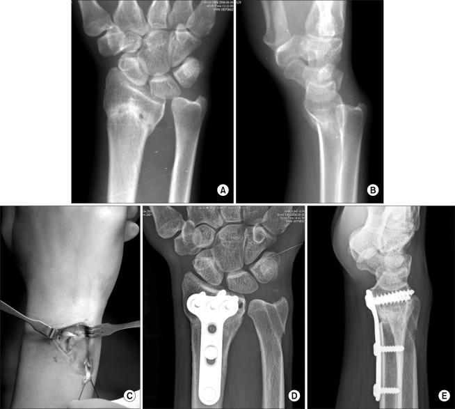
A multi-panel medical image sequence illustrating the management of distal radioulnar joint (DRUJ) instability. Subfigures A and B are preoperative anteroposterior and lateral X-rays of the wrist showing malunion of a distal radius fracture and associated DRUJ subluxation. Subfigure C is an intraoperative clinical photograph showing a palmaris longus tendon graft being used for ligament reconstruction. Subfigures D and E are postoperative anteroposterior and lateral radiographs showing surgical correction. These images demonstrate internal fixation with a volar locking plate and screws on the distal radius following a corrective osteotomy. Restored anatomical alignment of the radius and ulnar head is visible. Key diagnostic features include the presence of radiolucent bone tunnels in the distal radius and ulnar head, indicating the pathways for the ligament reconstruction. The image provides educational value for orthopedic surgery, specifically in treating distal radius malunion and chronic DRUJ instability through combined bony and soft tissue reconstruction.
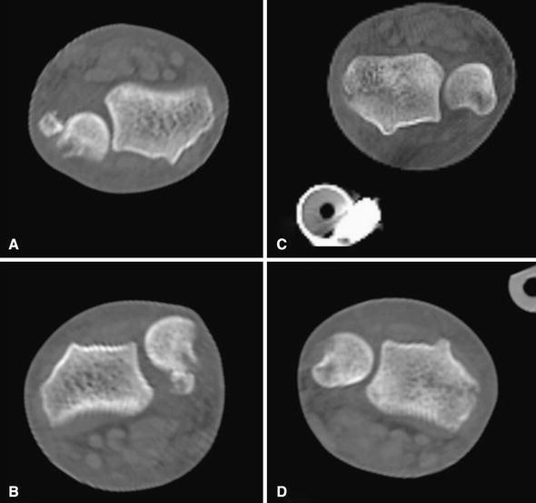
Axial plane computed tomography (CT) scans of bilateral wrists comparing the congruity of the distal radioulnar joint (DRUJ) in different forearm rotations. Figures A and B represent the right wrist in supination and pronation, respectively, demonstrating persistent dorsal subluxation of the ulnar head relative to the sigmoid notch of the radius. This finding is characteristic of an unstable DRUJ following a Galeazzi-type injury. Figures C and D show the left wrist in supination and pronation, illustrating near-normal anatomical congruity where the ulnar head remains appropriately seated within the radial notch throughout the rotational range. Image C also contains a cross-section of an external fixator pin. These diagnostic images are used to assess post-surgical stability and alignment in orthopedic trauma cases involving radius fractures with associated DRUJ disruption.
"distal radioulnar joint" subluxation treatment
Distal Radioulnar Joint (DRUJ) Subluxation - Left
Anatomy & Biomechanics
- Triangular fibrocartilage complex (TFCC) - the primary stabilizer; its deep fibers (foveal attachment) are especially important
- Anterior and posterior radioulnar ligaments (parts of the joint capsule)
- Ulnar collateral ligament - attaches from ulnar styloid tip to pisiform/triquetrum
- Pronator quadratus muscle - spans the volar distal forearm and acts as a dynamic stabilizer
- Bony geometry - the sigmoid notch of the radius articulates with the ulnar head
Types of DRUJ Subluxation
| Type | Direction | Forearm Position | Mechanism |
|---|---|---|---|
| Dorsal (most common) | Ulnar head displaced dorsally | Pronation | Fall on outstretched hand; associated with distal radius fractures, Galeazzi fracture |
| Volar | Ulnar head displaced volarly | Supination | Less common; hypersupination injury |
Clinical Assessment
History
- Mechanism: FOOSH (fall on outstretched hand), twisting injury, or associated with distal radius fracture
- Pain at ulnar wrist, weakness of grip/rotation, clicking/clunking
- Acute vs. chronic (>6-8 weeks = chronic)
Examination Signs
- Piano key sign: With wrist pronated, the prominent dorsal ulnar head is springy/reducible with downward pressure - pathognomonic of dorsal DRUJ subluxation
- Ballottement/Shuck test: With the radial wrist stabilized, anteroposterior stress applied to the ulnar side detects excessive motion
- Press test: Patient pushes body weight up off a chair arms - reproduction of pain indicates DRUJ instability or ulnar impaction (low specificity)
- Fovea sign: Tenderness in the fovea (between ulnar styloid and FCU tendon) points to deep TFCC/ulnocarpal ligament injury
- Ulnar impaction test: Hyperextension + ulnar deviation + axial compression recreates pain
Imaging
| Modality | Findings |
|---|---|
| PA + lateral X-ray | Widening of DRUJ space on PA; ulnar head displaced on lateral. Assess ulnar variance. |
| CT (axial, bilateral) | Gold standard - compare both DRUJs in neutral, pronation, and supination. Mino criteria: subluxation if >25% of ulnar head width lies outside the lines drawn along volar and dorsal cortical borders of distal radius |
| MRI | Sensitivity/specificity ~100% for TFCC tears; best for soft tissue assessment. Note: many asymptomatic patients >40 yrs have positive MRI findings |
| CT arthrography | Highly sensitive for central TFCC tears |
Management
Acute DRUJ Subluxation (<2 months)
- Dorsal subluxation - supinate the forearm + direct pressure on the prominent ulnar head
- Volar subluxation - pronate the forearm
- Immobilize in a long arm cast for 4-6 weeks in the position of stability (supination for dorsal, pronation for volar)
- An excellent result is usually expected when reduced early
- Open reduction + TFCC repair
- Entrapped extensor carpi ulnaris (ECU) is the most common cause of irreducible dorsal dislocation - dorsal approach to free the ECU, then TFCC repair or transosseous K-wire pinning
Chronic DRUJ Instability (>2 months)
- Open reduction + TFCC repair - if <2 months and closed reduction failed
- Ligament reconstruction - rarely indicated; requires undeformed bones as prerequisite. Techniques include ECU tenodesis, FCU tenodesis, or tendon graft-based procedures
- Distal ulna resection (Darrach procedure) - for chronic instability with significant degenerative change; relieves pain but sacrifices ulnar support
- Ulnar head replacement / DRUJ arthroplasty - in select cases with arthritis
- Sauvé-Kapandji procedure - radioulnar arthrodesis with proximal pseudarthrosis; preserves the ulnar support of the carpus; recommended for fixed DRUJ subluxation with concomitant instability
Associated Injuries (Acute)
- Galeazzi fracture - radial shaft fracture + DRUJ disruption (DRUJ instability must always be assessed after Galeazzi fixation)
- Distal radius fracture - any fracture of the forearm can coexist with DRUJ dislocation; if unrecognized, the TFCC will not heal and recurrent instability follows
- TFCC tear - central vs. peripheral tears have different management (peripheral tears are more amenable to repair)
- Ulnar styloid fracture - large/base fractures destabilize the DRUJ
Key Points
- Failure to diagnose acute DRUJ dislocation/subluxation leads to chronic instability and eventual DRUJ osteoarthritis
- The piano key sign is the bedside hallmark of dorsal subluxation
- CT (bilateral, in multiple rotations) is the best imaging for quantifying subluxation
- Acute injuries reduced within 2 months have an excellent prognosis; chronic cases often require reconstructive surgery
- Ligament reconstruction is only viable if the bones are undeformed - this makes early diagnosis critical
Out ,: grossly intact male
What would you like me to do with this?