
Pic of recesses of lesser sac
Reading File
The lesser sac (omental bursa) has several recesses - the superior, inferior (greater), splenic, and caudate recesses. Let me find the best anatomical images from the medical library.
Searching sources...
Finding Sources
Finding Sources
Reading File
Finding Sources
I found several good sources with images. Let me read the Color Atlas section and fetch the key images in parallel.
Reading File
Reading File
Loading Image
Now let me get the Color Atlas image showing the omental bursa recesses specifically.
Reading File
I need to find the actual Color Atlas image of the omental bursa recesses. Let me look around that section for the figure.
Reading File
Found the key image. Let me fetch it along with another from the Grainger & Allison radiology text.
Loading Image
Excellent! Here are the images and a complete explanation of the recesses of the lesser sac (omental bursa):
Recesses of the Lesser Sac (Omental Bursa)
Fig. 21.47 - Peritoneal Cavity Diagram (Grainger & Allison's Diagnostic Radiology)
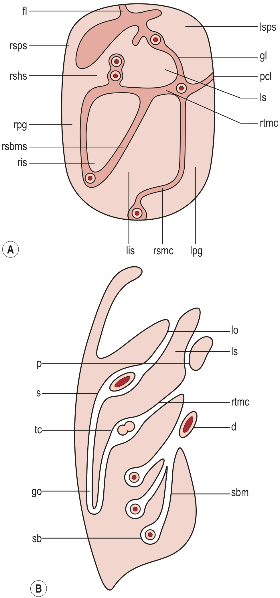
(A) Coronal diagram showing division of peritoneal cavity; (B) Midsagittal diagram - note ls = lesser sac lying posterior to the stomach (s), bounded anteriorly by the lesser omentum (lo) and posteriorly by the pancreas (p), above the root of transverse mesocolon (rtmc).
Fig. 4.41 - Topography of the Omental Bursa (Color Atlas of Human Anatomy)
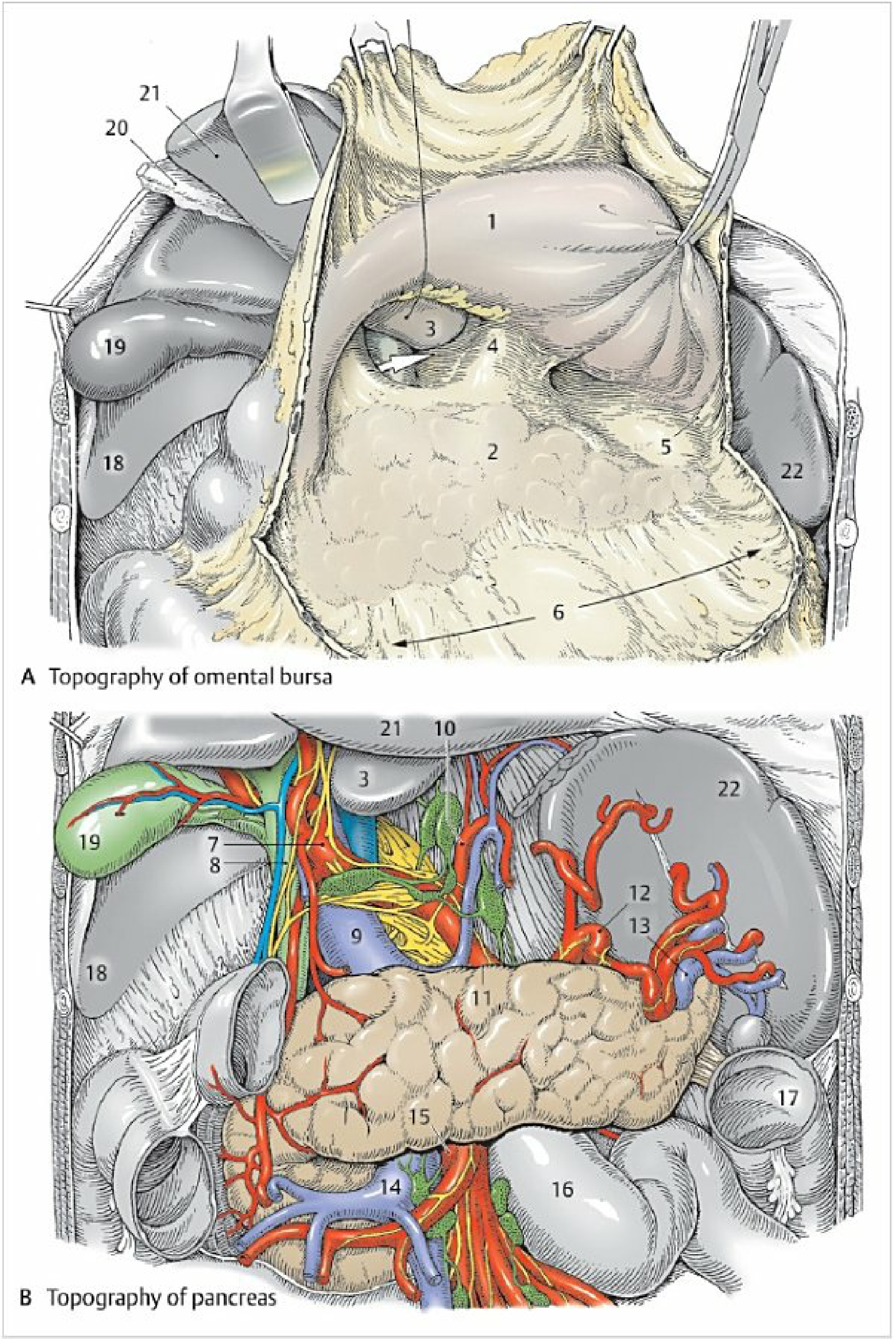
Panel A - Topography of the omental bursa (stomach and lesser omentum reflected superiorly):
| Label | Structure |
|---|---|
| 1 | Stomach (reflected, showing posterior wall) |
| 2 | Pancreas (posterior wall of omental bursa) |
| 3 | Papillary process of caudate lobe |
| 4 | Gastropancreatic fold (divides vestibule from main cavity) |
| 5 | Splenic recess (extending left between splenic ligaments and stomach) |
| 6 | Inferior recess (extending downward between greater curvature and transverse colon) |
| Arrow | Omental foramen (epiploic foramen of Winslow) |
| 18 | Right lobe of liver |
Recesses of the Lesser Sac - Summary
The omental bursa is subdivided into a vestibule and a main cavity with three named recesses:
-
Superior recess - extends upward between the esophagus and inferior vena cava as far as the gastric fundus; surrounds the caudate lobe of the liver. It is separated from the inferior recess by the peritoneal reflection over the left gastric artery.
-
Splenic recess (label 5 above) - extends to the left between the splenorenal and gastrosplenic ligaments toward the splenic hilum.
-
Inferior recess (label 6 above) - the largest; extends downward between the greater curvature of the stomach and the transverse colon (within the layers of the greater omentum in early life).
The gastropancreatic fold (containing the left gastric artery) separates the vestibule from the main cavity. Entry to the entire bursa is via the single omental foramen (of Winslow), whose anterior wall is the hepatoduodenal ligament containing the portal triad.
Sources: Color Atlas of Human Anatomy Vol. 2 (Thieme), Fig. 4.41; Grainger & Allison's Diagnostic Radiology, Fig. 21.47
I want superior recess diagram
Searching sources...
Finding Sources
Finding Sources
Reading File
Finding Sources
Let me search for images specifically showing the superior recess in anatomy atlases and embryology texts.
Finding Sources
Reading File
Reading File
Found two key images - the Fischer's Mastery of Surgery liver/peritoneal recesses figure and the Fischer's peritoneal cavity figure. Let me fetch these in parallel.
Loading Image
Loading Image
These are excellent diagrams. Here they are:
Superior Recess of the Omental Bursa
Fig. 162.1 - Liver Surfaces & Peritoneal Recesses (Fischer's Mastery of Surgery)
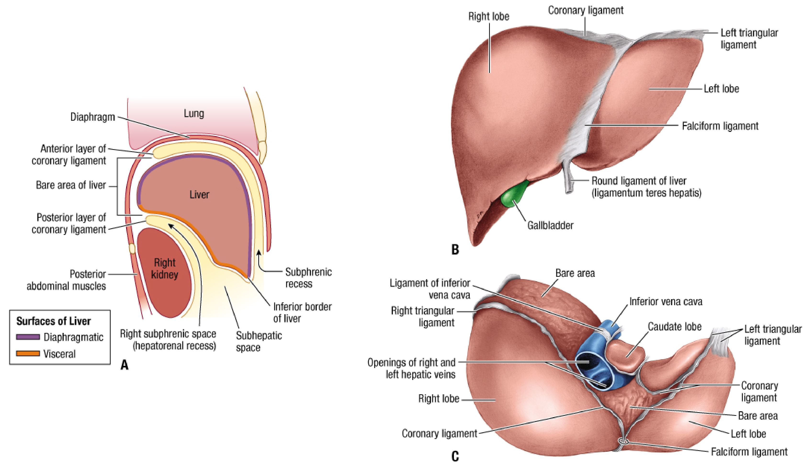
Panel C is the key view - posterior view of the liver showing the caudate lobe (highlighted in blue) which projects into the superior recess. The left triangular ligament forms the upper boundary of the superior recess, and the inferior vena cava lies to its right.
Fig. 162.2A - Sagittal Section showing the Superior Recess (Fischer's Mastery of Surgery)
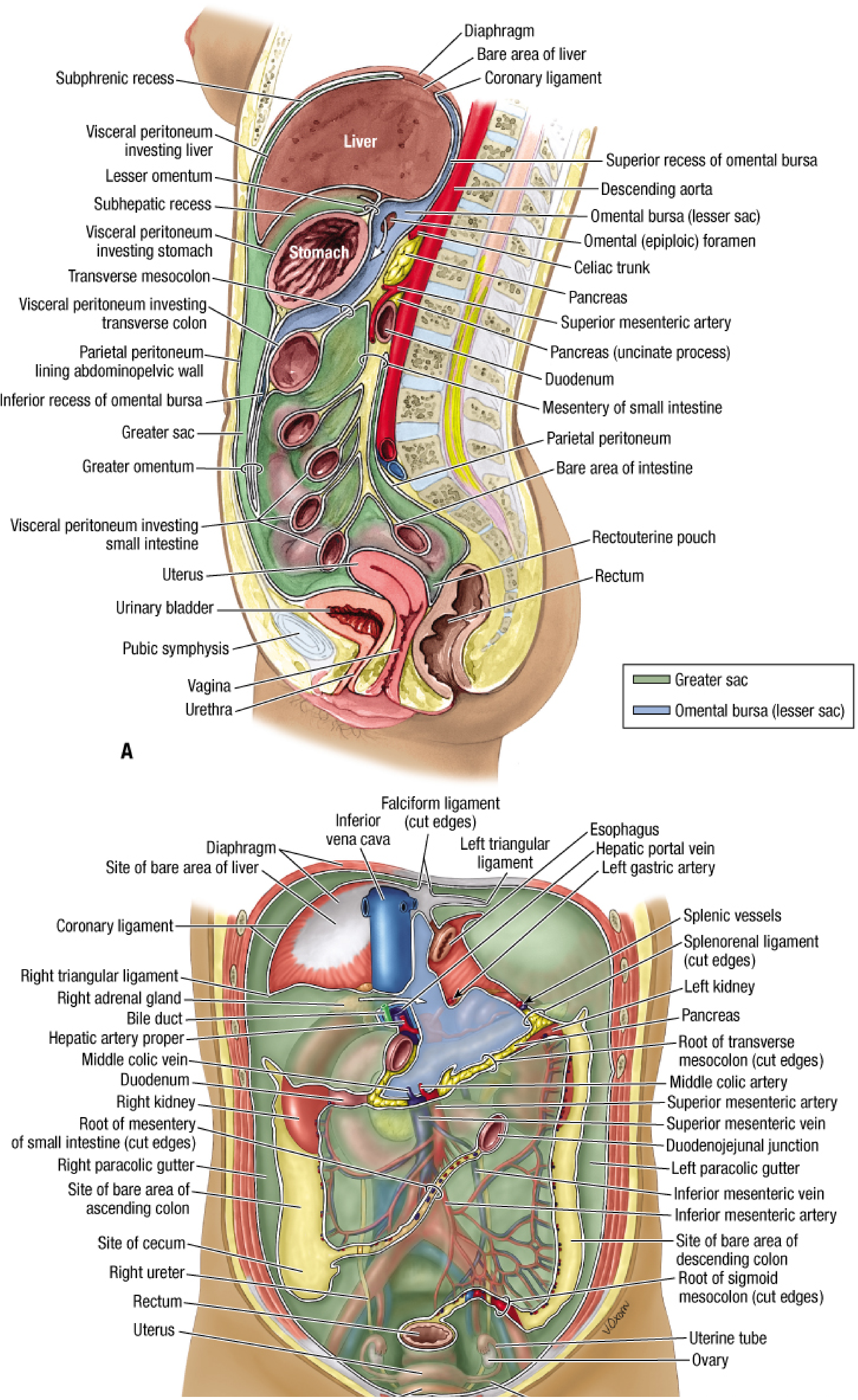
This is the most informative diagram for the superior recess. In Panel A (midsagittal section), the superior recess of the omental bursa is clearly labelled (top right, shown in blue), positioned:
- Posteriorly behind the liver (above the caudate lobe)
- Anteriorly bounded by the lesser omentum
- Above the omental (epiploic) foramen
- Behind the descending aorta and in front of the caudate lobe
The entire omental bursa (lesser sac) is shown in blue, with the superior recess extending upward toward the diaphragm/esophagus, clearly distinguished from the inferior recess (lower left, also labelled).
Key Points - Superior Recess
| Feature | Detail |
|---|---|
| Position | Behind left lobe of liver, above caudate lobe |
| Upper boundary | Left triangular ligament |
| Right boundary | IVC (ligament of inferior vena cava) |
| Contents | Surrounds/wraps around the caudate lobe |
| Communication | Opens into vestibule via the gastropancreatic fold region |
| Surgical note | Accessible by dividing the left triangular ligament |
The superior recess is smaller than the inferior recess and is separated from it by the peritoneal fold over the left gastric artery (gastropancreatic fold).
Source: Fischer's Mastery of Surgery 8th ed., Figs. 162.1 & 162.2
This is a shared conversation. Sign in to Orris to start your own chat.