Iam a radiologist teach me tvs scan
transvaginal ultrasound scan TVS technique radiology teaching guide
TVS transvaginal scan technique probe orientation anatomy findings
TVS transvaginal scan normal anatomy uterus ovaries findings radiology
TVS transvaginal scan technique anatomy normal findings uterus ovaries endometrium
Teach me tvs clinically on pt i want to do scan of pts
TVS transvaginal scan step by step clinical technique patient preparation probe insertion scanning protocol
TVS transvaginal scan patient preparation probe insertion technique scanning planes uterus ovaries measurements normal values
TVS normal measurements uterus endometrium ovary size values radiology reference
TVS transvaginal scan normal findings pathology ectopic pregnancy fibroids ovarian cyst PCOS endometriosis
Show tvs scan pictures practically
TVS transvaginal scan images uterus ovary endometrium normal anatomy labeled ultrasound pictures
TVS transvaginal scan ovary follicle ectopic pregnancy fibroid ovarian cyst ultrasound image labeled
transvaginal ultrasound normal ovary with follicle labeled image
TVS transvaginal ultrasound early intrauterine pregnancy gestational sac yolk sac image
TVS Scan - Practical Picture Guide for Radiologists
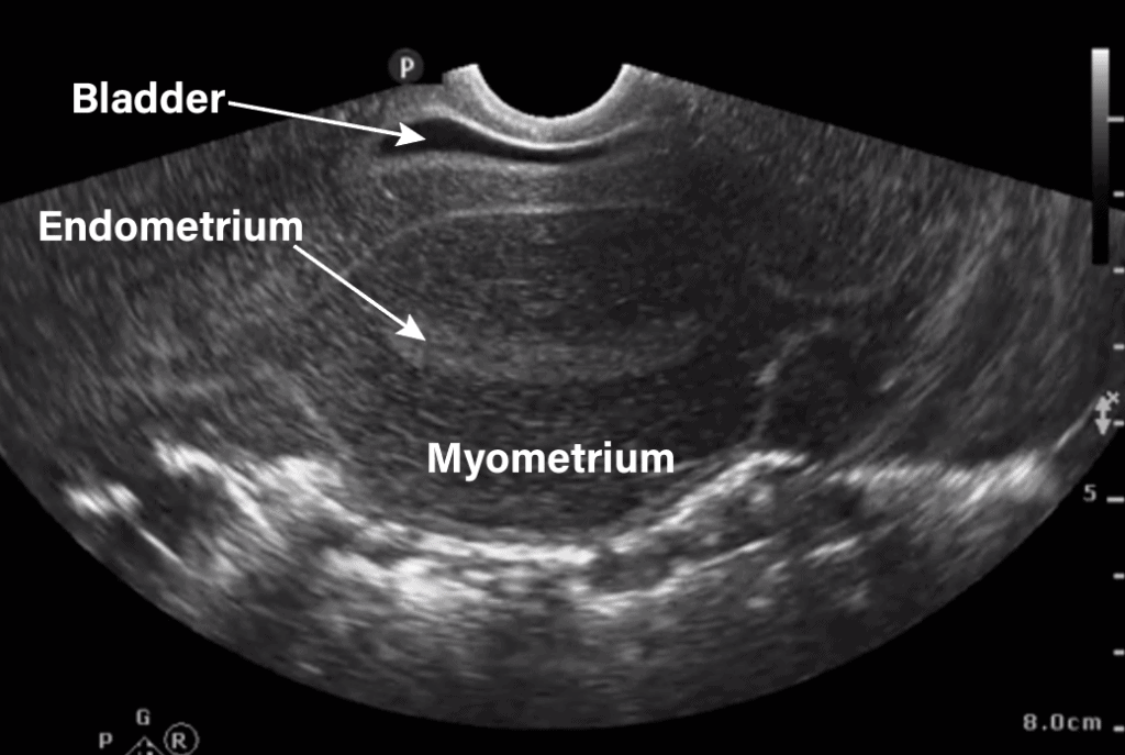
🔬 IMAGE 1 - Uterus: Sagittal (Longitudinal) View

| Structure | Appearance |
|---|---|
| Endometrium | Bright (hyperechoic) central stripe |
| Myometrium | Homogeneous mid-grey surrounding tissue |
| Cervix | Posterior, lower segment |
| Bladder | Anechoic (black) anteriorly - should be EMPTY |
| Pouch of Douglas | Posterior to uterus - check for free fluid |
| Rectum | Posterior to PoD |
Key tip: The endometrial stripe should sit in the CENTER of your image. Adjust depth of probe insertion until you achieve this.
🔬 IMAGE 2 - Uterus: Transverse (Coronal) View
- Uterine body in cross-section
- Endometrium as a central bright oval/stripe
- Symmetrical myometrium on both sides
- Measure uterine width in this plane
🔬 IMAGE 3 - Labeled Anatomy: Transabdominal Reference (for orientation)
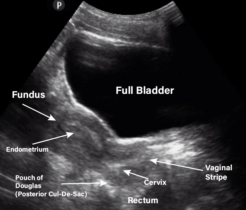
- Full bladder displaces uterus posteriorly (for TAS). For TVS - bladder should be empty.
- Vaginal stripe - bright linear echo from probe tip
- Pouch of Douglas - look for free fluid here
🔬 IMAGE 4 - Normal Ovary with Follicle
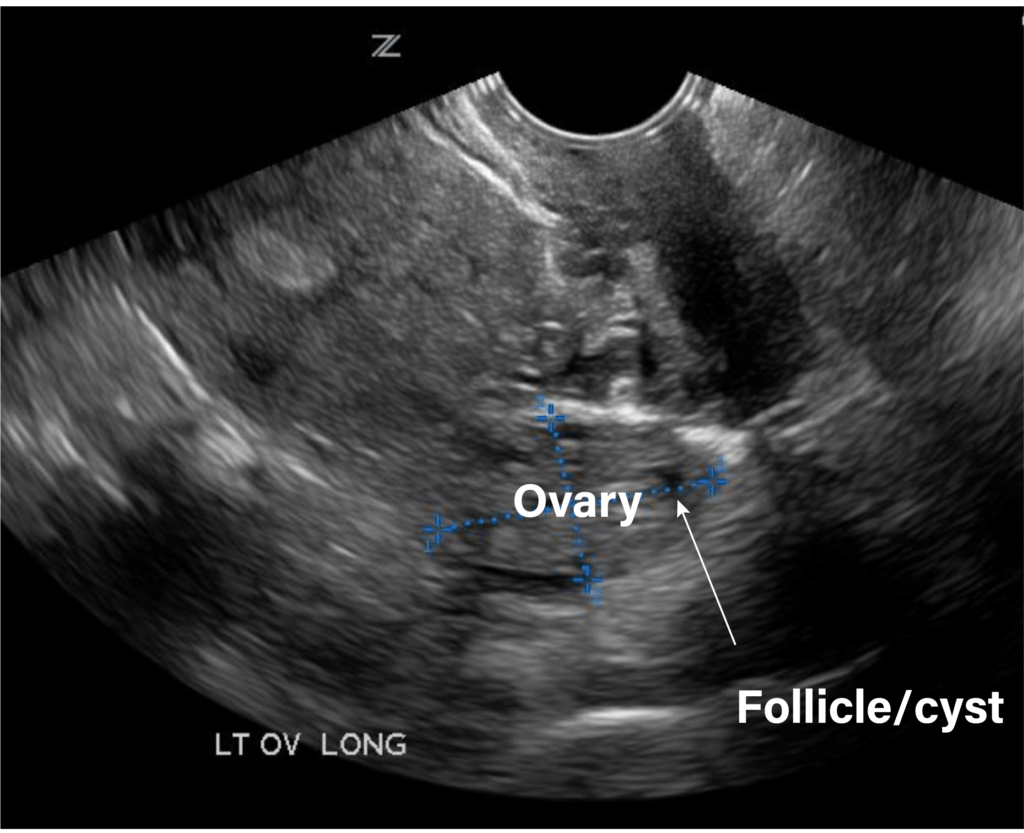
- Ovary: medium echogenicity, oval structure with calipers measuring it
- Follicle/cyst: anechoic (black) round structure within or adjacent to ovary
- Measure 3 dimensions: length x width x depth
- Volume formula: 0.52 × L × W × D
| Age group | Normal volume |
|---|---|
| Premenopausal | Up to 10 ml (avg. ~6 ml) |
| Postmenopausal | < 3 ml |
| Dominant follicle (pre-ovulatory) | 18-27 mm diameter |
🔬 IMAGE 5 - Ovary with Color Doppler / Follicle Blood Flow
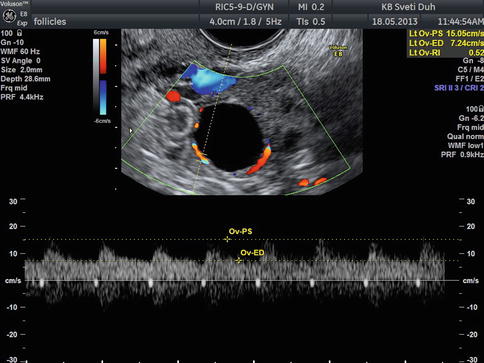
- Ring of color around a dominant follicle = normal perifollicular flow
- "Ring of fire" = high color flow around adnexal ring → think ectopic pregnancy or corpus luteum
- Corpus luteum RI ≈ 0.43-0.49 (low resistance)
🔬 IMAGE 6 - Intrauterine Pregnancy (IUP) - Gestational Sac + Yolk Sac
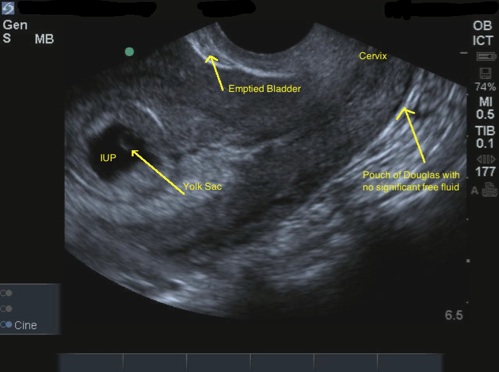
- IUP - gestational sac within uterus
- Yolk sac - small bright ring inside the sac → this CONFIRMS IUP
- Emptied bladder (top right) - correct TVS preparation
- Pouch of Douglas - no free fluid = reassuring
Critical rule: Yolk sac inside the sac = confirmed IUP. Gestational sac alone is NOT enough (can be pseudosac in ectopic pregnancy).
🔬 IMAGE 7 - Very Early IUP (5-6 weeks)
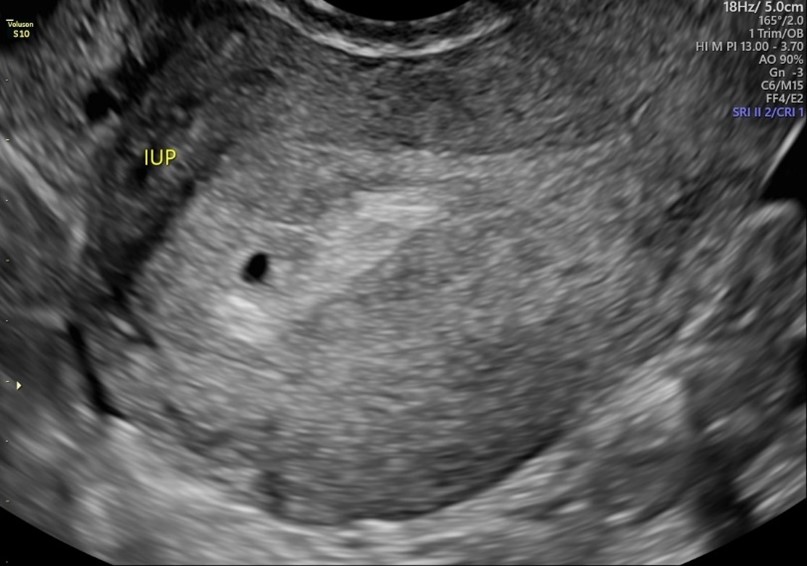
- Small anechoic gestational sac eccentrically placed in endometrium
- Surrounded by echogenic decidual reaction
- At this stage (5 weeks) - you may only see the sac
- By 5w5d - yolk sac visible. By 6 weeks - fetal pole + cardiac activity
| Finding | β-hCG level |
|---|---|
| Gestational sac visible on TVS | ≥ 1,000-1,500 IU/L |
| Yolk sac | ≥ 2,000 IU/L |
| Fetal pole with cardiac activity | ≥ 5,000 IU/L |
Summary: Standard TVS Scanning Protocol
STEP 1 - Sagittal uterus
   → Identify: endometrium, myometrium, cervix, PoD, bladder
   → Measure: uterine length (fundus to cervix) & AP diameter
   → Measure: endometrial thickness (AP, single layer method)

STEP 2 - Transverse uterus
   → Rotate 90° counterclockwise
   → Fan superior to inferior
   → Measure: uterine width

STEP 3 - Right ovary
   → Fan probe to right side of pelvis
   → Tilt handle toward patient's left
   → Measure in 3 planes, calculate volume

STEP 4 - Left ovary
   → Mirror technique
   → Same measurements

STEP 5 - Pouch of Douglas
   → Check sagittal view
   → Note any free fluid (physiological = small amount post-ovulation)

STEP 6 - Adnexa
   → Sweep laterally both sides
   → Look for masses, tubes, free fluid
Key Normal Measurements - Quick Reference Card
| Structure | Normal Value |
|---|---|
| Uterus length (nulliparous) | 6-8 cm |
| Uterus length (multiparous) | 8-10 cm |
| Uterus AP diameter | 3-5 cm |
| Endometrium - proliferative phase | 4-8 mm |
| Endometrium - secretory phase | 8-14 mm |
| Endometrium - postmenopausal | ≤ 4-5 mm (> 5 mm = investigate) |
| Ovary - premenopausal volume | < 10 ml |
| Ovary - postmenopausal volume | < 3 ml |
| Dominant follicle at ovulation | 18-27 mm |
| Free fluid in PoD | Small amount = normal peri-ovulatory |
What to Look for in Common Clinical Scenarios
| Indication | Key TVS Finding |
|---|---|
| Pelvic pain (1st trimester) | IUP vs ectopic - yolk sac location |
| PV bleeding | Endometrial thickness, gestational sac |
| Infertility | Follicle count, endometrial pattern, fibroids |
| PCOS | ≥12 follicles per ovary 2-9mm, ovarian volume >10ml |
| Postmenopausal bleeding | Endometrial thickness >5mm → biopsy |
| Fibroid (leiomyoma) | Hypoechoic whorled mass in myometrium |
| Ovarian cyst | Simple = anechoic, thin-walled. Complex = septae/solid areas |
| Ectopic pregnancy | Empty uterus + adnexal ring + free fluid in PoD |