
Stage of wallerian degeneration and prerequisites of neuronal regeneration
Reading File
Finding Sources
Finding Sources
Finding Sources
Reading File
Reading File
Reading File
Reading File
Wallerian Degeneration and Nerve Regeneration
Stages of Wallerian Degeneration
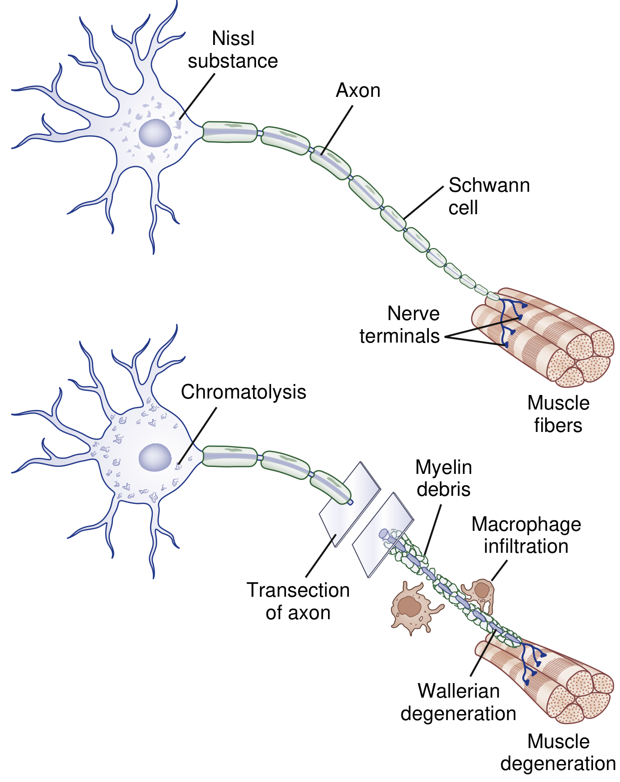
Wallerian degeneration is the sequence of axonal and myelin breakdown that occurs distal to a site of nerve transection or severe injury (Sunderland grade II-V injuries). It is a "dying forward" process, in contrast to axonal ("dying-back") degeneration - Adams and Victor's Principles of Neurology, p. 1290.
A. Distal segment changes (complete within ~1 week)
- Axotomy/disruption (hours) - Injury disrupts retrograde and anterograde axoplasmic flow. Rapid influx of extracellular Ca²⁺ and Na⁺ through the damaged axolemma triggers a cascade resembling programmed cell death.
- Axonal and myelin fragmentation (day 1-3) - The axon and its myelin sheath distal to the transection begin to break down. Axonal injury also recruits leukocytes and triggers cytokine signaling that induces neurotrophins, chemokines, and proteolytic enzymes.
- Schwann cell transition (~day 3) - Schwann cells retract from the nodes of Ranvier and, under upregulation of c-Jun, switch phenotype from myelin-maintaining cells to "repair cells."
- Debris clearance - Activated Schwann cells and recruited macrophages digest the fragmented myelin (myelin ovoids) and axonal debris, clearing the endoneurial tube.
- Schwann cell proliferation and Büngner band formation - Proliferating Schwann cells, bounded by the intact basal lamina, line up to form the bands of Büngner, which will scaffold the regenerating axon.
B. Proximal segment changes
- Limited axonal breakdown extends proximally only up to the first node of Ranvier (unless the injury is very proximal, e.g., a proximal limb amputation, which may cause cell body apoptosis).
- The neuronal cell body undergoes chromatolysis: dispersal/breakup of the rough endoplasmic reticulum (Nissl substance), eccentric displacement of the nucleus, and a shift in gene transcription from "axon maintenance" mode to "axon regeneration/protein synthesis" mode.
- Bradley and Daroff's Neurology in Clinical Practice, p. 1334-1335
- Adams and Victor's Principles of Neurology, 12th Ed., p. 1289-1290
Prerequisites for Neuronal (Axonal) Regeneration
For a peripheral axon to regenerate successfully after Wallerian degeneration, several conditions must be met:
- Survival of the neuronal cell body - chromatolysis must remain reversible; if the cell body dies, no regeneration is possible.
- Completion of Wallerian degeneration and debris clearance - macrophages/Schwann cells must clear myelin and axonal debris so the endoneurial tube is patent for the advancing growth cone.
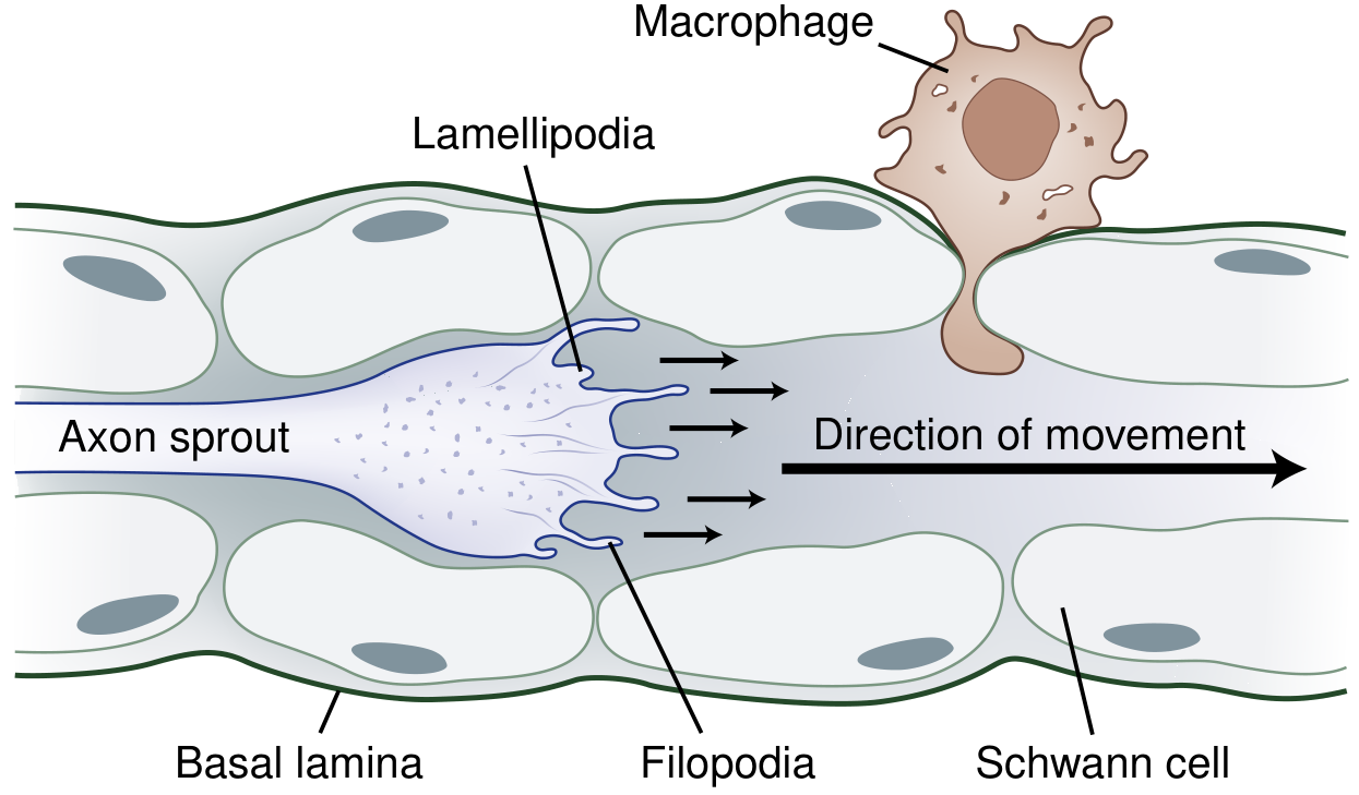
- Schwann cell support - dedifferentiated, proliferating Schwann cells forming bands of Büngner are essential to guide and nourish the regenerating sprout; they also secrete trophic factors.
- Growth cone formation - the sprouting axon tip develops filopodia and lamellipodia that sense the local environment and drive elongation (about 1-2 mm/day, faster from proximal lesions).
- Guidance (neurotropism) - directional molecules such as semaphorins, ephrins, netrins, and slits attract or repel the growth cone to prevent misdirected regrowth; plasminogen activators secreted by the cone dissolve debris blocking the tubule.
- Trophic/growth factor milieu - adequate local levels of nerve growth factor, brain-derived neurotrophic factor, neurotrophins 3/4, GDNF-family ligands (neurturin, artemin, persephin), insulin-like growth factor, IL-6, leukemia inhibitory factor, ciliary neurotrophic factor, and fibroblast growth factors.
- Intact endoneurial tube/basal lamina - if the tube is preserved, the regrowing axon is channeled correctly; if severely disrupted, axons stray into surrounding connective tissue, risking disorganized sprouting or neuroma formation (a source of neuropathic pain from ectopic sodium-channel accumulation).
- Limited gap and timely regrowth - the proximal and distal stumps must be close enough, and regeneration must occur promptly; if the axon takes longer than about 4 months to cross the gap, the distal endoneurial tube shrinks (to ~3 micrometers or less), impeding full recovery. Some reinnervation can still occur up to 2 years post-injury.
- A viable, receptive end organ - the target muscle or sensory receptor must not have atrophied irreversibly (denervated muscle undergoes progressive atrophy and endomysial/perimysial collagen deposition over time, reducing the chance of functional reinnervation the longer denervation persists).
- Bradley and Daroff's Neurology in Clinical Practice, p. 1335-1336
Note on regeneration type by injury severity:
- Grade I injury (demyelination only, axon intact) -> remyelination, recovery in weeks-months.
- Partial axonal loss -> collateral sprouting from surviving axons (nodal/terminal sprouts), recovery in 3-6 months.
- Complete/severe axonal injury (grade II-V) -> true proximal-to-distal regeneration only after Wallerian degeneration is complete, requiring all the prerequisites above.
Stage of wallerian degeneration and prerequisites of neuronal regeneration 5 mark question in easy word
Here's a simplified version for a 5-mark exam answer, using content already covered:
Wallerian Degeneration - Stages (in simple words)
Wallerian degeneration is the breakdown of the axon and myelin sheath that happens below (distal to) the point where a nerve is cut or badly injured.
Stage 1 - Axon injury
The nerve is cut. The part of the axon below the cut loses its connection to the cell body, so it can no longer get nutrients.
Stage 2 - Axon and myelin breakdown (Day 1-3)
The axon and its myelin sheath (covering) start to break into small pieces. Calcium rushes into the cell and starts destroying the axon from inside.
Stage 3 - Schwann cell change
By day 3, Schwann cells (which normally make myelin) pull away from the nerve and change job - instead of making myelin, they now start clearing debris and helping repair.
Stage 4 - Clearing of debris
Macrophages (cleaner cells) come to the injury site and "eat up" (phagocytose) the broken myelin and axon pieces. This clears the path for a new axon to grow later.
Stage 5 - Changes near the cell body (proximal side)
A small part of the axon just above the cut also breaks down (up to the nearest node of Ranvier). The nerve cell body swells and undergoes chromatolysis - its Nissl granules scatter and nucleus moves to one side. This means the cell has switched from its normal job to "repair mode," getting ready to regrow the axon.
Prerequisites for Neuronal Regeneration (in simple words)
For the nerve to regrow properly, these conditions are needed:
- Cell body must survive - if the neuron's cell body dies, regeneration cannot happen.
- Debris must be cleared - macrophages and Schwann cells must clean up the old myelin and axon bits first.
- Schwann cells must help - they multiply and line up to form a tube-like path (bands of Büngner) that guides the new axon.
- A growth cone must form - the tip of the new axon sprout that moves forward and senses its surroundings.
- Guidance signals must be present - chemical signals direct the growing axon in the right direction.
- Growth factors must be available - substances like nerve growth factor (NGF) nourish and support the growing axon.
- The nerve tube (endoneurial tube) must be intact - if it is damaged, the axon may go in the wrong direction or form a painful lump (neuroma).
- The gap must be small and regrowth must happen in time - the two cut ends must not be too far apart, and regrowth must occur quickly (nerve grows about 1-2 mm per day); if it takes too long, the path shrinks and closes.
- The target (muscle/organ) must still be healthy - if the muscle has wasted away too much from long-term disuse, reconnecting the nerve will not restore function.
In short: Wallerian degeneration clears away the damaged nerve, and regeneration can only succeed if the cell body is alive, Schwann cells guide the new growth, debris is removed, growth factors are present, and the target muscle/organ is still viable.
This is a shared conversation. Sign in to Orris to start your own chat.