
Can diabetic cause eye bleeding
Yes, diabetes can definitely cause eye bleeding. This happens through a condition called diabetic retinopathy, where high blood sugar damages the tiny blood vessels in the retina. These weakened vessels can leak blood or fluid into the eye. In advanced stages, new fragile blood vessels grow and bleed into the vitreous (the gel inside the eye), causing significant vision loss.
Searching sources...
Reading File
Finding Sources
Searching PubMed
"diabetic retinopathy" AND "vitreous hemorrhage"
Reading File
Reading File
Reading File
Reading File
Finding Sources
Reading File
I now have comprehensive information from Kanski's Clinical Ophthalmology (10th edition) and Wills Eye Manual. Here is the full answer:
Can Diabetes Cause Eye Bleeding?
Yes, absolutely. Eye bleeding is one of the most serious complications of diabetes. It occurs through a condition called diabetic retinopathy (DR), which affects the blood vessels of the retina at the back of the eye.
How It Happens - The Mechanism
Diabetic retinopathy is primarily a microangiopathy - high blood glucose levels damage the tiny blood vessels (microvasculature) in the retina. This causes:
- Pericyte loss - the cells that maintain vessel wall integrity are destroyed
- Basement membrane thickening - weakens vessel walls
- VEGF (Vascular Endothelial Growth Factor) overproduction - drives abnormal, fragile new blood vessel growth (neovascularization)
These damaged and abnormal vessels are prone to leaking blood into and around the retina and into the vitreous (the jelly-like fluid filling the eye).
- Kanski's Clinical Ophthalmology, 10th Ed., p. 507
Stages of Diabetic Retinopathy and Bleeding
| Stage | Bleeding Seen |
|---|---|
| Background DR (BDR) | Dot and blot haemorrhages - small bleeds within the retina layers |
| Preproliferative DR (PPDR) | Moderate to severe retinal haemorrhages; indicates worsening ischaemia |
| Proliferative DR (PDR) | Preretinal haemorrhage and vitreous haemorrhage (VH) - blood floods the eye cavity |
| Advanced Diabetic Eye Disease | Significant persistent vitreous haemorrhage; tractional retinal detachment; neovascular glaucoma |
- Kanski's Clinical Ophthalmology, 10th Ed., pp. 507-522
What is Vitreous Haemorrhage (VH)?
This is the most dramatic form of diabetic eye bleeding. In proliferative DR, fragile new blood vessels grow on the retina and optic disc (neovascularization). These vessels bleed directly into the vitreous cavity, causing:
- Sudden painless vision loss
- Floaters or dark shadows
- Complete vision blackout in severe cases
High-risk PDR is defined as having ANY of:
-
Vitreous haemorrhage or preretinal haemorrhage
-
Active neovascularization
-
New vessels near the optic disc (NVD >1/3 disc area)
-
Kanski's Clinical Ophthalmology, 10th Ed., p. 508
How Common Is It?
- About 40% of all diabetics develop some form of retinopathy
- After 10 years with Type 2 diabetes, 67% have DR, and 10% progress to proliferative disease
- DR is the leading cause of new blindness in most industrialised countries
- Type 1 diabetics tend to have more aggressive PDR with earlier and more severe bleeding
Treatment for Diabetic Eye Bleeding
| Treatment | When Used |
|---|---|
| Panretinal photocoagulation (PRP) laser | High-risk PDR - destroys ischaemic retina to reduce VEGF |
| Anti-VEGF injections (ranibizumab, bevacizumab, aflibercept) | Alternative to PRP; preferred when DME is present or VH limits view |
| Vitrectomy (surgery) | Dense non-clearing VH, recurrent bleeding, tractional retinal detachment |
Recent meta-analyses comparing anti-VEGF vs PRP confirm anti-VEGF injections are as effective as laser for proliferative DR, with better macular outcomes (PMID 39128789). A 2026 network meta-analysis further supports perioperative anti-VEGF therapy for PDR surgery (PMID 41620203).
Warning Signs to See a Doctor Immediately
- Sudden vision loss
- A flood of floaters or dark spots
- A "veil" or red haze over your vision
- Any painless sudden change in vision
These could indicate a vitreous haemorrhage and need urgent ophthalmology review.
Prevention
- Tight blood sugar control (HbA1c <7%) is the strongest protection
- Blood pressure and cholesterol control
- Annual eye screening - early detection before bleeding occurs can prevent blindness
How it cause bleeding
The bleeding happens through a step-by-step process: high blood sugar damages retinal blood vessels, they weaken and leak, then the body tries to grow new vessels that are even more fragile and bleed heavily. Let me pull the detailed pathogenesis from the textbooks.
Diving deeper...
Finding Sources
Finding Sources
Reading File
Reading File
Reading File
Loading Image
Loading Image
Reading File
Loading Image
Here is a detailed step-by-step explanation of exactly how diabetes causes eye bleeding, with real retinal images from Kanski's Clinical Ophthalmology (10th Edition):
How Diabetes Causes Eye Bleeding - Step by Step
Step 1: High Blood Sugar Damages the Vessel Wall
The retina is supplied by millions of tiny capillaries. Each capillary has two key cell types:
- Endothelial cells - line the inside of the vessel
- Pericytes - wrap around the outside and give the vessel structural strength and control blood flow
Chronically high glucose directly poisons and kills the pericytes. This is the very first event. Without pericytes, the vessel wall becomes weak and unstable - like a pipe losing its outer support.
You can see degenerate pericytes in this histology image from a trypsin digest preparation:

Step 2: Weakened Vessels Balloon Out into Microaneurysms
With pericyte loss, the walls of capillaries bulge outward, forming tiny balloon-like swellings called microaneurysms. These are the earliest visible sign of diabetic retinopathy.
These microaneurysms are leaky - they allow blood and fluid to seep through their thin, damaged walls into the retinal tissue. This causes:
- Dot haemorrhages - small round bleeds from capillary microaneurysms
- Blot haemorrhages - slightly larger bleeds from the venous end of capillaries in the middle retinal layers
- Retinal oedema - fluid accumulation causing swelling
Here is a histology specimen showing capillary looping that forms microaneurysms (Indian ink-injected retina):
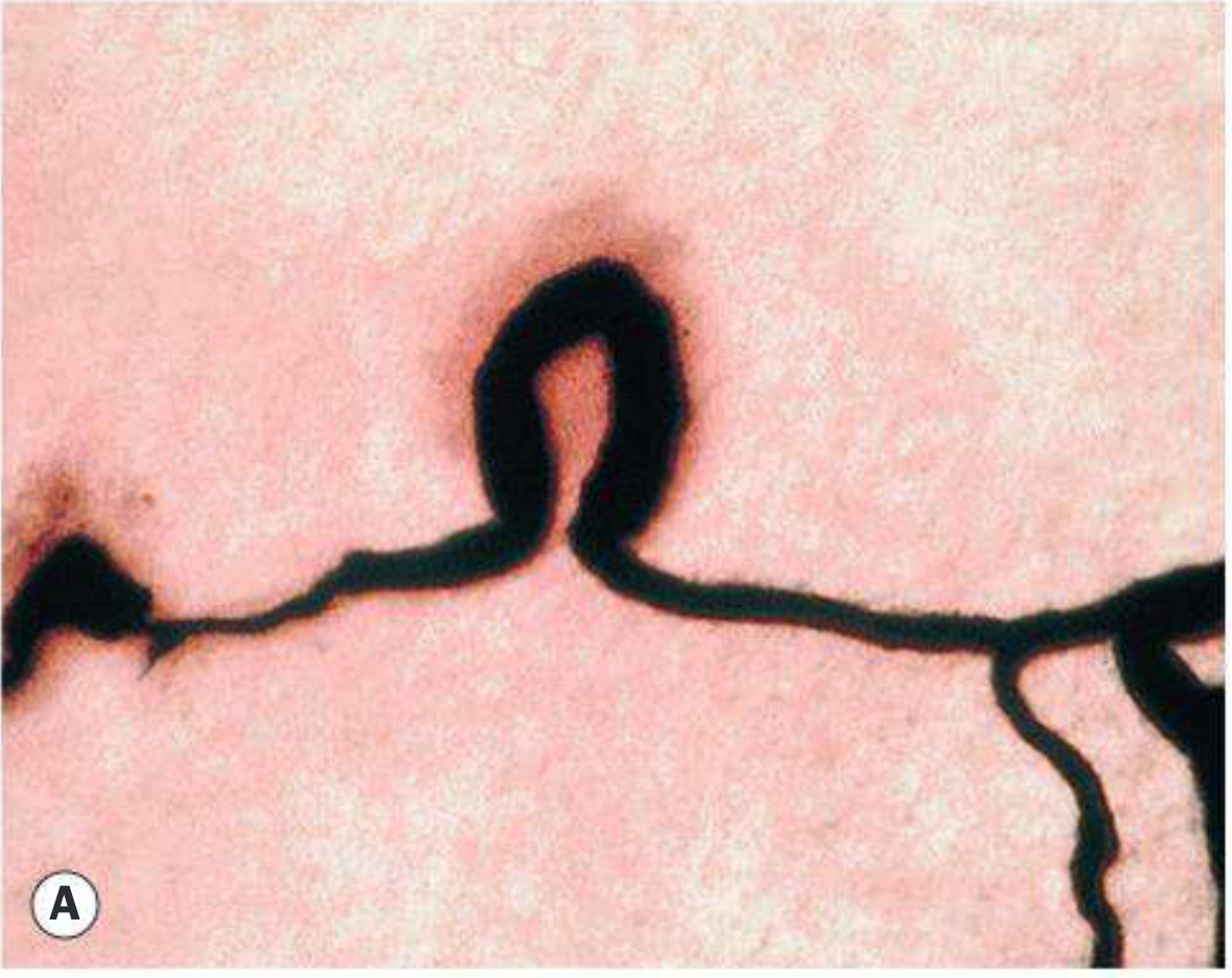
- Kanski's Clinical Ophthalmology, 10th Ed., p. 508
Step 3: Capillaries Close Off - Retina Becomes Ischaemic
As more and more vessels are damaged, they eventually close off completely (capillary non-perfusion). Large patches of retina are now starved of oxygen - this is called retinal ischaemia.
The dying retina sends out a distress signal: it massively overproduces a chemical called VEGF (Vascular Endothelial Growth Factor).
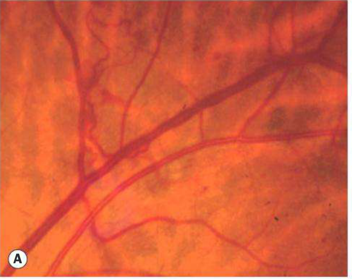
Step 4: VEGF Triggers Abnormal New Blood Vessel Growth (Neovascularization)
VEGF is the key driver of bleeding. It tells the body to grow new blood vessels to rescue the ischaemic retina. However, these new vessels are structurally abnormal:
- They have no pericyte support at all
- Their walls are extremely thin and fragile
- They grow in the wrong places - on the surface of the retina and into the vitreous gel
- They are accompanied by fibrous tissue that contracts and pulls on the retina
This is called Proliferative Diabetic Retinopathy (PDR).
Here is a fundus photo showing new vessels growing on the retinal surface (neovascularization elsewhere, NVE):
Step 5: These Fragile New Vessels Rupture and Bleed
Because these new vessels have no structural integrity, they rupture easily - sometimes from just a Valsalva manoeuvre (coughing, straining) or during sleep. The blood pours into two locations:
| Location | Name | Effect |
|---|---|---|
| Between retina and vitreous | Preretinal haemorrhage | A pool of blood sits in front of the retina |
| Into the vitreous cavity itself | Vitreous haemorrhage (VH) | Blood floods the jelly of the eye - sudden severe vision loss |
The patient sees this as a sudden red haze, shower of dark floaters, or complete loss of vision - depending on how much blood is released.
Summary Diagram of the Process
High Blood Sugar
↓
Pericyte death → Vessel wall weakens
↓
Microaneurysms → Dot/blot haemorrhages (early bleeding)
↓
Capillary closure → Retinal ischaemia
↓
VEGF overproduction → Neovascularization (fragile new vessels)
↓
New vessels rupture → Preretinal + Vitreous Haemorrhage (severe bleeding)
↓
Possible blindness
The earlier small bleeds (dot and blot) are a warning sign that the more dangerous stage - vitreous haemorrhage - may be coming. This is why annual diabetic eye screening is so important: to catch and treat the disease before the vessels grow and bleed catastrophically.
- Kanski's Clinical Ophthalmology, 10th Ed., pp. 506-522
Diabetic acute complications
The main acute complications of diabetes are DKA (diabetic ketoacidosis), HHS (hyperosmolar hyperglycaemic state), and hypoglycaemia. These are medical emergencies that can be fatal if untreated.
Searching sources...
Reading File
Finding Sources
Finding Sources
Finding Sources
Finding Sources
Finding Sources
Reading File
Reading File
Finding Sources
Finding Sources
Loading Image
Reading File
Finding Sources
Reading File
Reading File
Excellent - I now have comprehensive content from Harrison's (22nd Ed.) and Tintinalli's Emergency Medicine. Here is the full answer:
Acute Complications of Diabetes
There are three major acute (sudden/emergency) complications of diabetes:
1. Diabetic Ketoacidosis (DKA)
Mainly in Type 1 DM (but can occur in Type 2)
What happens?
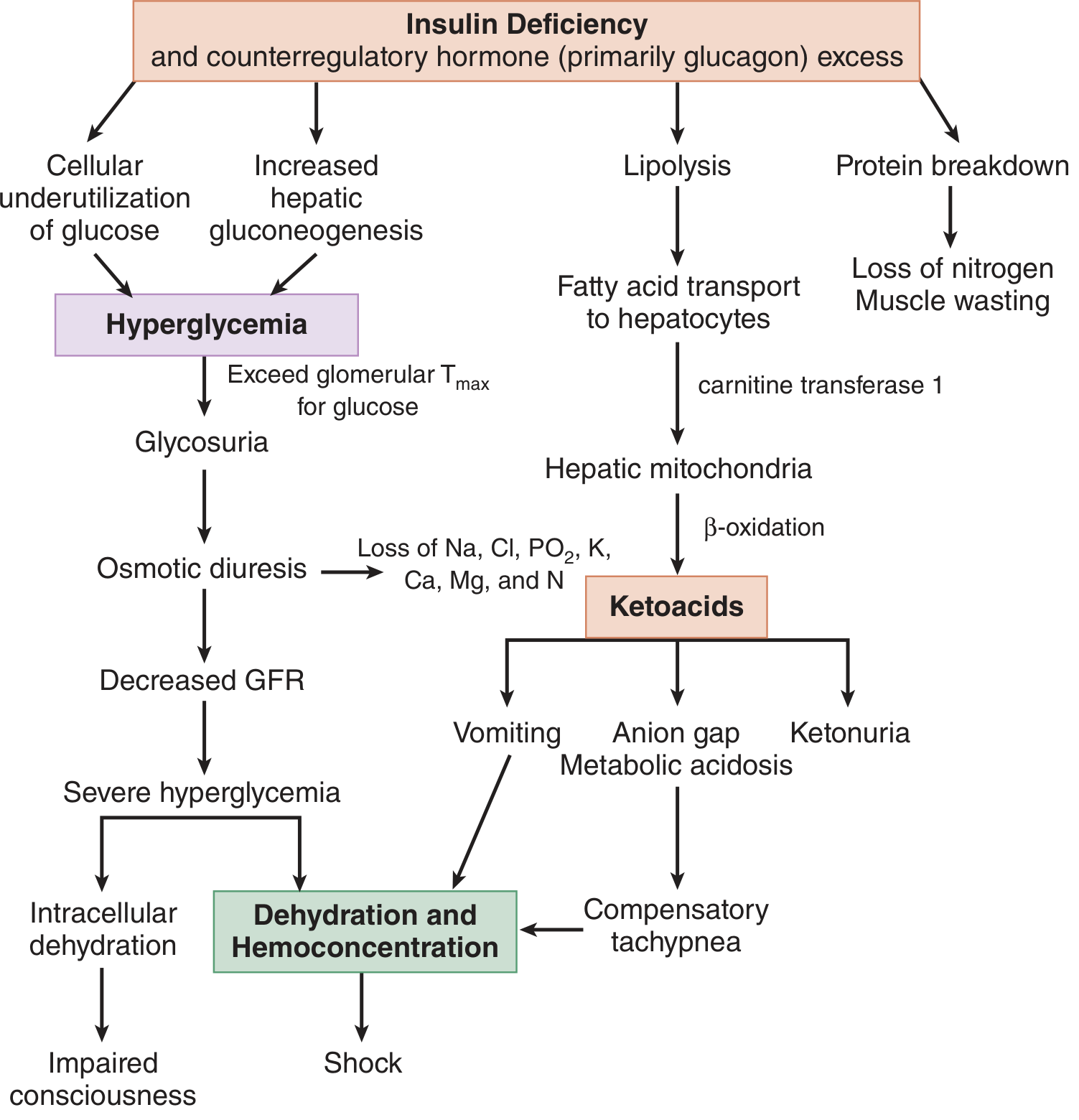
When insulin is severely deficient, the body cannot use glucose for energy. It switches to burning fat, which produces toxic acids called ketone bodies (beta-hydroxybutyrate and acetoacetate). These accumulate in the blood, causing a dangerous metabolic acidosis.
The DKA pathway is shown in this diagram from Tintinalli's Emergency Medicine:
Common triggers
- Missed insulin doses / pump failure
- Infection (most common trigger)
- Surgery, trauma, MI, stroke
- Steroids, cocaine, antipsychotics
- New-onset Type 1 DM at diagnosis
Symptoms
| Symptom | Why it happens |
|---|---|
| Nausea, vomiting, abdominal pain | Prostaglandins from fat breakdown |
| Kussmaul breathing (deep, rapid) | Body trying to blow off CO2 to compensate for acid |
| Fruity/acetone breath | Acetone on the breath |
| Polyuria and polydipsia | Osmotic diuresis from high glucose |
| Dehydration, hypotension, tachycardia | Massive fluid and electrolyte loss |
| Altered consciousness / coma | Hyperosmolarity + acidosis |
Diagnosis (all three must be present)
- Blood glucose >250 mg/dL (13.9 mmol/L)
- Anion gap >10-12 mEq/L
- pH <7.3 + bicarbonate <15 mEq/L + ketonemia/ketonuria
| Severity | pH | HCO3 | Mental Status |
|---|---|---|---|
| Mild | 7.25-7.3 | 15-18 | Alert |
| Moderate | 7.0-7.24 | 10-15 | Drowsy |
| Severe | <7.0 | <10 | Stuporous/Comatose |
Treatment
-
IV fluids - Normal saline (0.9%) to restore volume
-
Insulin infusion (IV regular insulin 0.1 units/kg/hr)
-
Potassium replacement (critical - insulin drives K into cells, causing dangerous hypokalaemia)
-
Treat the underlying trigger (antibiotics if infected)
-
Monitor glucose, electrolytes, pH hourly
-
Harrison's Principles of Internal Medicine, 22nd Ed., p. 3262
-
Tintinalli's Emergency Medicine, p. 1476
2. Hyperosmolar Hyperglycaemic State (HHS)
Mainly in Type 2 DM, elderly patients
How it differs from DKA
HHS has no ketoacidosis. There is still some residual insulin present, which is enough to prevent ketone formation but not enough to control blood glucose. Blood sugar climbs to extreme levels over days to weeks, pulling water out of cells (hyperosmolarity).
| Feature | DKA | HHS |
|---|---|---|
| Blood glucose | >250 mg/dL | >600 mg/dL (can reach 1000+) |
| Ketones | High | Absent or trace |
| Acidosis | Yes (pH <7.3) | No (pH >7.3) |
| Osmolality | Mildly elevated | Severely elevated (>320 mOsm/L) |
| Onset | Hours to 1 day | Days to weeks |
| Who | Type 1, younger | Type 2, elderly |
| Mortality | 1-5% | Up to 15% |
Symptoms
- Weeks of polyuria, weight loss, reduced fluid intake
- Severe mental confusion, lethargy, or coma (more prominent than DKA)
- Profound dehydration, hypotension, tachycardia
- No Kussmaul breathing, no vomiting, no abdominal pain (key difference from DKA)
- Often triggered by: MI, stroke, pneumonia, sepsis
Treatment
-
Aggressive IV fluid replacement (1-3 L normal saline over first 2-3 hours)
-
Switch to 0.45% (half-normal) saline once stable
-
Insulin infusion (lower doses than DKA)
-
Potassium replacement
-
Treat the precipitating illness
-
Note: Correct osmolarity slowly - too rapid reversal can worsen neurological function
-
Harrison's Principles of Internal Medicine, 22nd Ed., p. 3262
3. Hypoglycaemia (Low Blood Sugar)
Most common acute complication - can happen in both Type 1 and Type 2 on insulin or sulphonylureas
What happens?
Blood glucose falls too low (<70 mg/dL / 3.9 mmol/L). The brain is entirely dependent on glucose and becomes immediately starved.
Hypoglycaemia is the most serious complication of diabetes therapy and occurs in >90% of patients on insulin at some point.
Symptoms - Two stages
Stage 1 - Adrenergic (Sympathetic) Warning Signs (glucose ~60-70 mg/dL):
- Sweating, trembling, palpitations
- Anxiety, hunger
- Pallor
Stage 2 - Neuroglycopaenic (Brain Starved) (glucose <50 mg/dL):
- Confusion, difficulty concentrating
- Slurred speech, drowsiness
- Seizures
- Coma and death if untreated
Common triggers
- Too much insulin / sulphonylurea dose
- Missed meal or delayed eating
- Excessive exercise
- Alcohol (blocks gluconeogenesis in the liver)
Treatment
| Severity | Treatment |
|---|---|
| Mild (conscious, can swallow) | 15-20g fast-acting carbohydrate (glucose tablets, fruit juice, regular soda) |
| Moderate | Repeat carbs, check glucose in 15 min (Rule of 15) |
| Severe (unconscious) | IV Dextrose (50% glucose) or IM/intranasal Glucagon |
Special concern - Hypoglycaemia Unawareness
Patients with long-standing diabetes or recurrent hypoglycaemia can lose their adrenergic warning symptoms. They skip straight to coma with no warning - extremely dangerous.
- Harrison's Principles of Internal Medicine, 22nd Ed.
Quick Comparison Table
| Feature | DKA | HHS | Hypoglycaemia |
|---|---|---|---|
| Blood glucose | >250 mg/dL | >600 mg/dL | <70 mg/dL |
| Ketones | +++ | Absent | Absent |
| Acidosis | Yes | No | No |
| Onset | Hours | Days-weeks | Minutes |
| Type of DM | Mainly T1 | Mainly T2 | Both |
| Key sign | Kussmaul breathing + fruity breath | Severe dehydration + confusion | Sweating + trembling |
| First treatment | IV fluids + insulin | IV fluids + insulin | Glucose/glucagon |
| Mortality | 1-5% | Up to 15% | Low if treated quickly |
All three are medical emergencies. Any diabetic patient found unconscious or acutely confused must have blood glucose checked immediately as the first step.
What tests diagnose DKA vs HHS vs hypoglycemia?
The key tests are blood glucose, blood gas (pH/bicarbonate), ketones, and serum osmolality. Together they separate all three conditions clearly.
Searching sources...
Finding Sources
Finding Sources
Reading File
Reading File
Reading File
Finding Sources
Finding Sources
Reading File
I now have all the precise lab values directly from Harrison's and Tintinalli's. Here is the complete diagnostic breakdown:
Tests That Diagnose DKA vs HHS vs Hypoglycaemia
The Master Comparison Table
This is taken directly from Harrison's Principles of Internal Medicine, 22nd Edition (Table 416-7):
| Lab Test | DKA | HHS | Hypoglycaemia |
|---|---|---|---|
| Blood Glucose | 11.1-33.3 mmol/L (250-600 mg/dL) | 33.3-66.6 mmol/L (600-1200 mg/dL) | <3.9 mmol/L (<70 mg/dL) |
| Arterial pH | 6.8-7.3 (acidotic) | >7.3 (normal) | Normal |
| Serum Bicarbonate (HCO3) | <18 mEq/L | >18 mEq/L (normal) | Normal |
| Anion Gap [Na-(Cl+HCO3)] | Elevated (↑) | Normal to slightly ↑ | Normal |
| Serum β-hydroxybutyrate | >3.0 mmol/L | <1.0 mmol/L | Normal |
| Urine/Serum Ketones | ++ (strongly positive) | +/- (absent or trace) | Absent |
| Serum Osmolality | >300 mOsm/L | Markedly >320 mOsm/L | Normal |
| Sodium | 125-135 mEq/L (low-normal) | 135-145 mEq/L (normal/high) | Normal |
| Creatinine | Slightly to moderately ↑ | Moderately ↑ | Normal |
| Potassium | Normal to ↑ (but body-depleted) | Normal | Normal |
| PCO2 | 20-30 mmHg (low - compensatory) | Normal | Normal |
- Harrison's Principles of Internal Medicine, 22nd Ed.
Test-by-Test Breakdown
1. Blood Glucose (Bedside Glucometer - FIRST TEST)
This is the immediate, first test done at the bedside in any confused or unconscious diabetic patient.
| Result | Interpretation |
|---|---|
| <70 mg/dL (<3.9 mmol/L) | Hypoglycaemia - treat immediately |
| 250-600 mg/dL | Likely DKA - proceed to blood gas + ketones |
| >600 mg/dL (can reach 1000+) | Likely HHS - check osmolality |
Important caveat - Euglycaemic DKA: DKA can occur with glucose <250 mg/dL in patients on SGLT-2 inhibitors (e.g. dapagliflozin, empagliflozin), pregnant patients, and those who received insulin recently. Never rule out DKA on glucose alone.
2. Blood Gas (Arterial or Venous) - KEY DIFFERENTIATOR
This is the test that separates DKA from HHS:
| Parameter | DKA | HHS |
|---|---|---|
| pH | <7.3 (acidotic) | >7.3 (normal) |
| HCO3 | <18 mEq/L (consumed by acid) | >18 mEq/L (normal) |
| PCO2 | 20-30 mmHg (low - Kussmaul compensation) | Normal |
HHS has a normal pH because there are no ketoacids. DKA is always acidotic.
3. Serum β-hydroxybutyrate (βHB) - BEST KETONE TEST
| Level | Meaning |
|---|---|
| >3.0 mmol/L | DKA confirmed |
| <1.0 mmol/L | Ketosis absent - favours HHS |
| Normal | Hypoglycaemia (no ketosis in insulin excess) |
Critical point: Urine dipstick ketone tests use the nitroprusside reaction, which only detects acetoacetate - NOT beta-hydroxybutyrate, which is the dominant ketone in DKA. This means urine ketone strips can falsely underestimate DKA severity. Serum βHB is the preferred test. - Tintinalli's Emergency Medicine
4. Anion Gap [Na - (Cl + HCO3)]
- Normal: 8-12 mEq/L
- DKA: Anion gap is elevated (ketoacids replace bicarbonate as unmeasured anions)
- HHS: Normal or only slightly elevated (no ketoacids)
- Hypoglycaemia: Normal
Mild: >10 | Moderate/Severe: >12 mEq/L
5. Serum Osmolality [2×Na + Glucose/18 + BUN/2.8]
| Condition | Osmolality |
|---|---|
| HHS | Severely elevated >320 mOsm/L (hallmark) |
| DKA | Mildly elevated >300 mOsm/L |
| Hypoglycaemia | Normal (~285-295 mOsm/L) |
Altered mental status in HHS correlates best with hyperosmolality (>320 mOsm/L), not with any acid-base change.
6. Serum Electrolytes (Sodium, Potassium)
Potassium is the most dangerous electrolyte abnormality in DKA:
- At presentation, K may appear normal or high because acidosis drives K out of cells into blood
- But total body K is severely depleted (lost in urine via osmotic diuresis)
- When insulin is given to treat DKA, K rapidly shifts back into cells → life-threatening hypokalaemia and cardiac arrhythmias
- Potassium must be replaced before starting insulin if K <3.5 mEq/L
Sodium in HHS:
- Measured sodium may appear normal or low despite extreme hyperglycaemia (glucose dilutes serum Na)
- Corrected sodium = measured Na + 1.6 mEq per every 100 mg/dL rise in glucose above normal
7. Whipple's Triad - Diagnosing Hypoglycaemia
For hypoglycaemia to be formally confirmed, all three criteria must be present (Whipple's Triad):
- Symptoms or signs consistent with hypoglycaemia (sweating, trembling, confusion)
- Low plasma glucose (<70 mg/dL in diabetic patients; <45 mg/dL in non-diabetic)
- Relief of symptoms after glucose is given
- Goldman-Cecil Medicine
The Diagnostic Algorithm in Practice
Diabetic patient - confused / unwell
↓
STEP 1: Bedside glucose
↓
< 70 mg/dL? 250-600? > 600?
HYPOGLYCAEMIA Go to step 2 Likely HHS
→ Give glucose ↓ Check osmolality
STEP 2: Blood gas
pH < 7.3?
HCO3 < 18?
↓
STEP 3: Ketones
βHB > 3.0?
↓
YES → DKA
NO + high osmolality → HHS
(may have mixed features)
Summary: The Three Deciding Tests
| Test | DKA | HHS | Hypoglycaemia |
|---|---|---|---|
| Glucose | 250-600 | >600 | <70 |
| pH / HCO3 | Low (<7.3/<18) | Normal | Normal |
| Serum βHB | >3 mmol/L | <1 mmol/L | Normal |
These three tests together give the diagnosis in virtually every case.
- Harrison's Principles of Internal Medicine, 22nd Ed., Table 416-7
- Tintinalli's Emergency Medicine, p. 1478
This is a shared conversation. Sign in to Orris to start your own chat.