
Is there any medical condition like tight calf muscle in medicine
Yes, tight calf muscles are a recognized clinical finding with several specific medical conditions associated with them. Let me look this up properly.
Searching sources...
Reading File
Finding Sources
Finding Sources
Searching the Web
medical conditions causing tight calf muscles gastrocnemius soleus
Finding Sources
Finding Sources
Finding Sources
Finding Sources
Finding Sources
Finding Sources
Finding Sources
Searching PubMed
gastrocnemius tightness equinus clinical conditions
Here is a thorough answer based on the medical textbooks and clinical knowledge:
Medical Conditions Associated with Tight Calf Muscles
Yes - "tight calf muscles" is a clinically significant finding, not just an everyday complaint. The medical umbrella term is gastrocnemius-soleus contracture or equinus deformity, and it appears in a wide range of conditions.
1. Gastrocnemius Contracture (Isolated)
This is the most common specific condition. The gastrocnemius muscle (which crosses both the knee and ankle) becomes pathologically short or stiff, limiting ankle dorsiflexion. Clinically assessed with the Silfverskiold test:
- With the knee extended: ankle dorsiflexion is reduced (gastrocnemius tight)
- With the knee flexed: dorsiflexion improves - this confirms isolated gastrocnemius contracture (as opposed to combined gastrocnemius + soleus tightness, which is a true Achilles contracture)
This distinction directly guides treatment: gastrocnemius recession vs. tendo-Achilles lengthening (TAL).
(Miller's Review of Orthopaedics 9th Ed; Campbell's Operative Orthopaedics 15th Ed)
2. Equinus Deformity
A fixed downward pointing of the foot (plantarflexion) caused by calf muscle contracture. It occurs in multiple settings:
- Spastic equinus - the most common foot deformity in cerebral palsy (CP); spasticity of the gastrocnemius-soleus causes progressive contracture and a toe-walking gait
- Equinovarus deformity - combination of equinus + inward turning of the foot (as in clubfoot or post-stroke spasticity)
- Surgical correction involves gastrocnemius recession or tendon lengthening (Campbell's Operative Orthopaedics)
3. Cerebral Palsy (Spastic Diplegia/Hemiplegia)
Tight calf muscles are a hallmark feature. The spastic gastrocnemius-soleus:
- Causes equinus gait (toe-walking)
- In "crouched gait," hamstring contracture also develops, but the calf component is nearly always present
- Treated with intramuscular botulinum toxin (Botox), serial casting, gastrocnemius recession, or TAL depending on severity (Miller's Review of Orthopaedics)
4. Acute Compartment Syndrome
This is an emergency. The calf is the most commonly affected site. Pressure builds in the enclosed fascial compartments of the leg, causing:
- Tense, tight, board-like calf muscles
- Severe pain out of proportion to injury, pain with passive stretch of calf muscles
- The "5 Ps" (Pain, Pallor, Pulselessness, Paresthesia, Paralysis) in advanced cases
- Treatment: urgent fasciotomy (Bailey and Love's Surgery; Pfenninger's Primary Care Procedures)
5. Chronic Exertional Compartment Syndrome (CECS)
A reversible form that affects athletes. The calf feels tight, painful, and cramping during exercise but resolves with rest. The anterior compartment is most often involved, but the deep posterior compartment (calf muscles) is also frequently affected. (Current Surgical Therapy)
6. Duchenne and Becker Muscular Dystrophy (DMD/BMD)
Tight, firm, rubbery calf muscles are a classic early sign. This is due to pseudohypertrophy - the muscle fibers are replaced by fat and fibrous tissue, making the calves look large and feel tight. Progressive contractures develop across multiple joints as the disease advances. (Bradley and Daroff's Neurology in Clinical Practice; Braunwald's Heart Disease)
7. Plantar Fasciitis (as a secondary association)
While not a primary calf disease, tightness of the gastrocnemius-soleus complex is one of the most well-recognized contributing factors to plantar fasciitis and heel pain syndromes. Reduced ankle dorsiflexion increases stress on the plantar fascia. (Bailey and Love's Surgery)
8. Clubfoot (Talipes Equinovarus)
A congenital deformity where the gastrocnemius-soleus contracture is a central anatomical component, producing the characteristic equinus (downward-pointing) foot position.
Summary Table
| Condition | Nature of Calf Tightness | Key Feature |
|---|---|---|
| Gastrocnemius contracture | Isolated shortening | Silfverskiold test positive |
| Cerebral palsy | Spastic contracture | Toe-walking, equinus gait |
| Acute compartment syndrome | Tense/swollen compartment | Surgical emergency |
| Chronic exertional CS | Exercise-induced tightness | Resolves with rest |
| Duchenne/Becker MD | Pseudohypertrophy + fibrosis | Large, rubbery calves |
| Equinovarus/Clubfoot | Congenital contracture | Fixed foot deformity |
| Plantar fasciitis | Contributing factor | Heel pain |
In summary, what feels like a "tight calf" is a symptom that can represent anything from a benign postural/biomechanical problem to a surgical emergency (compartment syndrome) or a sign of serious neuromuscular disease. Clinical evaluation - including the Silfverskiold test, range of motion measurement, and neuromuscular examination - is key to determining the underlying cause.
What is gastrocnemius recession
Gastrocnemius recession is a surgical procedure that lengthens the gastrocnemius muscle/tendon to correct equinus deformity and restore ankle dorsiflexion. Let me pull the detailed clinical information from the textbooks.
Searching sources...
Finding Sources
Finding Sources
Finding Sources
Finding Sources
Reading File
Reading File
Reading File
Finding Sources
Finding Sources
Reading File
Loading Image
Loading Image
Finding Sources
Excellent - I now have detailed textbook content and surgical illustrations. Here is a comprehensive answer:
Gastrocnemius Recession
Gastrocnemius recession is a surgical procedure that selectively lengthens the gastrocnemius muscle or its aponeurosis (tendinous sheet) to relieve an isolated gastrocnemius contracture - restoring ankle dorsiflexion without cutting the Achilles tendon.
Background: Why Is It Needed?
The gastrocnemius muscle is unique because it crosses two joints - the knee (origin at the femoral condyles) and the ankle (via the Achilles tendon). When it becomes pathologically tight/shortened, it restricts ankle dorsiflexion - the ability to bring the foot upward.
The key diagnostic test is the Silfverskiold (Silverskiöld) test:
- Ankle dorsiflexion restricted with knee extended, BUT
- Dorsiflexion improves with the knee flexed (relaxing the gastrocnemius origin)
This pattern = isolated gastrocnemius contracture - treated with gastrocnemius recession
- If dorsiflexion is restricted with knee both extended and flexed = both gastrocnemius + soleus tight - treated with full Achilles tendon lengthening (TAL)
(Miller's Review of Orthopaedics, 9th Ed)
Anatomical Zones - Where the Cut Is Made
The gastrocnemius-soleus complex is divided into 3 anatomical zones, which determine the approach for different procedures:
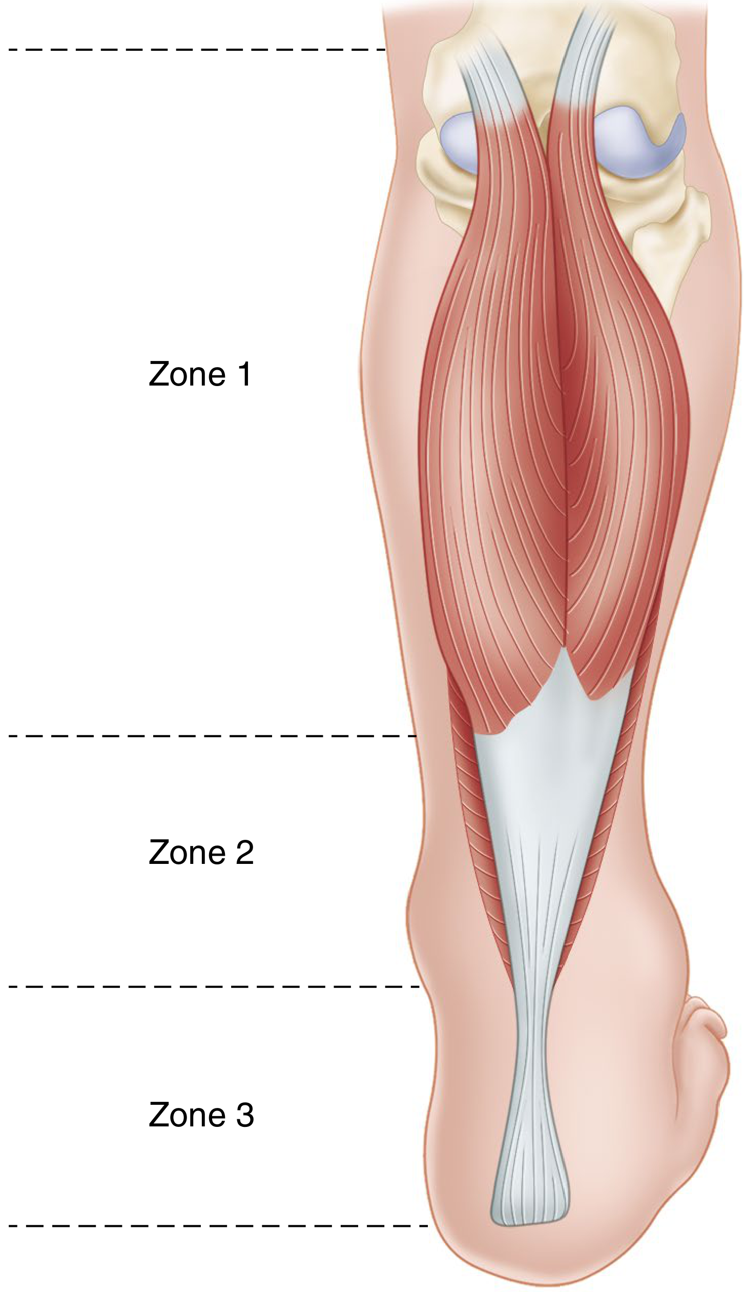
- Zone 1 (proximal): the gastrocnemius muscle belly, below its knee origin
- Zone 2 (middle): the musculotendinous junction where muscle meets aponeurosis - this is where most recession procedures are performed
- Zone 3 (distal): the Achilles tendon itself - lengthening here is a full TAL, not a recession
(Campbell's Operative Orthopaedics, 15th Ed 2026)
Named Procedures
| Procedure | What Is Cut | Level |
|---|---|---|
| Strayer procedure | Gastrocnemius aponeurosis divided transversely at musculotendinous junction | Zone 2 |
| Vulpius procedure | V- or inverted-V shaped cut through the aponeurosis | Zone 2 |
| Baker modification | Tongue-in-groove lengthening of the gastrocnemius aponeurosis | Zone 2 |
| Baumann procedure | Intramuscular lengthening (used in cerebral palsy) | Zone 1 |
The Strayer/Vulpius are most common. The Baker tongue-in-groove technique provides a more controlled, stable lengthening.
Surgical Technique (Strayer/Aponeurosis Recession)
(Campbell's Operative Orthopaedics, Technique 35.8)
The operative steps are illustrated here:
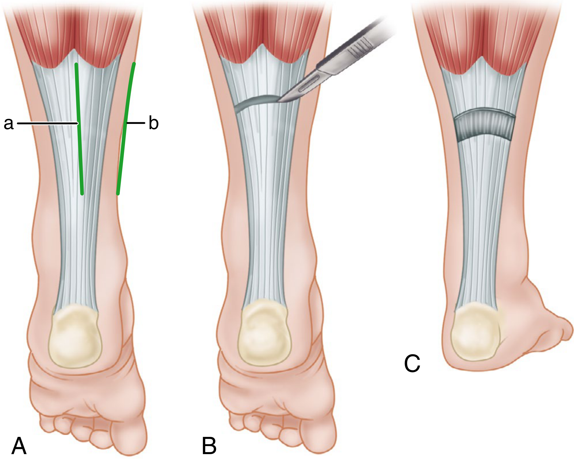
- A - Posteromedial longitudinal incision (a = skin incision line; b = gastrocnemius aponeurosis outline) over the posterior calf at the musculotendinous junction
- B - Scalpel divides the aponeurosis with a transverse cut, lateral-to-medial
- C - Foot placed in dorsiflexion; the tendon ends naturally separate, achieving lengthening without suturing
Additional steps:
- The raphes (fibrous connections) of the gastrocnemius-soleus and the plantaris tendon are completely released
- The ankle is brought to slight dorsiflexion, which separates the cut tendon ends
- The soleus aponeurosis may be partially incised if additional length is needed
Postoperative care: Short leg cast for 4 weeks (knee extension encouraged), then ankle-foot orthosis (AFO) in neutral, weight bearing initiated. Full-time brace wear initially, modified based on growth and physiotherapy progress.
Indications (When Is It Used?)
- Cerebral palsy - spastic equinus gait (toe-walking) in ambulatory children - preferred over TAL because over-lengthening risk is lower
- Idiopathic toe-walking in children
- Equinovarus deformity (clubfoot, post-stroke) as part of combined reconstruction
- Total ankle arthroplasty - improves tibiotalar dorsiflexion when gastrocnemius contracture is present
- Diabetic forefoot ulcers - reduces plantar pressure; gastrocnemius recession increases midfoot ulcer healing rates
- Anterior tibialis tendon reconstruction - added to take tension off the repair
- Flatfoot reconstruction - as an adjunct to address the equinus component
Gastrocnemius Recession vs. Achilles Tendon Lengthening
| Feature | Gastrocnemius Recession | TAL (Achilles Lengthening) |
|---|---|---|
| Level of cut | Musculotendinous junction (Zone 2) | Achilles tendon (Zone 3) |
| Indication | Isolated gastrocnemius contracture | Combined gastroc + soleus contracture |
| Over-lengthening risk | Lower | Higher |
| Weakness in push-off | Less likely | More likely if over-lengthened |
| Preferred in | Ambulatory children | Severe deformity, non-ambulatory patients |
(Campbell's Operative Orthopaedics, 15th Ed)
Key Complication to Watch For
Over-lengthening is the main risk - it causes:
- Weakness in push-off during gait
- Crouch gait (excessive knee/hip flexion) if done in the wrong setting
- Unmasking of toe flexion contractures (flexor digitorum/hallucis longus), which may need simultaneous Z-lengthening
This is why gastrocnemius recession is preferred over TAL in ambulatory children - the risk of over-lengthening is significantly lower.
Is calf lengthening exercises helpful
Yes, calf stretching/lengthening exercises are well-supported as first-line conservative treatment before considering surgery. Let me check the textbooks and recent evidence.
Searching sources...
Finding Sources
Searching PubMed
gastrocnemius stretching exercises equinus contracture outcomes
Searching the Web
calf stretching exercises evidence gastrocnemius tightness plantar fasciitis equinus
Finding Sources
Searching PubMed
calf stretching plantar fasciitis heel pain
Finding Sources
Searching PubMed
stretching exercise gastrocnemius soleus dorsiflexion range of motion
Reading Article
PMID: 16926259
Reading Article
PMID: 34766860
Finding Sources
Finding Sources
Finding Sources
Finding Sources
Finding Sources
Reading File
Finding Sources
Great evidence gathered. Here is a thorough, evidence-based answer:
Are Calf Lengthening (Stretching) Exercises Helpful?
Yes - they are helpful, and they are the recommended first-line treatment for most conditions caused by gastrocnemius tightness. However, the degree of benefit depends on the specific condition, how consistently the exercises are done, and how severe the contracture is.
What the Evidence Shows
1. Do stretches actually increase ankle dorsiflexion?
A systematic review and meta-analysis (Radford et al., Br J Sports Med 2006) pooled 5 RCTs and found:
- Static calf stretching for ≤15 minutes → +2.07° dorsiflexion (95% CI: 0.86–3.27°)
- Stretching for 15–30 minutes → +3.03° (95% CI: 0.31–5.75°)
- Stretching for >30 minutes → +2.49° (95% CI: 0.16–4.82°)
The gain is small but statistically significant at all durations. Whether a 2–3 degree change is clinically meaningful depends on the condition being treated.
2. For Plantar Fasciitis
Calf (gastrocnemius-soleus) stretching is the cornerstone of conservative management, recommended in every treatment protocol. From the Textbook of Family Medicine (9th Ed):
"Most treatment plans include plantar fascia stretching, ice or ice massage, heel cushioning, and analgesic medication... other options include night splints, physical therapy, and orthotic devices."
A study comparing gastrocnemius-soleus stretching vs. tendo-Achilles stretching found gastrocnemius-soleus stretching gave a greater reduction in pain scores after 4 weeks (mean reduction 2.50 ± 0.86 vs. 1.71 ± 1.0 on a 10 cm pain scale, P < 0.05). (PMC8956500)
However, a 2022 systematic review (Arshad et al., Foot Ankle Int) found that when conservative stretching fails, gastrocnemius recession surgery produces greater improvement than continued stretching - reinforcing that exercises come first, surgery is the fallback.
3. For Achilles Tendinopathy
The Textbook of Family Medicine notes: "An eccentric training program is effective in treating chronic Achilles tendinopathy" - this is the Alfredson protocol of eccentric heel drops (a form of loaded calf lengthening), which has very strong evidence behind it.
The Main Calf Stretching Exercises Used Clinically
A. Gastrocnemius Stretch (Wall Stretch - Knee Straight)
- Stand facing a wall, one foot behind the other
- Keep the back knee straight and heel flat on the floor
- Lean forward until a stretch is felt in the calf
- Targets the gastrocnemius (crosses the knee, so it must be straight)
- Hold 30 seconds, repeat 3× per session
B. Soleus Stretch (Knee Bent)
- Same position, but bend the back knee slightly
- This relaxes the gastrocnemius and isolates the soleus (which does not cross the knee)
- Important when both gastrocnemius AND soleus are tight
- Hold 30 seconds, repeat 3×
C. Step Stretch (Eccentric/Weight-Bearing)
- Stand on the edge of a step with heels hanging off
- Lower heels slowly below step level, hold, then rise back up
- Combines passive lengthening + eccentric strengthening
- Strongest evidence in Achilles tendinopathy (Alfredson protocol)
D. Night Splints
- Ankle held in dorsiflexion during sleep (passive prolonged stretch)
- Particularly helpful for plantar fasciitis - prevents the fascia and calf from tightening overnight
- Explains why first-step morning pain is the hallmark symptom of plantar fasciitis
E. Serial Casting (Clinician-Applied)
- Plaster/fibreglass cast holds ankle in progressively increasing dorsiflexion
- Used in cerebral palsy and severe contractures where active exercises alone are insufficient
- A step up from home exercise before surgery is considered
When Do Exercises Work Best?
| Condition | Role of Stretching | Expected Outcome |
|---|---|---|
| Plantar fasciitis | First-line, core treatment | Good - ~80% resolve without surgery |
| Achilles tendinopathy | Eccentric loading is gold standard | Very good with consistency |
| Mild gastrocnemius contracture | First-line conservative | Moderate - works best when flexible |
| Cerebral palsy spastic equinus | Adjunct to botox/casting | Maintains gains, does not reverse spasticity |
| Severe fixed contracture | Insufficient alone | Surgery usually needed eventually |
| Diabetic foot / equinus | Reduces plantar pressure | Adjunct; surgery better for recurrent ulcers |
Limitations of Stretching Alone
- The dorsiflexion gained (2–3°) is small - may not be enough in moderate-to-severe contractures
- Spastic calf (cerebral palsy, stroke) - the underlying neurological drive to tighten is never corrected by stretching; gains from stretching are temporary without adjuncts (botulinum toxin, AFO bracing)
- Requires consistency - benefits are lost quickly if stretching stops
- Over-aggressive stretching of a recently injured Achilles tendon can be counterproductive
Bottom Line
Calf lengthening exercises are genuinely effective as first-line treatment and should always be tried before surgery. They reduce pain, improve dorsiflexion, and address the biomechanical root cause in conditions like plantar fasciitis and Achilles tendinopathy. The key is doing the right stretch (knee straight for gastrocnemius, knee bent for soleus), holding long enough (at least 30 seconds), and being consistent (multiple times daily). When 6–12 months of dedicated conservative treatment fails, that is the point at which surgical options like gastrocnemius recession are considered.
This is a shared conversation. Sign in to Orris to start your own chat.