
Malaria stages flowchart
Reading File
Now let me search the medical library for malaria lifecycle/stages content and images in parallel.
Finding Sources
Finding Sources
Found two textbook sources with diagrams. Let me read both sections in parallel to get the full content and image URLs.
Reading File
Reading File
Excellent! I have both images and detailed content. Let me fetch both diagrams in parallel and read more of the Robbins pathology text.
Loading Image
Loading Image
Reading File
I now have two excellent textbook diagrams and comprehensive content. Here is the full malaria stages flowchart explanation with both diagrams:
Malaria Life Cycle - Stages Flowchart
Here are two authoritative diagrams from major medical textbooks:
Diagram 1 - Complete Life Cycle (Goodman & Gilman's Pharmacological Basis of Therapeutics)
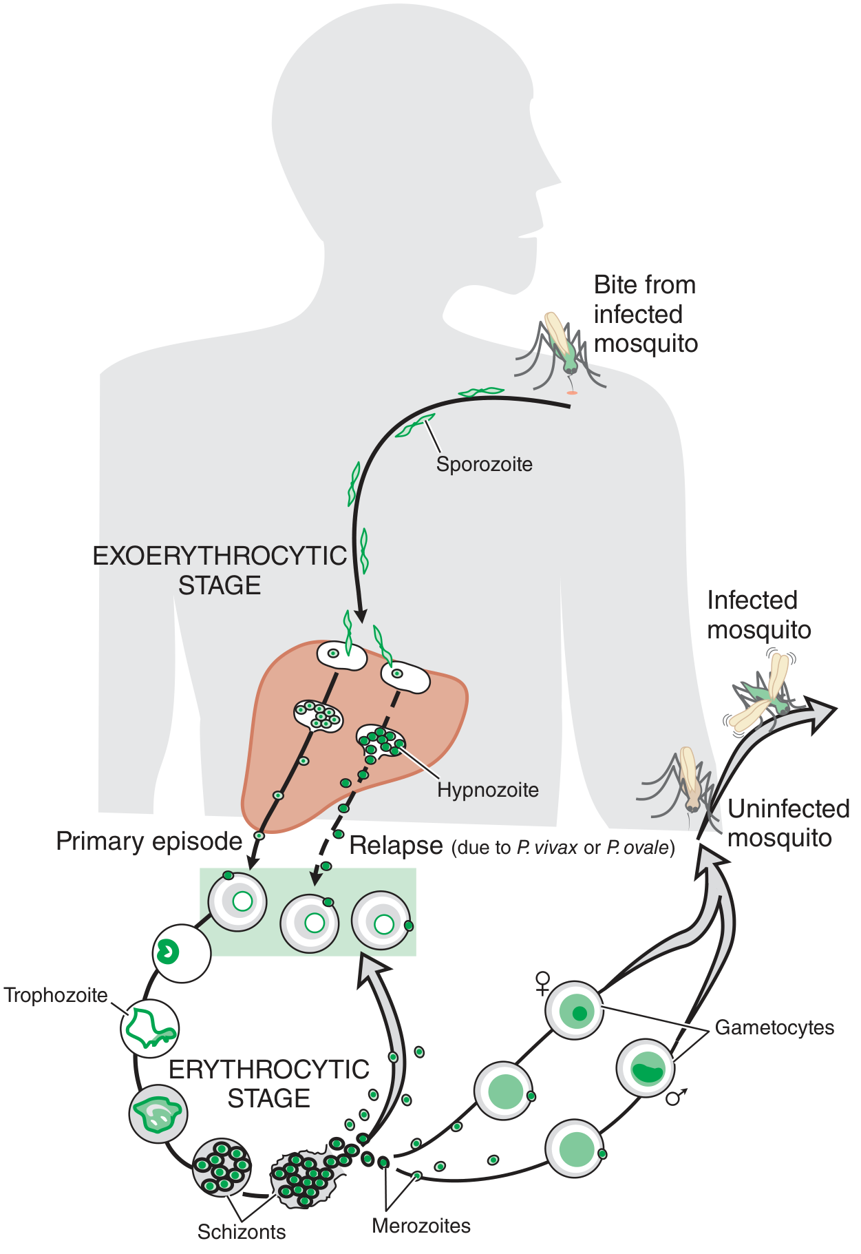
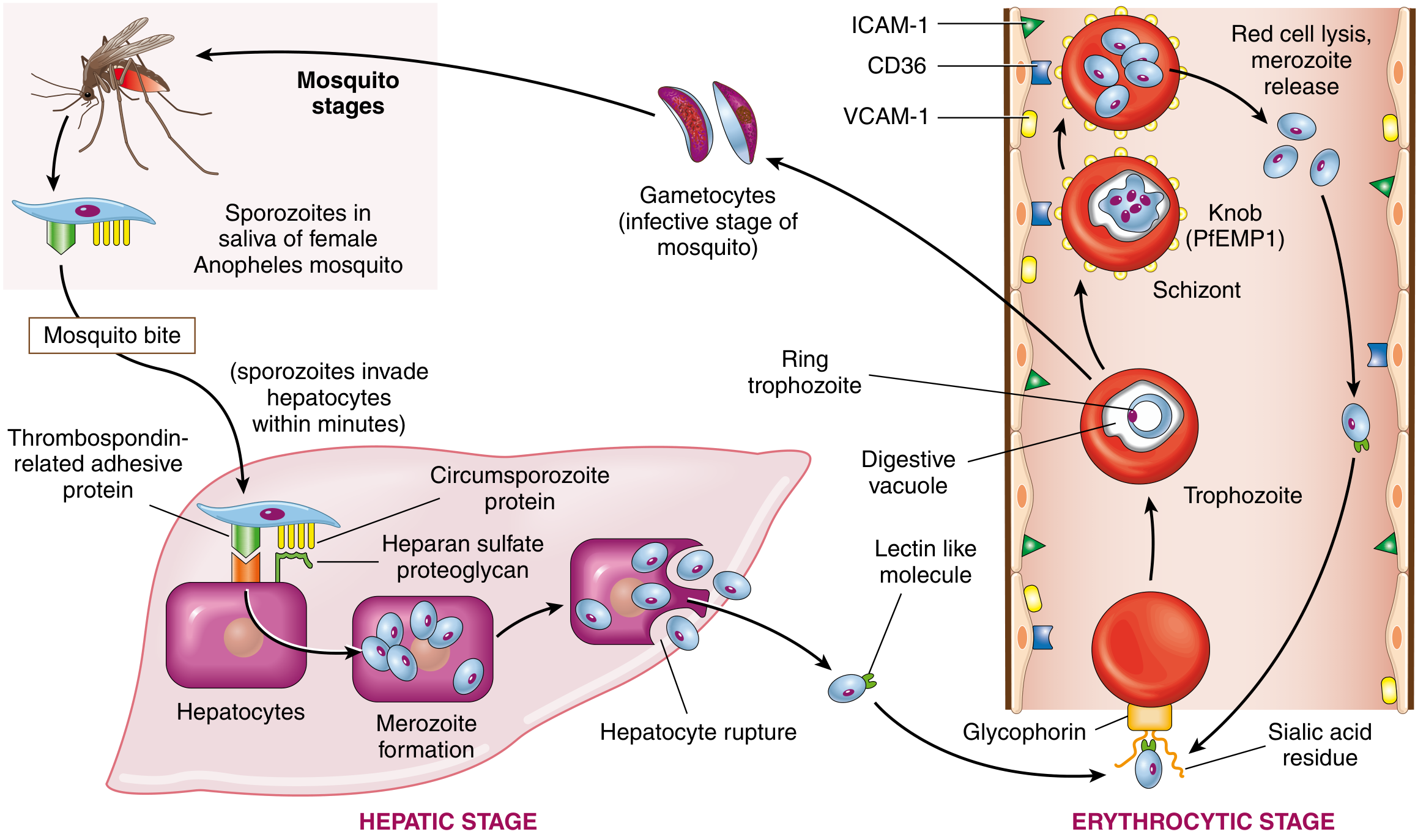
Diagram 2 - Detailed Cellular Mechanisms (Robbins & Cotran Pathologic Basis of Disease)
The Three Main Stages
Stage 1 - Mosquito Bite (Sporozoite Inoculation)
- Female Anopheles mosquito takes a blood meal
- Sporozoites are released from salivary glands into the human bloodstream
- Within minutes, sporozoites travel to the liver, binding to hepatocytes via circumsporozoite protein + heparan sulfate proteoglycan and thrombospondin-related adhesive protein
Stage 2 - Hepatic (Exo-erythrocytic) Stage
| Step | Event |
|---|---|
| Invasion | Sporozoites enter hepatocytes |
| Multiplication | Asexual schizogony - parasites multiply massively inside liver cells |
| Merozoite release | Each infected hepatocyte ruptures, releasing up to 30,000 merozoites |
| Timing (P. falciparum) | Hepatocyte rupture occurs at 8-12 weeks |
Key difference for relapse: P. vivax and P. ovale form dormant hypnozoites in hepatocytes. These reactivate weeks to months later, causing relapses. P. falciparum and P. malariae do NOT form hypnozoites.
Stage 3 - Erythrocytic (Blood) Stage - The Cyclic Stage
This is the stage that causes clinical symptoms:
Merozoite → invades RBC
    ↓  (binds Glycophorin via sialic acid residues)
Ring trophozoite (early)
    ↓
Trophozoite (late, with digestive vacuole - digests hemoglobin)
    ↓
Schizont (multinucleated, forms knobs/PfEMP1 on RBC surface)
    ↓
RBC ruptures → releases new Merozoites
    ↓
→ Reinvade new RBCs (cycle repeats every 48-72 hrs)
    OR
→ Some become Gametocytes (sexual forms)
Fever periodicity:
- P. vivax / P. ovale: 48-hour cycle (tertian fever)
- P. malariae: 72-hour cycle (quartan fever)
- P. falciparum: irregular (can infect RBCs of all ages)
Stage 4 - Mosquito (Sexual) Stage - Completing the Cycle
| Step | Event |
|---|---|
| Gametocyte uptake | Uninfected Anopheles bites an infected human, ingesting gametocytes |
| Fertilization | Male + female gametocytes form a zygote in the mosquito midgut |
| Ookinete | Motile zygote penetrates midgut wall |
| Oocyst | Forms on outer gut wall, undergoes sporogony |
| Sporozoite release | Mature oocyst bursts, sporozoites migrate to salivary glands |
| Transmission | Ready to infect the next human host |
Why P. falciparum is Most Dangerous
- Infects RBCs of all ages (others only infect young or old RBCs - a smaller pool)
- Sequestration: infected RBCs express PfEMP1 knobs that bind endothelial receptors (ICAM-1, CD36, VCAM-1), blocking microvascular flow - causes cerebral malaria
- High parasitemia leading to severe anemia, renal failure, pulmonary edema
Host Genetic Resistance (Protective Mutations)
These mutations reduce susceptibility to malaria and are common in endemic regions:
- HbS (sickle cell trait) and HbC
- Thalassemias (alpha and beta)
- G6PD deficiency
- Absence of Duffy antigen (DARC) - protects against P. vivax (90% of West Africans lack it)
Sources: Robbins, Cotran & Kumar - Pathologic Basis of Disease, p. 372-373 | Goodman & Gilman's Pharmacological Basis of Therapeutics, p. 1312
Act regimen acts against which stage of malarial parasite
ACT (Artemisinin-based Combination Therapy) acts primarily against the asexual erythrocytic (blood) stage - the ring trophozoite stage specifically. Artemisinins are the fastest-acting antimalarials known.
Searching sources...
Finding Sources
Finding Sources
Reading File
The textbook has a very clear table and mechanism description. Let me read just a bit more to capture the full table and partner drug data.
Reading File
All the data needed is in hand. Here is the complete answer:
ACT - Which Stages Does It Target?
Direct Answer
ACT acts primarily against the asexual erythrocytic (blood) stage, and the artemisinin component also has significant activity against gametocytes. It does NOT act against sporozoites, primary liver schizonts, or hypnozoites.
Stage-by-Stage Breakdown (Drug Activity Table)
| Stage | Artemisinin | Notes |
|---|---|---|
| Sporozoite | - | No activity |
| Primary liver schizont (exo-erythrocytic) | - | No activity |
| Hypnozoite (P. vivax/P. ovale dormant liver forms) | - | No activity - needs primaquine |
| Asexual erythrocytic (ring, trophozoite, schizont) | + | PRIMARY target |
| Gametocyte (sexual stage) | + | Reduces transmission |
Mechanism of Action Against the Erythrocytic Stage
Artemisinin is activated by intraparasitic heme inside the highly acidic digestive vacuole of the trophozoite (where hemoglobin is being digested). This interaction cleaves the drug's endoperoxide bridge, generating free radicals that:
- Promiscuously alkylate parasite proteins
- Oxidize macromolecules within the parasite
- Cause a 4-log₁₀ (10,000-fold) reduction in parasite burden per 48-hour erythrocytic cycle
This is why artemisinins work only against blood-stage parasites - they depend on hemoglobin digestion (which only happens in the erythrocytic stage) to become activated.
Why ACT is Used as Combination Therapy
Artemisinins alone require >7 days to eliminate all parasites (they have a very short half-life of 1-2 hours). When paired with a slower-acting, longer-lasting partner drug, a 3-day course is sufficient. The two components work synergistically:
| Component | Speed | Half-life | Kills |
|---|---|---|---|
| Artemisinin (dihydroartemisinin, artesunate, artemether) | Fast | 1-2 hours | Rapidly clears 99.99% of parasites per cycle |
| Partner drug (lumefantrine, piperaquine, amodiaquine, etc.) | Slow | Days to weeks | Eliminates residual parasites, prevents recrudescence |
Comparison with Other Antimalarials (Stage Activity)
| Drug | Sporozoite | Liver (primary) | Hypnozoite | Asexual Blood | Gametocyte |
|---|---|---|---|---|---|
| Artemisinin | - | - | - | + | + |
| Chloroquine | - | - | - | + | +/- |
| Mefloquine | - | - | - | + | - |
| Atovaquone/Proguanil | - | + | - | + | +/- |
| Primaquine | - | + | + | - | + |
| Tafenoquine | + | + | + | + | + |
Key takeaway: ACT treats and clears active blood-stage infection + reduces mosquito transmission via gametocytes. To prevent relapse in P. vivax or P. ovale (due to hypnozoites), you must add primaquine (or tafenoquine) to the ACT regimen.
- Goodman & Gilman's Pharmacological Basis of Therapeutics, p. 1312-1313
Teacg me if a patient comes with low bicarbonate what dose of bicarb should I give Teach me the bicarb dose for bicarbonate deficiency
The classic formula for bicarbonate replacement is: HCO₃ deficit (mEq) = 0.4 × weight (kg) × (desired HCO₃ - measured HCO₃). Give half the calculated deficit slowly, then reassess.
Searching sources...
Finding Sources
Finding Sources
Finding Sources
Finding Sources
Good finds. Let me read the Schwartz Surgery section (which mentioned the formula directly) and Harrison's section in parallel.
Reading File
Reading File
Good - got the Schwartz formula. Now let me read more of Harrison's for the bicarb treatment section, and also check Brenner & Rector (nephrology) for the adult formula.
Reading File
Finding Sources
Finding Sources
Reading File
Excellent - Harrison's gives the specific DKA protocol and the general target. Now let me get the standard adult formula from a nephrology or ICU text.
Finding Sources
Finding Sources
I have enough authoritative content from Schwartz's Surgery and Harrison's for a complete, well-structured answer. Here it is:
Bicarbonate (NaHCO₃) Dosing for Low Bicarbonate / Metabolic Acidosis
Step 1 - First Ask: WHY Is the Bicarbonate Low?
Before giving bicarb, identify the cause - because not all low-HCO₃ states need bicarb therapy:
| Type | Examples | Bicarb Needed? |
|---|---|---|
| High-AG acidosis with metabolizable anions | DKA, lactic acidosis | Usually NO - treat the cause; anions will regenerate HCO₃ |
| High-AG acidosis with non-metabolizable anions | Renal failure (uremic), toxins (ethylene glycol, methanol) | YES |
| Normal-AG (hyperchloremic) acidosis | Diarrhea, RTA, ureteral diversion | YES |
| DKA with severe acidemia | pH <7.00 | YES (cautiously, limited target) |
Step 2 - The Formula
Standard HCO₃ Deficit Formula (Adults)
HCO₃ deficit (mEq) = Volume of Distribution × Weight (kg) × (Desired HCO₃ − Measured HCO₃)
The volume of distribution factor varies by age:
| Patient | VD Factor |
|---|---|
| Adults | 0.4 (some sources use 0.5) |
| Smaller children | 0.4 |
| Older children | 0.3 |
| Newborns / critically ill neonates | 0.5 |
Worked Example
- Patient: 70 kg adult
- Measured HCO₃: 12 mEq/L
- Desired HCO₃: 22 mEq/L (target, not full normal - see below)
- Formula: 0.4 × 70 × (22 − 12) = 0.4 × 70 × 10 = 280 mEq
Step 3 - How to Give It (Critical Rules)
Rule 1: Give HALF first, then reassess
Give ½ of the calculated deficit → recheck ABG/electrolytes → recalculate if needed.
Never give the full deficit at once - this prevents overshoot (metabolic alkalosis, hypokalemia, paradoxical CSF acidosis).
Rule 2: Target HCO₃ 22 mEq/L, NOT 24-25 mEq/L
Harrison's explicitly states: "increase plasma [HCO₃⁻] to a target of 22 mmol/L. Overcorrection should be avoided."
Rule 3: Give it SLOWLY as an IV infusion
- Dilute to 0.5 mEq/mL (isotonic bicarb) - avoid full-strength (hyperosmolar, risks IVH in neonates)
- Infuse over 30-60 minutes minimum, never as a fast IV push except in cardiac arrest
Rule 4: Monitor electrolytes during infusion
- Risk of hypokalemia (bicarb drives K⁺ into cells)
- Risk of hypocalcemia (alkalosis reduces ionized Ca²⁺)
- Risk of hypernatremia (each mEq NaHCO₃ contains 1 mEq Na⁺)
Special Situations
DKA (pH <7.00 only)
Per Harrison's:
- 50 mEq NaHCO₃ diluted in 300 mL NS, infused over 30-45 minutes
- Target: HCO₃ 10-12 mEq/L, pH ~7.20 - do NOT normalize
- Monitor K⁺ closely as urine output reestablishes
Cardiac Arrest / CPR
- Give ½ the corrective dose as IV bolus, other half given slowly
Chronic Metabolic Acidosis (e.g. CKD, RTA)
- Oral NaHCO₃ tablets 650 mg twice daily, titrated upward
- Or Shohl's solution (sodium citrate/citric acid)
- Goal HCO₃ ≥22 mEq/L
Quick Reference Summary
HCO₃ deficit (mEq) = 0.4 × Wt(kg) × (22 − measured HCO₃)

Give HALF the dose first → reassess → give rest if needed
Rate: slow IV infusion (dilute to 0.5 mEq/mL)
Watch: K⁺, Ca²⁺, Na⁺, pH
Target: HCO₃ = 22 mEq/L (not full normal)
When NOT to Give Bicarb
- Lactic acidosis or DKA with pH >7.00 - treat the underlying cause
- Respiratory acidosis - bicarb is wrong here; improve ventilation
- Post-cardiac arrest (routine use not recommended)
Sources: Schwartz's Principles of Surgery 11th Ed, p. 1735 | Harrison's Principles of Internal Medicine 22nd Ed, p. 759-761
The bicarb is given with NS?
Good catch - this is an important practical point. No, NaHCO₃ should NOT be mixed with NS (Normal Saline / 0.9% NaCl) as the primary diluent for routine bicarb infusions. The DKA example in Harrison's was a specific exception.
Verifying details...
Finding Sources
Finding Sources
Finding Sources
Searching the Web
sodium bicarbonate IV diluent D5W vs normal saline compatibility mixing
Now I have a clear, complete answer from both the FDA prescribing information and clinical sources. Here is the precise answer:
What Do You Mix NaHCO₃ With?
Short Answer
NS (0.9% NaCl) is technically compatible with NaHCO₃ - but D5W is the preferred diluent in most clinical protocols. The Harrison's DKA example using NS was a specific, context-driven choice. Here is what each situation calls for:
Standard Diluents - What to Use
| Situation | Preferred Diluent | Why |
|---|---|---|
| Routine metabolic acidosis infusion | D5W (5% dextrose in water) | Preferred; isotonic bicarb drip; dextrose metabolized leaving free water to dilute |
| DKA correction | NS (as per Harrison's) | Volume resuscitation is also needed in DKA; saline serves dual purpose |
| Urinary alkalinization (e.g. salicylate OD, methotrexate) | 150 mEq NaHCO₃ in 850 mL D5W = 1L bag | Standard protocol |
| Cardiac arrest | Undiluted 8.4% push | Emergency only |
Classic "Isotonic Bicarb Drip" Recipe:
150 mL of 8.4% NaHCO₃ added to 850 mL D5W → 1 litre bag ≈ 150 mEq/L (isotonic, ~300 mOsm)
This is the standard ICU/EM drip for bicarbonate infusion.
Why Not Just NS?
NS is compatible (FDA confirms 8.4% can be diluted in either D5W or 0.9% NaCl), but there are reasons D5W is preferred:
- Sodium load - NS + NaHCO₃ together give a very high Na⁺ load (both are ~154 mEq/L Na⁺). This risks hypernatremia and volume overload
- Chloride load - NS adds chloride, which can worsen hyperchloremic acidosis - counterproductive
- D5W is electrolyte-free - the dextrose is quickly metabolized, effectively delivering free water that dilutes the bicarb without adding Na⁺ or Cl⁻
What NaHCO₃ Must NEVER Be Mixed With
This is critical for safety:
| Incompatible substance | Problem |
|---|---|
| Calcium salts (Ca gluconate, CaCl₂) | Precipitates as CaCO₃ - never mix in same line |
| Magnesium sulfate | Precipitation |
| Lactated Ringer's | Incompatible (contains calcium) |
| Sodium lactate | Incompatible |
| Dobutamine, noradrenaline | Degradation/inactivation |
| Insulin | Incompatible |
| Vancomycin | Precipitates |
Key clinical rule: Never run NaHCO₃ through the same IV line as calcium or magnesium - always flush the line with NS between them if using the same access.
Concentration to Use
| Line type | Concentration |
|---|---|
| Peripheral IV | 0.5 mEq/mL (4.2% or diluted 8.4%) |
| Central line | Up to 1 mEq/mL (8.4% undiluted or minimally diluted) |
Full-strength 8.4% (1 mEq/mL) through a peripheral vein risks thrombophlebitis and tissue necrosis if it extravasates - always dilute for peripheral access.
Summary
✅ Mix with: D5W (preferred) or ½NS
✅ Mix with: NS if volume replacement also needed (e.g. DKA)
❌ Never mix with: Calcium, Magnesium, Lactated Ringer's
❌ Peripheral line: dilute to 0.5 mEq/mL max
Treatment of drug induced lupus
The primary treatment for drug-induced lupus (DIL) is stopping the offending drug - symptoms usually resolve within weeks to months after discontinuation. NSAIDs and hydroxychloroquine manage mild symptoms; corticosteroids are used for severe cases.
Searching sources...
Finding Sources
Finding Sources
Excellent sources found. Let me read the three most relevant - Harrison's (most comprehensive), Firestein's Rheumatology, and Rosen's Emergency Medicine in parallel.
Reading File
Reading File
Reading File
Now let me get the Harrison's block 41 entry for additional treatment details, and also check the TNF-alpha induced DIL section in Murray & Nadel.
Reading File
Reading File
I have comprehensive content from multiple authoritative textbooks. Here is the complete answer:
Treatment of Drug-Induced Lupus (DIL)
Step 1 - STOP the Offending Drug (Cornerstone of Treatment)
This is the single most important step. In most cases, symptoms resolve spontaneously after drug withdrawal - no further treatment is needed.
"The diagnosis is typically clinical and confirmed by resolution of symptoms with the withdrawal of the offending medication." - Rosen's Emergency Medicine
Timeline of resolution:
- Symptoms: resolve within weeks to months after stopping the drug
- Anti-histone antibodies: may persist for months to years even after symptoms resolve
- ANA: can remain positive long-term - a persistently positive ANA alone is NOT a reason to restart or avoid treatment
Step 2 - Symptomatic Treatment (While Awaiting Resolution)
| Symptom / Severity | Treatment |
|---|---|
| Arthralgia, myalgia, fever, mild serositis | NSAIDs (first-line for mild symptoms) |
| Moderate symptoms (persistent rash, pleuritis, pericarditis) | Hydroxychloroquine (antimalarial - used as in SLE) |
| Severe symptoms (significant serositis, cytopenias, vasculitis) | Corticosteroids (prednisone, moderate-to-high dose) |
| Immunosuppression needed (rare, severe) | Azathioprine, mycophenolate (if corticosteroid-sparing needed) |
Step-by-Step Management Algorithm
Patient presents with lupus-like features
              ↓
Review all medications - identify likely culprit
              ↓
STOP the offending drug
              ↓
        Mild symptoms?          Moderate/severe?
        ↓                       ↓
      NSAIDs                Hydroxychloroquine
      (e.g. ibuprofen,      ± NSAIDs
      naproxen)             ↓
                        Not improving?
                            ↓
                    Corticosteroids
                    (prednisolone 0.5-1 mg/kg/day)
                            ↓
                    Taper as symptoms resolve
Key Clinical Features That Guide Treatment
| Feature | DIL | SLE |
|---|---|---|
| Renal involvement | Rare - usually no nephritis | Common |
| CNS involvement | Rare | Common |
| Anti-histone Ab | +75-95% (hallmark) | 50% |
| Anti-dsDNA Ab | Rare | Common (specific for SLE) |
| Anti-Smith Ab | Rare | Specific for SLE |
| Hypocomplementemia | Uncommon | Common |
| Resolution after stopping drug | Yes - confirms diagnosis | No |
The rarity of renal and CNS involvement means aggressive immunosuppression (cyclophosphamide, belimumab) is almost never needed in true DIL.
Common Causative Drugs to Know
| Category | High-risk Drugs |
|---|---|
| Antiarrhythmics | Procainamide (up to 30% risk - highest), quinidine |
| Antihypertensives | Hydralazine (first drug ever identified, 1954), methyldopa |
| Antibiotics | Minocycline, isoniazid, rifampin, nitrofurantoin |
| Anticonvulsants | Phenytoin, carbamazepine, ethosuximide |
| Biologics | TNF-α inhibitors (infliximab, etanercept) - important in modern practice |
| Others | Sulfasalazine, chlorpromazine, lithium, propylthiouracil, ACE inhibitors, beta-blockers, PPIs, HCTZ, immune checkpoint inhibitors, trastuzumab |
Note: Biologic-induced DIL (especially from TNF-α inhibitors) can be more persistent and may require longer courses of corticosteroids even after stopping the biologic.
Drug-Induced Subacute Cutaneous Lupus (SCLE) - Special Subset
A related but distinct entity with prominent skin involvement (annular or papulosquamous photosensitive rash). Key drugs:
- Proton pump inhibitors, diltiazem, HCTZ, terbinafine, interferons, paclitaxel, docetaxel, aromatase inhibitors
Treatment: stop drug + topical corticosteroids + hydroxychloroquine + sun protection.
What Does NOT Need Treatment
- Asymptomatic positive ANA alone in a patient on a known causative drug - no need to stop the drug just for a positive ANA unless symptoms develop
- Persistent positive ANA after drug cessation without symptoms - observe only
Sources: Harrison's Principles of Internal Medicine 22nd Ed, p. 2879 | Firestein & Kelley's Textbook of Rheumatology | Rosen's Emergency Medicine
This is a shared conversation. Sign in to Orris to start your own chat.