
Management of plantar warts
plantar warts treatment
plantar wart verruca plantaris clinical photo
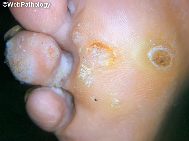
Clinical photography of a plantar foot displaying a verruca plantaris (plantar wart). Modality is a high‑quality gross image used in dermatology documentation. Precise location is the plantar surface of the foot (sole), with a focal, endophytic lesion that is rough and hyperkeratotic. Visual features include a circular to oval papule with a central crater or indent, irregular surface texture, and surrounding callus‑like thickening. Punctate black dots representing thrombosed capillaries may be seen within the lesion, while surrounding epidermis shows white scaling and fissuring. The lesion is typically mildly elevated above the surrounding skin and may be tender with pressure; it often coexists with other warts on the same sole. The clinical impression is verruca plantaris caused by human papillomavirus infection, with characteristic epidermal hyperplasia, papillomatosis, and hyperkeratosis evident on gross inspection. Differential diagnoses include callus/corn, plantar keratoderma, and foreign body reaction; histology would show koilocytosis and viral cytopathic effects if examined. This image supports clinical decision making, differential diagnosis, and treatment planning (topical salicylic acid, cryotherapy, or laser), and serves as an educational reference for students, residents, and clinicians evaluating plantar lesions. Clinical correlation includes assessing for additional lesions and patient history of HPV exposure; follow-up imaging is not required.
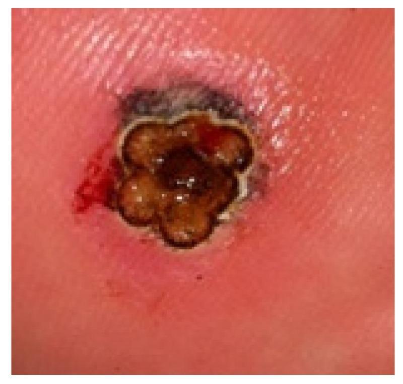
This clinical photograph displays a large plantar wart (verruca plantaris) measuring over 4 mm in diameter on the weight-bearing surface of the foot. The lesion is characterized by a raised, hyperkeratotic appearance with a clustered, multi-lobulated morphology resembling a 'mosaic' pattern. The central tissue exhibits dark brown to black discoloration, likely due to the application of India ink used in laser therapy protocols to enhance thermal absorption. A distinct, pale rim or halo surrounds the lobules, followed by a wider zone of erythematous, inflamed perilesional skin. Minor punctate bleeding is visible on the lateral margin, typical of exposed capillaries (thrombosed capillaries) following the removal of overlying hyperkeratosis. This image serves as a clinical example of a recalcitrant plantar wart prepared for laser treatment, illustrating the anatomical challenges of treating thick epidermal lesions on the plantar surface.
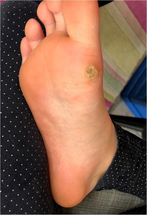
This clinical photograph displays a solitary, deep plantar wart (verruca plantaris) located on the weight-bearing plantar surface of the human foot, specifically over the metatarsal region. The lesion is well-circumscribed and approximately circular, characterized by a central area of significant hyperkeratosis. The surface texture is rough, verrucous, and uneven, exhibiting a beige to yellowish-tan discoloration that contrasts with the surrounding healthy pink skin. A slightly raised, indurated outer rim demarcates the lesion from the adjacent tissue. The visual findings are consistent with a myrmecia-type wart typically associated with Human Papillomavirus type 1 (HPV 1). Key educational features include the disruption of normal dermatoglyphics and the thick keratotic plug, which are hallmark diagnostic signs of plantar verrucae in dermatology.
PMID: 38243639
Management of Plantar Warts (Verruca Plantaris)
Overview
Diagnosis
- Endophytic hyperkeratotic papule on the plantar surface
- Punctate black dots = thrombosed capillaries (pathognomonic)
- Disruption of dermatoglyphics (skin lines stop at the wart edge) - distinguishes from a callus/corn, where lines are accentuated
- Tenderness on lateral compression rather than direct pressure
- Trimming surface keratin reveals capillaries more prominently
Indications for Treatment
- Pain or interference with gait/function
- Social embarrassment
- Risk of spread or enlargement
- Patient preference
Treatment Options
First-Line: Keratolysis with Salicylic Acid
- Best first-line option - all evidence agrees aggressive keratolysis with salicylic acid is superior to watchful waiting
- Available as 17-40% topical preparations (paints, gels, plasters), with up to 50-60% used on plantar surfaces
- Technique: soak foot in warm water for 5 minutes, pare down the wart with a pumice stone/emery board, apply salicylic acid, cover with occlusive dressing
- Daily application; cure rates vary from 50%-75% with sustained use
- Salicylic acid combined with 5-FU cream yields high cure rates in plantar warts
- Safer than cryotherapy and preferred in immunocompetent patients who can sustain long-term self-treatment
Second-Line: Cryotherapy (Liquid Nitrogen)
- Reasonable first-line option in office settings; note that no RCT has demonstrated cryotherapy to be superior to placebo for plantar warts specifically
- Cure rate: 20%-50% with repeated applications over several months
- Technique: sustained 10-second freeze with a spray gun; produces a freeze halo of 2-3 mm; a blister should form after 1-2 days
- Single freeze-thaw cycle may be as effective as double cycles
- Treat every 2-3 weeks (as the old blister peels off, to prevent regrowth)
- Liquid nitrogen is preferred over other cryogens (lower temperature = deeper freeze)
- Complications: hypopigmentation, depigmentation, post-procedural pain for several days, and (rarely) digital nerve damage if too deep
- Contraindicated/caution: Raynaud's phenomenon, cryoglobulinemia, Fanconi anemia, poor peripheral circulation
Combination Therapy
- Salicylic acid + cryotherapy is more efficacious than cryotherapy alone (supported by multiple RCTs and Fitzpatrick's Dermatology)
- Cantharidin 1% + podophylotoxin 20% + salicylic acid 30% (CPS) - ranked highest in a 2024 Bayesian network meta-analysis of 33 RCTs (SUCRA = 0.94), significantly superior to no treatment (Xu et al., 2024)
Immunotherapy (for Recalcitrant Warts)
- Intralesional Candida antigen: 0.1 mL injected into 1-3 lesions every 3-4 weeks; up to 80% cure rate reported; side effects include fever, chills, and flu-like symptoms 6-8 hours post-injection (resolving in 24-48 hours)
- Dinitrochlorobenzene (DNCB) / Squaric acid dibutyl ester (SADBE) / Diphenylcyclopropenone (DPCP): topical contact sensitizers applied to forearm first, then wart; induce delayed-type hypersensitivity
- Imiquimod 5% cream: off-label; requires daily application under occlusion for plantar warts (the standard 3x/week condylomata regimen is insufficient); elicits marked inflammatory reaction
- HPV quadrivalent vaccine: limited evidence; one report showed resolution of plantar warts in young patients after vaccination; role in non-genital warts is not established Andrews' Diseases of the Skin, p. 468
Antiproliferative/Antimitotic Therapy
- Intralesional bleomycin (1 U/mL): reserved for recalcitrant warts in adults
- Inject until lesion blanches; multipuncture (Shelley) technique or air-jet injector also used
- Small warts (<5 mm): 0.1 mL; larger warts: 0.2 mL; repeat every 3 weeks
- Wart turns black; eschar separates in 2-4 weeks
- Rarely requires >1-2 treatments for common warts; less effective for plantar warts
- Side effects: pain (may need local anesthesia), rarely scarring, Raynaud phenomenon of treated fingers, flagellate hyperpigmentation, digital necrosis (rare)
- Microneedles + bleomycin (MNB) is an emerging delivery method ranked second-best in the 2024 NMA
- Topical 5-fluorouracil (5-FU): variable results alone; combined with salicylic acid, high cure rates in plantar warts
- Intralesional 5-FU: used with variable results; adding to cryotherapy shows no additional benefit over cryo alone
Laser Therapy
- CO2 laser (ablative): direct ablation of the wart; effective but painful, requires anesthesia, risk of scarring
- Pulsed-dye laser (PDL, 585/595 nm): targets the thrombosed capillaries; efficacy >65% in reported series; less scarring than CO2
- Combination (PDL + bleomycin or Candida antigen): useful for particularly refractory periungual and plantar warts Andrews' Diseases of the Skin, p. 468
- Nd:YAG and Er:YAG lasers also used in some centers
Direct Antiviral Therapy
- Topical cidofovir (1-3%, rarely up to 5%): for difficult-to-treat cases and immunosuppressed patients; compounded preparation applied once or twice daily; extremely expensive; local irritation and erosion may occur
- Intralesional cidofovir (up to 5%) also used
Surgical/Destructive Options
- Curettage and electrodesiccation: generally avoided for plantar warts given high recurrence rates and risk of painful scar on weight-bearing surface
- Sharp excision: similarly associated with painful scarring; not routinely recommended
Duct Tape Occlusion Therapy
- Simple, low-risk option: apply duct tape directly over the wart; change every 6 days; soak and debride in between
- Evidence is mixed; some RCTs show no benefit over placebo, while others (particularly in children) showed rates comparable to cryotherapy
- Considered a reasonable conservative home therapy
Step-Up Treatment Algorithm
Observation (especially children)
  ↓  [if treatment desired]
Salicylic acid (daily) ± paring/debridement    [1st line]
  ↓  [persistent at 3 months]
Salicylic acid + cryotherapy (q2-3 weeks)      [2nd line]
  ↓  [recalcitrant]
Immunotherapy (Candida antigen IL / DPCP)      [3rd line]
  ↓  [still recalcitrant]
Intralesional bleomycin or antimitotic CPS     [4th line]
  ↓  [refractory]
Laser (PDL or CO2) ± bleomycin combination    [5th line]
  ↓  [severely refractory / immunosuppressed]
Topical cidofovir                              [specialist]
Special Considerations
| Situation | Approach |
|---|---|
| Children <12 yrs (HPV-1) | High spontaneous resolution; prefer observation or topical SA; avoid aggressive destructive therapy |
| Immunosuppressed patients | Warts may be numerous and recalcitrant; cidofovir, systemic retinoids (acitretin, isotretinoin) may be needed |
| Mosaic warts | Multiple coalescent lesions; particularly difficult; combination modalities needed |
| Pregnancy | Avoid bleomycin, podophylotoxin, 5-FU; salicylic acid and cryotherapy are preferred |
| Diabetes/poor circulation | Avoid aggressive cryotherapy; risk of ulceration |
Key Evidence Update (2024)
- CPS (cantharidin 1% + podophylotoxin 20% + salicylic acid 30%) - highest probability of achieving complete response (SUCRA 0.94)
- Microneedles + bleomycin and intralesional bleomycin also significantly superior to no treatment
- Importantly: traditional SA and cryotherapy alone were NOT statistically superior to no treatment for plantar warts in this NMA - though they remain widely used first-line due to safety and accessibility
Clinical note: This NMA finding challenges the traditional first-line status of SA and cryotherapy for plantar warts. In practice, these remain reasonable starting points given their safety profiles, availability, and low cost. For patients with recalcitrant disease, the evidence now favors antimitotic combination approaches.
- Andrews' Diseases of the Skin: Clinical Dermatology, Chapter on Viral Diseases, pp. 467-468
- Fitzpatrick's Dermatology, Vol. 1-2, Chapter 91 (Nail Unit Warts)
- Pfenninger and Fowler's Procedures for Primary Care, Third Edition
- Xu Y et al. "Topical antimitotic treatments for plantar warts are more beneficial: A Bayesian network meta-analysis of RCTs." J Evid Based Med. 2024 (PMID 38243639)

What are these infection
friction melanosis frictional hyperpigmentation foot skin
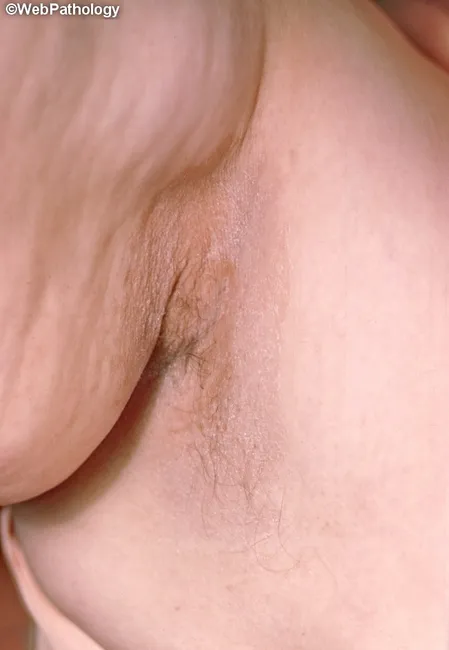
Clinical photograph of axillary skin demonstrates a localized brown pigmented patch within the axillary crease. The lesion appears as a hyperpigmented macule/patch with mild surface texturing and subtle desquamation; adjacent skin is lighter and mostly unremarkable. There is no obvious edema, vesiculation, or ulceration. Hair density is normal around the lesion, and the contour is relatively flat with irregular, but non-destructive, margins. The image relies on standard external photography without dermoscopy or histologic data. Anatomically, the finding localizes to the integument of the axilla (intertriginous region) and involves the epidermis with possible deep dermal pigmentary changes. The phenotype could reflect frictional melanosis, post-inflammatory hyperpigmentation, or acanthosis nigricans spectrum; point toward endocrinologic associations if thickened, velvety textures were present. Differential considerations include tinea versicolor involvement, contact dermatitis-associated hyperpigmentation, and pigmented scars. Clinically, recognition of axillary pigmentation patterns aids in assessment of metabolic risk (insulin resistance, obesity) and inflammatory skin processes. This image is suitable for dermato-educational contexts, reinforcing terminology such as axillary hyperpigmentation, intertriginous skin changes, acanthosis nigricans spectrum, friction melanosis, and post-inflammatory pigmentation, while supporting search queries in dermatology repositories and medical image databases. Consider follow-up evaluation for progression and response to treatment; seek dermatology input if uncertain, or biopsy.
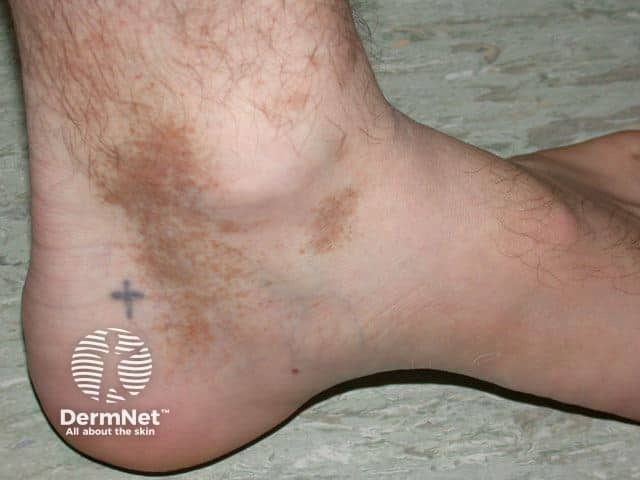
Imaging modality and technique: High-resolution clinical photograph of the left ankle/foot region, color-balanced, macro view. The subject shows multiple hyperpigmented brown patches over the posterolateral ankle and calcaneal region with irregular, slightly scaly borders. The skin between patches appears normal to mildly erythematous without edema. The largest patch spans approximately 2–3 cm in diameter, with peripheral hypopigmented rim in places and variable pigmentation intensity across the patch. The involved skin is otherwise non-edematous, with visible hair follicles and normal surface texture. The lesion distribution is unilateral, confined to the lateral malleolar area, consistent with dermatitis or fungal pigmentary change. No vesicles or pustules are evident. Clinically, this pattern could reflect post-inflammatory hyperpigmentation, tinea (pityriasis) versicolor or dermatophyte infection, or pigmented contact dermatitis. Differential diagnoses include superficial fungal infection (tinea corporis/cruris variant limited to ankle), lichen simplex chronicus with pigment change, stasis dermatitis, or melanosis associated with friction. Correlating history (exposure to humid environments, prior inflammation, trauma, footwear friction) and primary symptoms such as pruritus will guide workup. Useful tests include KOH microscopy, fungal culture, Wood lamp examination, or biopsy if lesions persist or progress. This image documents pigmentary change with potential inflammatory or infectious etiologies, highlighting the need for dermoscopic or microbiologic evaluation and targeted therapy.
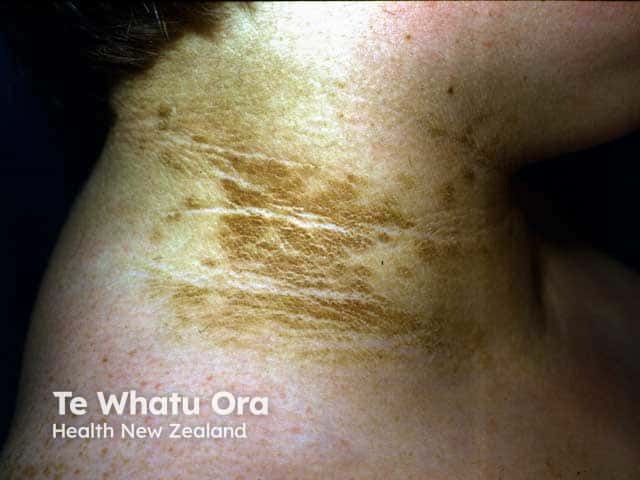
Clinical photography of the neck demonstrating acanthosis nigricans. This close-up dermatology image captures hyperpigmented, velvety plaques concentrated in the posterior and lateral cervical region. The epidermal thickening presents as dark-brown to slate-gray patches with diffuse, corrugated surface and subtle fissuring along skin folds. Palpation would reveal a soft, velvety texture consistent with hyperkeratosis. The condition commonly coalesces in intertriginous areas and may extend toward the base of the skull, with sparing of normal nonfolded skin. The appearance is often symmetrical and may be accompanied by mild irritation or friction. Underlying etiologies include insulin resistance, obesity, type 2 diabetes mellitus, and endocrinopathies; in older adults, malignant acanthosis nigricans can signal GI or other visceral malignancies. Differential diagnoses include frictional melanosis, post-inflammatory hyperpigmentation, and tinea versicolor. Clinically, this finding is a cutaneous marker prompting metabolic risk assessment and screening for metabolic syndrome, diabetes, and hormonal disorders. This image serves educational purposes for dermatology and internal medicine, illustrating classic morphology velvety, hyperpigmented plaques in the neck, along with the importance of clinical correlation and follow-up. Notably, treatment focuses on addressing the underlying cause and improving skin texture with topical keratolytics and barrier repair. Photograph notes: correlate with BMI, fasting glucose, insulin resistance, lipid profiles carefully.
Image Analysis: This Is Not an Infection
Most Likely Diagnoses (in order of probability)
1. Friction Melanosis / Frictional Hyperpigmentation (Most Likely)
- Very common in people who wear tight sandals, straps, or ill-fitting shoes, especially in warm climates
- The pigmentation can have a curvilinear, band-like configuration exactly matching contact points of footwear
- No active inflammation, no itch required - can be completely asymptomatic
- More common and more visible in darker skin types (Fitzpatrick types III-VI)
2. Post-Inflammatory Hyperpigmentation (PIH) from Contact Dermatitis
- Rubber accelerators or antioxidants in shoe soles/insoles
- Leather tanning agents (chromium, formaldehyde)
- Adhesive glues in footwear
- Synthetic fabric dyes
3. Lichen Simplex Chronicus (LSC)
- Well-demarcated, hyperpigmented plaque with exaggerated skin markings (lichenification)
- Intense pruritus is the usual driver
- Common on accessible areas where people rub habitually
4. Macular Amyloidosis (Less Common but Worth Considering)
Key Differentiating Questions to Ask
| Question | Points toward |
|---|---|
| Does the dark area match exactly where a shoe strap sits? | Friction melanosis |
| Was there redness, blisters, or intense itch before the discoloration appeared? | PIH from contact dermatitis |
| Is the area currently very itchy, and does the patient rub/scratch it? | Lichen simplex chronicus |
| Is the pattern distinctly "rippled" like a washboard? | Macular amyloidosis |
| Any new or unusual footwear used before this appeared? | Contact dermatitis (shoe) |
Bottom Line
Disclaimer: This is an educational visual assessment only and does not replace an in-person clinical examination by a licensed physician.