
grades of placenta with usg images
Reading File
Placental grading (Grannum classification) describes progressive calcification and maturation of the placenta on ultrasound, with grades 0-III. Let me search the medical library and web for detailed content and USG images.
Searching sources...
Finding Sources
Searching the Web
placenta grading Grannum classification ultrasound images grade 0 1 2 3
Finding Sources
Reading Web Page
https://radiopaedia.org/articles/placental-grading?lang=us
Loading Image
Searching the Web
placenta grade 0 grade 1 grade 2 grade 3 ultrasound sonography actual USG scan images
Loading Image
Loading Image
Here is a complete overview of placental grading (Grannum Classification) with USG images.
Placental Grading - Grannum Classification (1979)
Placental grading is an ultrasound system introduced by Grannum et al. (1979) to assess placental maturity. Grading is based on changes in three zones as pregnancy advances:
- Chorionic plate (fetal surface)
- Placental substance (parenchyma)
- Basal layer (maternal surface)
Schematic Diagram of All Grades
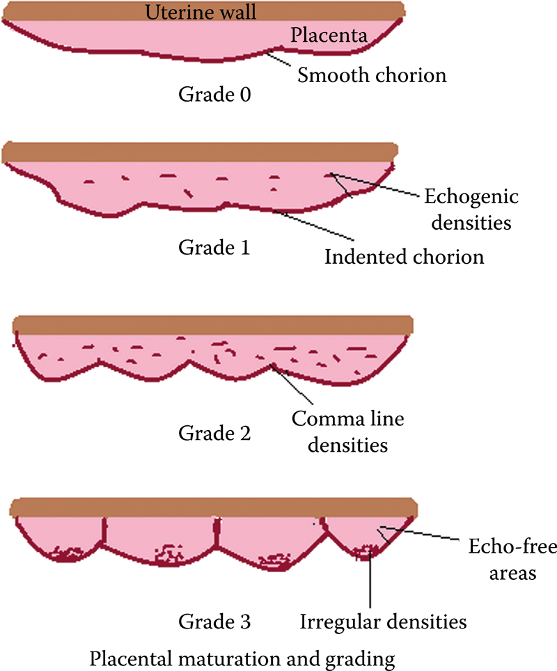
USG Images + Diagrams Side by Side (Grades 0-III)
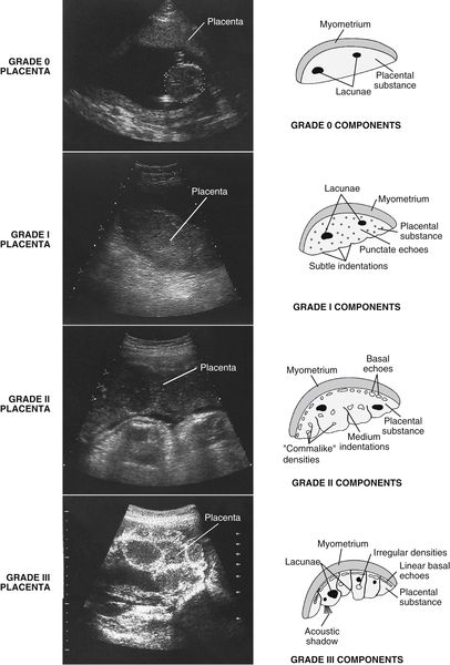
Grade-by-Grade Breakdown
Grade 0 - Late 1st Trimester to Early 2nd Trimester (< 18 weeks)
| Zone | Appearance |
|---|---|
| Chorionic plate | Smooth, straight, well-defined unbroken line |
| Placental substance | Uniform, homogeneous echotexture - no echogenic areas |
| Basal layer | No echogenicities |
- Placenta appears as a uniform, homogeneous structure
- No calcifications
- Normal in early pregnancy
Grade I - 18 to 29 Weeks
| Zone | Appearance |
|---|---|
| Chorionic plate | Well-defined but with fine undulations/subtle indentations |
| Placental substance | Scattered echogenic areas (punctate echoes) - loss of homogeneity begins |
| Basal layer | No echogenicities yet |
- Earliest sign of placental maturation
- Occasional hyperechoic areas in parenchyma

Grade II - 30 to 38 Weeks
| Zone | Appearance |
|---|---|
| Chorionic plate | More marked indentations (deeper than Grade I, but do NOT reach basal layer) |
| Placental substance | Comma-shaped / linear echogenic densities incompletely dividing the placenta |
| Basal layer | Basal stippling - small linear echoes ("dot-dash" configuration) parallel to basal plate |
- Basal echoes are the hallmark of Grade II
- The comma-type densities are a key feature
Grade III - >= 39 Weeks (Mature Placenta)
| Zone | Appearance |
|---|---|
| Chorionic plate | Interrupted by indentations that extend all the way to the basal layer (complete division into cotyledons) |
| Placental substance | Divided into compartments (cotyledons); central areas may show echo-free/fallout zones |
| Basal layer | Large, dense echogenic areas; may cast acoustic shadows |
- Represents the late, mature placenta
- Cotyledon demarcation is visible
- Echo-free (anechoic) areas within cotyledons
Summary Table
| Grade | Gestational Age | Chorionic Plate | Placental Substance | Basal Layer |
|---|---|---|---|---|
| 0 | < 18 wks | Smooth, straight | Homogeneous, uniform | Absent |
| I | 18-29 wks | Fine undulations | Scattered echogenic foci | Absent |
| II | 30-38 wks | Deeper indentations (not reaching basal) | Comma-shaped densities | Dot-dash / basal stippling |
| III | >= 39 wks | Interrupted, reaches basal layer | Echo-free cotyledon areas | Large dense echoes ± shadowing |
Clinical Significance
Premature grading (grade ahead of gestational age) is associated with:
- IUGR (intrauterine growth restriction)
- Preeclampsia
- Oligohydramnios
- Low birth weight
- Conditions: chronic hypertension, SLE, diabetes, smoking
Delayed grading (staying at Grade 0 after 33 weeks) may be associated with:
- Gestational diabetes
- Rh sensitization
Grade III before 37 weeks = "hypermature placenta" - concerning for placental insufficiency.
Useful Video Resource
Full video walkthrough of placental grading with real USG scans:
Reference: Grannum PA et al. The ultrasonic changes in the maturing placenta and their relation to fetal pulmonic maturity. Am J Obstet Gynecol. 1979. | Radiopaedia - Placental Grading
i need grade 0 usg image
Searching the Web
grade 0 placenta ultrasound USG image homogeneous smooth chorionic plate
Loading Image
Loading Image
Loading Image
Here are the Grade 0 Placenta USG images:
Grade 0 Placenta - USG
Image 1 - Annotated USG with Schematic

Key features visible:
- Smooth, straight, and unbroken chorionic plate (top surface)
- Uniform, homogeneous placental substance throughout
- No calcifications, no echogenic foci
- No basal layer echogenicities
Image 2 - Classic Grade 0 USG Scan
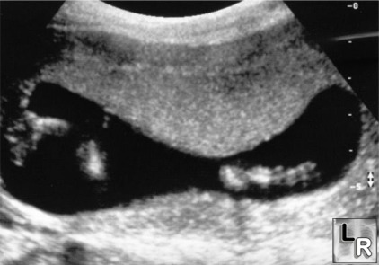
What to look for:
- Placenta appears as a uniformly grey, featureless structure
- No bright spots or calcifications anywhere
- Chorionic plate (fetal surface) is a smooth, flat line with no indentations
- Seen in < 18 weeks gestation (late 1st trimester to early 2nd trimester)
Quick Recap - Grade 0 Features
| Zone | Appearance |
|---|---|
| Chorionic plate | Smooth, straight, unbroken dense line |
| Placental substance | Uniform homogeneous echotexture - no echogenic areas |
| Basal layer | No echogenicities |
| Gestational age | < 18 weeks |
need grade 2 placenta usg image
Searching the Web
grade 2 placenta ultrasound USG image comma densities basal stippling
Loading Image
Loading Image
Here are the Grade 2 Placenta USG images:
Grade 2 Placenta - USG (30-38 Weeks)
Image 1 - Annotated USG with Schematic

Key features labeled:
- Basal layer echogenic areas - bright dots/dashes along the maternal surface (basal stippling - hallmark of Grade II)
- Comma-like indentations - chorionic plate shows deeper indentations into the substance, but they do NOT reach the basal layer
Image 2 - Classic Grade 2 USG Scan
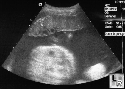
What to look for on this scan:
- Bright (hyperechoic) area visible at one edge = basal calcification
- Chorionic plate shows undulations/indentations
- Placental parenchyma appears slightly heterogeneous with comma-shaped echogenic densities
Grade II - Key USG Features at a Glance
| Zone | Finding |
|---|---|
| Chorionic plate | Deeper indentations (do NOT reach basal layer) |
| Placental substance | Comma-shaped / linear echogenic densities |
| Basal layer | Basal stippling - "dot-dash" echogenic foci (hallmark feature) |
| Gestational age | 30-38 weeks |
The basal echoes (basal stippling) are the defining hallmark of Grade II - their presence instantly distinguishes it from Grade I.
Show me Grade 1 and Grade 3 placenta USG images
Fetching both simultaneously!
Loading Image
Loading Image
Loading Image
Loading Image
All four images retrieved. Here they are:
Grade 1 Placenta USG (18-29 Weeks)
Image 1 - Annotated USG with Schematic

Key features labeled:
- Scattered echogenic areas - few randomly distributed bright foci within the parenchyma (2-4 mm)
- Unbroken chorionic line with subtle undulations - still mostly smooth but just beginning to indent
- No basal layer echogenicities yet
Image 2 - Classic Grade 1 USG Scan
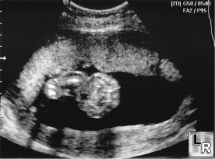
What to look for:
- Placenta is mostly homogeneous but with a few scattered bright (hyperechoic) spots
- Chorionic plate shows very mild waviness
- No calcification at the basal layer
Grade 1 - Key Features
| Zone | Finding |
|---|---|
| Chorionic plate | Unbroken, fine undulations (subtle) |
| Placental substance | Few scattered echogenic foci (punctate) |
| Basal layer | None |
| Gestational age | 18-29 weeks |
Grade 3 Placenta USG (≥ 39 Weeks)
Image 3 - Annotated USG with Schematic

Key features labeled:
- Echo-spared (anechoic) areas - fallout zones in the center of each cotyledon
- Inter-cotyledon septa - the chorionic plate indentations extend ALL the way down to the basal layer, dividing placenta into lobules
- Basal layer echogenic areas - large, dense, bright calcifications at the maternal surface
Image 4 - Classic Grade 3 USG Scan
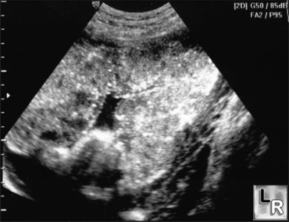
What to look for:
- Placenta looks very heterogeneous and "chunky"
- Bright calcifications scattered throughout + at basal layer
- Dark (anechoic) fallout zones within cotyledon compartments
- Acoustic shadowing may be present behind large calcifications
Grade 3 - Key Features
| Zone | Finding |
|---|---|
| Chorionic plate | Interrupted - indentations reach basal layer (cotyledon formation) |
| Placental substance | Divided into compartments; echo-free fallout areas centrally |
| Basal layer | Large dense echogenic foci ± acoustic shadowing |
| Gestational age | ≥ 39 weeks |
Quick Comparison - All 4 Grades Side by Side
| Grade | Age | Chorionic Plate | Parenchyma | Basal Layer |
|---|---|---|---|---|
| 0 | <18 wks | Smooth, straight | Homogeneous | Absent |
| I | 18-29 wks | Subtle undulations | Scattered echogenic foci | Absent |
| II | 30-38 wks | Deeper indentations (not reaching basal) | Comma-shaped densities | Dot-dash stippling ✦ |
| III | ≥39 wks | Interrupted, reaches basal layer | Cotyledons + echo-free fallout | Large dense calcifications |
refereances of these images
Here are the references for all the images shared across this conversation:
Image References
Source 1 - Ultrasound Chikitsa (Annotated USG + Schematics)
Images used: Grade 0, Grade 1, Grade 2, Grade 3 annotated diagrams with real USG scans
- Author: Dr. Kalpesh Khatal, DMRD
- Institution: Chikitsa Diagnostic Centre, Chembur, Mumbai, India
- Module: Teaching Module - Placental Grading
- Website: Ultrasound Chikitsa - Placental Grading
- Image URL base:
https://www.ultrasoundchikitsa.com/images/Teaching Modules/Placental Grading/
Source 2 - Learning Radiology (Classic USG Scans)
Images used: Grade 0, Grade 1, Grade 2, Grade 3 plain USG scans
- Website: LearningRadiology.com
- Section: Obstetric Ultrasound / Placental Grading
- URL: www.learningradiology.com
- Image URLs:
- Grade 0:
https://www.learningradiology.com/images/guimages/grade0placenta.jpg - Grade 1:
https://www.learningradiology.com/images/guimages/grade1placenta.jpg - Grade 2:
https://www.learningradiology.com/images/guimages/grade2placenta.jpg - Grade 3:
https://www.learningradiology.com/images/guimages/grade3placenta.jpg
- Grade 0:
Source 3 - Radiology Key (Schematic diagram + side-by-side USG)
Image used: All-grades schematic diagram and side-by-side USG comparison
- Book: Essentials of Abdomino-Pelvic Sonography
- Chapter: Placenta (Chapter 21, Figures 21.1 and 21.2)
- Website: Radiology Key - Placenta
- Image URL:
http://radiologykey.com/wp-content/uploads/2018/11/fig21_1.jpg
Primary Classification Reference
The grading system itself is based on:
Grannum PA, Berkowitz RL, Hobbins JC. The ultrasonic changes in the maturing placenta and their relation to fetal pulmonic maturity. American Journal of Obstetrics and Gynecology. 1979;133(8):915-922.
Additional Reference
Radiopaedia - Placental Grading (Grannum Classification) Weerakkody Y, Jones J. Last revised Sep 2021. https://radiopaedia.org/articles/placental-grading DOI: https://doi.org/10.53347/rID-13909
This is a shared conversation. Sign in to Orris to start your own chat.