
Explain briefly what dermatofibroma is. Include brief pathophysiology, risk factors, clinical features (symptoms and signs separately), diagnostic findings, complications, and initial management. Use Bailey and Love Short Practice of Surgery 28th Edition, Sabiston Textbook of Surgery 21st Edition, and Shwartz's Principles of Surgery 11th Edition, and for each part state what reference the information was extracted out of.
dermatofibroma clinical appearance skin lesion
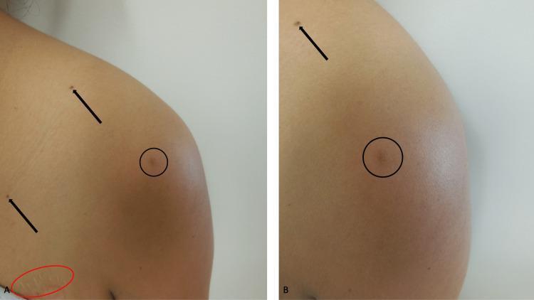
Clinical photograph (two-panel comparison) showing a dermatofibroma and associated skin findings on the left shoulder and chest of a 37-year-old woman. Panel A (distant view) and Panel B (close-up view) feature a primary lesion indicated by a black circle: a 5 x 5-millimeter, tan-colored, slightly raised dermal nodule. The lesion is firm and clinically consistent with a fibrocollagenous variant of dermatofibroma. Secondary findings highlighted with black arrows include small, dark, well-demarcated pigmented macules consistent with junctional nevi located on the shoulder and chest. In Panel A, a red oval demarcates a linear, hypopigmented area on the upper chest representing a healed surgical scar. The surrounding skin is otherwise normal in appearance. This image is used to demonstrate the clinical presentation of benign skin neoplasms in an immunosuppressed patient (post-liver transplant), emphasizing the morphology and distribution of dermatofibromas and nevi.
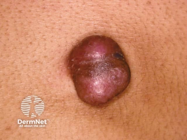
Clinical skin lesion photograph showing a solitary, dome-shaped cutaneous nodule with a smooth, glossy surface and a reddish-purple hue. The lesion sits within normal skin and appears firm, with minimal surface change and no overt ulceration in this frontal close-up view. The image captures a typical presentation of a slowly enlarging dermal/subcutaneous mass on the trunk or proximal limbs, where dermatofibrosarcoma protuberans (DFSP) commonly arises. DFSP characteristically presents as a firm plaque that evolves into a protuberant nodule; color ranges from skin-colored to violaceous. Although a single photograph cannot confirm histology, this lesion warrants biopsy given its clinical behavior and color, to differentiate DFSP from benign mimickers such as dermatofibroma, keloid, and fibrous histiocytoma. Histopathology (on biopsy) would reveal a storiform/spindle-cell proliferation with infiltration into subcutaneous fat (honeycomb pattern) and typically CD34 positivity. Clinically, DFSP is locally aggressive with a high risk of recurrence if margins are inadequate; treatment is wide local excision or Mohs micrographic surgery with clear margins and appropriate follow-up. This image is relevant for dermatology education, biopsy decision-making, differential diagnosis practice, and correlating clinical appearance with subsequent histopathology and treatment planning. Early diagnostic biopsy improves prognosis by guiding definitive surgical planning and surveillance for recurrence.
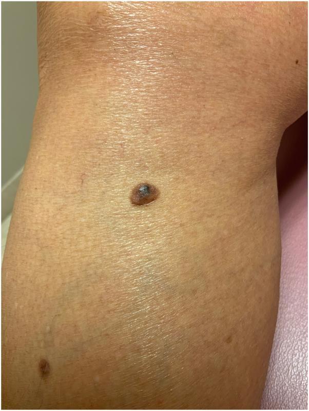
Clinical photograph of a lower extremity, specifically the leg, featuring a primary pigmented lesion characteristic of a dermatofibroma. The lesion is a solitary, well-circumscribed papule showing a distinctive variegated appearance: a dark, hyperpigmented, almost black central area surrounded by a lighter brown peripheral rim. The borders are well-defined and appear slightly elevated. The skin surface over the lesion exhibits some subtle texture changes. In the surrounding cutaneous field, several smaller, less prominent, and lighter-colored pigmented macules are visible, providing a comparison to the primary lesion's morphology. This image serves as a clinical example of a dermatofibroma, illustrating the classic central hyperpigmentation and firm, elevated borders often encountered in this benign fibrous histiocytoma of the dermis.
Dermatofibroma
Note on sources: Bailey and Love's Short Practice of Surgery 28th Edition does not contain a dedicated section on dermatofibroma in the available library text. The information below is drawn from Schwartz's Principles of Surgery 11th Edition and Sabiston Textbook of Surgery 21st Edition as the primary surgical references, supplemented where noted by Robbins & Cotran Pathologic Basis of Disease (10th Ed) and Andrews' Diseases of the Skin for pathophysiology, histology, and clinical detail. Each section is labelled with its source.
Definition
- Schwartz's Principles of Surgery 11th Ed, Chapter on Soft Tissue Tumors
- Sabiston Textbook of Surgery 21st Ed, Chapter 119
Pathophysiology
- Reactive/traumatic theory: Some cases follow antecedent minor trauma (insect bites, blunt injury), suggesting an aberrant response to injury and inflammation - analogous to excess collagen deposition in a hypertrophic scar or keloid.
- Neoplastic theory: Several fusion genes encoding portions of protein kinases - including the receptor tyrosine kinase ALK and protein kinase C (PKC) - have been identified in a subset of cases, supporting a true neoplastic process.
- Robbins & Cotran Pathologic Basis of Disease 10th Ed
- Andrews' Diseases of the Skin, Clinical Dermatology
Risk Factors
- Female sex and middle age (rarely seen in children)
- Minor skin trauma (insect bites, blunt injury) - likely triggers in susceptible individuals
- Immunosuppression: Systemic lupus erythematosus, treatment with prednisone or other immunosuppressive drugs, chronic myelogenous leukemia, and HIV infection have all been associated with the development of multiple dermatofibromas
Clinical Features
Symptoms
- Usually asymptomatic - the majority of patients have no pain or functional complaint
- Mild tenderness may be present in some lesions
- Lesions may slowly increase or decrease slightly in size over time; once a stable size is reached, growth typically ceases
Signs
- Location: Most commonly on the lower extremities (anterior legs); also seen above the elbows or on the sides of the trunk
- Appearance: A firm, pink to reddish-brown papule or nodule, sometimes with a yellowish hue; typically 0.5-1 cm in diameter (range 4-20 mm; giant lesions >5 cm occur rarely)
- Surface: The lesion is adherent to the overlying epidermis - the skin may be thinned or indented (a "dell-like depression" or atrophic surface)
- Dimple sign (Fitzpatrick sign): Gentle lateral pinching of the lesion causes it to pucker or dimple inward (retract into the skin) rather than protrude outward - this is the classic and pathognomonic clinical sign
- Borders: Sharply circumscribed; more prominent on palpation than on inspection
- Texture: Hard/firm on palpation
- In Sabiston: described as "a firm erythematous plaque, sometimes having central umbilication"
Diagnostic Findings
Clinical Diagnosis
- The clinical appearance and location (lower extremities, dimple sign) are usually sufficient for diagnosis
- Dimple sign on clinical examination is characteristic
Histopathology
- Benign spindle-shaped cells arranged in close whorls within the mid-dermis, forming a non-encapsulated mass
- Cells have features of fibroblasts and myofibroblasts, likely of primitive mesenchymal origin
- Collagen trapping at the periphery - the tumor surrounds individual collagen bundles
- Overlying epidermal changes: Acanthosis with downward elongation of hyperpigmented rete ridges (pseudoepitheliomatous hyperplasia); basal cell carcinoma-like epidermal changes are common overlying finding
- Extension into subcutaneous fat may occur in deeper lesions
- Touton giant cells containing hemosiderin are pathognomonic when present
- Foam cells, lipid deposits, and hemosiderin may be seen in some variants
Immunohistochemistry
- Positive: Factor XIIIa, CD10
- Negative: MAC387, S-100, CD34 (CD34 positivity should raise concern for dermatofibrosarcoma protuberans)
When to Biopsy
- Progressive enlargement beyond 2-3 cm in diameter should prompt excisional biopsy to exclude malignant fibrous histiocytoma or dermatofibrosarcoma protuberans (DFSP)
- Sabiston specifically notes: "Surgery is required primarily for diagnosis" when the lesion is uncertain
Complications
- Basal cell carcinoma can rarely develop within or overlying a dermatofibroma (Schwartz's explicitly notes this)
- Malignant transformation is exceptionally rare but possible with deep penetrating variants
- Lesions are otherwise generally indolent with no significant malignant potential in their typical form
- Deep/penetrating variants may extend into subcutaneous fat or fascia; these carry a higher local recurrence risk after incomplete excision
Initial Management
- Most lesions require no treatment - they are benign, asymptomatic, and indolent
- Simple reassurance is the appropriate initial approach for a typical, asymptomatic lesion (Andrews')
- Treatment is indicated when:
- Cosmetic concern
- Histologic diagnosis is required (diagnostic uncertainty or enlarging lesion)
- Symptomatic (pain, irritation)
- Surgical excision is the recommended treatment when intervention is needed (Schwartz's, Sabiston)
- Cryotherapy and laser treatment are alternatives when cosmetic removal is desired without deep excision (Schwartz's)
- For lesions enlarging beyond 2-3 cm: excisional biopsy is indicated to exclude malignancy
Summary Table
| Domain | Key Point | Source |
|---|---|---|
| Pathophysiology | Reactive vs. neoplastic; ALK/PKC fusion genes; dermal dendritic cell origin | Robbins & Cotran |
| Risk factors | Female, middle age, trauma, immunosuppression (SLE, HIV, steroids, CML) | Andrews'; Schwartz's |
| Symptoms | Usually asymptomatic; occasional tenderness | Schwartz's; Andrews' |
| Signs | Firm reddish-brown papule, lower leg; dimple sign; adherent to epidermis | Schwartz's; Sabiston; Andrews' |
| Diagnosis | Clinical + dimple sign; histology: spindle cells, collagen trapping, factor XIIIa+/CD34- | Andrews'; Robbins |
| Complications | Rare BCC development; deep variants may recur | Schwartz's |
| Management | Reassurance; excision for diagnosis/cosmesis; cryotherapy/laser as alternatives | Schwartz's; Sabiston; Andrews' |
Bailey and Love 28th Edition does not include a dedicated section on dermatofibroma in the textbook as indexed in this library. The surgical principles described in Schwartz's and Sabiston are consistent with standard surgical practice as taught in Bailey & Love's broader skin tumor chapters.
Explain briefly what melanoma is. Include brief pathophysiology, risk factors, clinical features (symptoms and signs separately), diagnostic findings, complications, and initial management. Use Bailey and Love Short Practice of Surgery 28th Edition, Sabiston Textbook of Surgery 21st Edition, and Shwartz's Principles of Surgery 11th Edition, and for each part state what reference the information was extracted out of.
melanoma ABCDE clinical features skin lesion
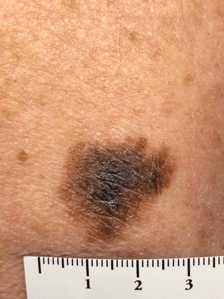
This clinical photograph shows a suspicious pigmented skin lesion on the left arm of an elderly patient, identified as melanoma in situ. The lesion is a large macule with a maximum diameter of approximately 2.5 to 3 cm as indicated by the adjacent metric ruler. It exhibits classic ABCDE features of melanoma, including significant asymmetry and highly irregular, notched borders. The color distribution is heterogeneous, displaying a variegated pattern of dark brown, tan, and focal areas of deep black pigment. The surface texture is uneven with subtle scaling and accentuation of skin markings, but without gross nodularity or ulceration. The surrounding skin shows signs of chronic actinic damage (dermatoheliosis), characterized by solar lentigines and textural changes. This image serves as a high-quality educational example of a primary cutaneous melanoma in its non-invasive stage, highlighting the importance of visual inspection and total skin examination in patients with high cumulative sun exposure.

This clinical photograph montage displays five separate skin lesions, primarily identified as melanocytic lesions or potential melanomas, sourced from the SIIM-ISIC Melanoma dataset. The series illustrates the ABCDE criteria used in dermatology (Asymmetry, Border irregularity, Color variegation, Diameter, and Evolution). The lesions range from dark brown to deep black and exhibit significant morphological diversity. Key visual features include: (1) an asymmetrical macule with indistinct, smudged borders; (2) a multi-lobed lesion with notched margins; (3) a highly irregular, pigmented plaque with a textured, verrucous surface; (4) a lesion showing varied shades of brown and tan with erythematous surrounding skin; and (5) a smaller, focal dark nodule. These images are utilized in the context of computer-aided diagnostic research for skin cancer identification, specifically focusing on lesion segmentation and classification using deep learning algorithms. The educational focus is on identifying visual hallmarks of malignancy, such as pigment variegation and irregular border definition.
Melanoma
Definition
- Bailey and Love's Short Practice of Surgery 28th Ed, Chapter 45 ("Cutaneous malignant melanoma")
- Sabiston Textbook of Surgery 21st Ed, Chapter 63 ("Melanoma")
Pathophysiology
- Melanoma has the highest mutational burden of any malignancy studied in humans
- Four genomic subtypes of cutaneous melanoma have been identified based on the Cancer Genome Atlas (2015): mutant BRAF (~50%), mutant RAS (~30%), mutant NF1 (~15%), and triple wild-type
- The most common mutation is BRAF V600E, followed by V600K. BRAF, RAS, and NF1 alterations all feed into the mitogen-activated protein kinase (MAPK/RAS-RAF-MEK-ERK) signaling pathway, which, when constitutively activated, drives unchecked cellular proliferation
- A second canonical pathway involved is the PI3K-AKT pathway
- Loss of tumor suppressor genes is also required: CDKN2A (coding for p16^INK4A and p14^ARF) is the most common pathogenic variant in hereditary melanoma. Loss of p16^INK4A allows CDK4 to drive cell cycle progression unchecked; loss of p14^ARF permits p53 degradation, disabling apoptosis
- Cumulative UV exposure drives lentigo maligna melanoma (LMM); intermittent/intense "flash-fry" UV exposure drives superficial spreading and nodular variants
- UV radiation (both UVA and UVB) causes a characteristic mutational signature found in over 90% of three of the four melanoma subtypes
- Malignant change begins in melanocytes at the basal epidermis. During the radial (horizontal) growth phase, cells spread along the dermoepidermal junction; during the vertical growth phase, cells invade the dermis, gaining access to blood vessels and lymphatics - this confers metastatic potential
- Melanoma development is strongly associated with the p16/CDK4,6/Rb and p14ARF/MDM2/p53 tumor suppressor pathways, and the RAF-MEK-ERK and PI3K-Akt oncogenic pathways
Risk Factors
- History of significant sunburns (especially >5 before age 16)
- More than 30 sun-acquired naevi
- Fair skin, red hair, living close to the equator
- Excessive UVR exposure (environmental or salon/tanning bed)
- Personal or family history of melanoma
- Genetic syndromes (rare)
- Male gender and solitary living associated with thicker melanomas at presentation
- Higher socioeconomic status in women positively correlated with melanoma development
- Fair complexion, blonde or red hair, blue eyes; sunburns easily; inability to tan (Fitzpatrick skin types I and II)
- Intermittent, intense UV radiation - especially tanning bed use (even without sunburn) doubles relative risk with >10 sessions
- Dysplastic nevi: 6-10% lifetime risk of melanoma
- Familial atypical multiple-mole melanoma (FAMMM) syndrome: numerous melanocytic nevi (often >100) with greatly elevated risk; CDKN2A is the most common pathogenic variant
- Congenital nevi, proportional to size; giant congenital nevi (>20 cm) carry 5-8% lifetime risk
- Immunosuppression
- Residence at high altitude or proximity to the equator
- Tanning bed exposure in adolescents/young adults (>10 sessions doubles risk)
- Intermittent childhood sunburns
- Personal and family history of melanoma
Clinical Features
Symptoms
- Change in size of a pre-existing lesion
- Change in shape or colour
- Increase in thickness (elevation, nodularity, or ulceration)
- Satellite lesions (pigment spreading into surrounding area)
- Tingling, itching, or serosanguineous discharge (usually late signs)
- Bleeding from or within a pigmented lesion
Signs
- A - Asymmetry: Asymmetric lesion
- B - Border: Irregular borders
- C - Colour: Colour variations within the lesion
- D - Diameter: Greater than 6 mm
- E - Evolution: Undergoing change (enlargement, ulceration, bleeding)
- Amelanotic lesions appear as raised pink, purple, or flesh-coloured papules and are often diagnosed late
- Superficial spreading melanoma (SSM): Most common (70%); usually arises in a pre-existing naevus; horizontal growth for years then rapid change
- Nodular melanoma (NM): 15% of melanomas; blue/black papule 1-2 cm in diameter; arises de novo; more common in men; typically on the trunk, head, or neck; up to 5% are amelanotic; sharply demarcated
- Lentigo maligna melanoma (LMM): Slow-growing, variegated brown macule on the face, neck, or hands of the elderly; more common in women; 5-10% of melanomas
- Acral lentiginous melanoma (ALM): Affects soles and palms; flat, irregular macule; 25% amelanotic; more common in Afro-Caribbean, Hispanic, and Asian populations
- Subungual melanoma: Hutchinson's sign - nail fold pigmentation widening progressively to form a triangular pigmented macule with nail dystrophy
- Desmoplastic melanoma: Head and neck region; propensity for perineural infiltration; often amelanotic
- Nodular melanoma is most likely to present at advanced stage due to early vertical growth pattern
- ALM is frequently misdiagnosed as a subungual haematoma; distinguishing feature: does not migrate distally with nail growth
Diagnostic Findings
Clinical/Dermoscopic Assessment
- History and examination directed at discovering the primary lesion and identifying local, regional, or distant spread
- Serial clinical and dermoscopic photography by a clinician with expertise in dermoscopy is mandatory when observation is chosen over biopsy
- Dermoscopy coupled with computer-aided learning is increasingly accurate
Biopsy
- Excision biopsy with 2-3 mm margin of skin and a cuff of subdermal fat is the standard initial approach (Bailey & Love)
- Schwartz's: excisional biopsy with 1-3 mm margins; incisional or punch biopsy for large lesions or cosmetically challenging areas
- Tissue specimen should include the full thickness of the lesion and adjacent normal skin
- Incision biopsy is occasionally appropriate (e.g. large facial lesions where full excision would be disfiguring)
Histopathology and Staging
- Breslow thickness (measured to the nearest 0.1 mm from the top of the granular layer to the deepest tumour cell) is the single most important prognostic indicator in node-negative disease
- Melanomas are classified as thin (<1 mm), intermediate (1-4 mm), or thick (>4 mm)
- Ulceration is a critically important independent prognostic factor; defined histologically by absence of an intact epithelium
- Mitotic rate ≥1/mm² worsens prognosis
- AJCC TNM staging (8th edition): T1-T4 based on Breslow thickness (cutoffs at 1.0, 2.0, and 4.0 mm); further subclassified by ulceration status; N stage by nodal involvement; M stage by distant metastasis
- Stage I-II: localized; Stage III: regional; Stage IV: distant metastatic
- For stage III/IV: elevated LDH is associated with worse prognosis
Imaging
- No routine chest X-ray or CT for staging unless there is positive regional node disease
- For clinical signs/symptoms of metastasis or stage III+ disease: CT chest/abdomen/pelvis, whole-body PET-CT, and brain MRI are recommended
Sentinel Lymph Node Biopsy (SLNB)
- Standard staging procedure for clinically node-negative melanoma; introduced in 1992
- Performed using preoperative lymphoscintigraphy with technetium-sulphur colloid and intraoperative isosulfan blue or methylene blue dye
- Sentinel node identified in 98-99% of cases using the combined technique
- Sentinel nodes processed with H&E and immunohistochemistry for S-100, HMB-45, and MART-1/Melan-A
- SLNB is recommended for T2a disease and greater (Bailey & Love), or for thin lesions with high-risk features (thickness >0.75 mm, ulceration, mitoses ≥1/mm²) (Schwartz's)
- Clinically suspicious nodes: fine-needle aspiration (FNA) has high sensitivity and specificity for detection in large lymph nodes (Schwartz's)
Complications
- Locoregional recurrence: Can develop as satellite lesions, in-transit metastases (dermal/subdermal deposits between the primary and regional lymph nodes), or nodal metastases
- Regional lymph node metastasis: Proportional to Breslow thickness; in the presence of regional node disease, the number of nodes affected is the most important prognostic factor
- Distant metastases: Melanoma can spread to virtually any organ - common sites include lung, liver, brain, bone, and skin. Stage IV disease carries a median survival of 6-8 months historically (Schwartz's)
- Perineural infiltration: Particularly characteristic of desmoplastic melanoma; leads to local recurrence if not widely excised (Bailey & Love)
- Second primary melanoma: 5% of all melanoma patients will develop a second primary melanoma (Bailey & Love)
- Bowel obstruction or intussusception: Can occur with amelanotic gastrointestinal melanoma (Bailey & Love)
- Death from metastatic disease: Melanoma accounts for over 75% of skin malignancy-related deaths despite representing <5% of skin malignancies (Bailey & Love)
- Ocular melanomas exclusively metastasize to the liver (Schwartz's)
Initial Management
Biopsy and Histologic Confirmation
- Initial excision biopsy with 2-3 mm margins (Bailey & Love); 1-3 mm margins (Schwartz's)
- Pathologic staging with Breslow thickness guides all subsequent treatment
Wide Local Excision (WLE) Margins
| Breslow Thickness | Recommended Excision Margin |
|---|---|
| Melanoma in situ | 5 mm (Bailey & Love) / 0.5-1 cm (Schwartz's) |
| <1 mm | 1 cm |
| 1-2 mm | 1-2 cm |
| >2 mm | 2 cm |
| >2 mm (Bailey & Love) | 2 cm only - wider margins show no added benefit |
Regional Lymph Node Management
- SLNB is offered to patients with T2a disease and greater (Bailey & Love), or tumours with Breslow >1 mm or with high-risk features (all three books)
- Therapeutic lymph node dissection for clinically positive nodes in the absence of distant metastasis is associated with 5-year survival of 30-50% (Schwartz's)
- Completion lymphadenectomy after a positive SLNB is controversial; prospective data (MSLT-1, MSLT-2) show no overall survival benefit from routine completion lymphadenectomy for micrometastasis <0.2 mm in a single node (Bailey & Love, Sabiston)
- For face/anterior scalp/ear primaries with positive SLNB: superficial parotidectomy plus modified radical neck dissection (Schwartz's)
Adjuvant Systemic Therapy
- BRAF V600 mutations are present in ~50% of melanomas
- BRAF inhibitors (vemurafenib, dabrafenib) block constitutively active BRAF signalling
- MEK inhibitor trametinib combined with dabrafenib counters acquired resistance via MAPK pathway reactivation; combined targeted therapy shows promising results in stage IV and as adjuvant treatment (Bailey & Love)
- Anti-CTLA-4 (ipilimumab): First immunotherapy approved for adjuvant melanoma (2015); EORTC 18071 trial showed improved 5-year OS (65.4% vs 54.4%) in resected stage III melanoma but with significant serious adverse events (54% vs 26%) (Sabiston)
- Anti-PD-1 agents (nivolumab, pembrolizumab): CheckMate 238 demonstrated superior recurrence-free survival with nivolumab vs ipilimumab in resected stage III/IV melanoma (5-year RFS 50% vs 39%; HR 0.72), with better tolerability (Sabiston)
- High-dose interferon-alpha has shown disease-free survival benefit in melanomas >4 mm with/without node involvement, but is poorly tolerated and does not improve overall survival (Schwartz's)
- High-dose interleukin-2 (IL-2), anti-PD-1 and anti-CTLA-4 antibodies, and BRAF inhibitors (sorafenib) have all shown survival benefit in metastatic disease in randomised trials (Schwartz's)
Observation and Surveillance
- AJCC staging directs optimum care and inclusion in clinical studies
- The only current curative treatment for melanoma is appropriate surgery
- Investigations for T3a+ disease should be directed by individual clinical presentation
- SNB is critical for T2a disease and greater to accurately upstage and direct treatment
Summary Table
| Domain | Key Points | Source(s) |
|---|---|---|
| Definition | Malignant tumour of melanocytes; <2% of skin cancers but majority of skin cancer deaths | Bailey & Love; Sabiston |
| Pathophysiology | BRAF/RAS/NF1 mutations → MAPK pathway activation; radial then vertical growth phase; UV-driven mutations | Sabiston; Bailey & Love; Schwartz's |
| Risk factors | UV/sunburn, fair skin, dysplastic nevi, FAMMM syndrome, family history, immunosuppression | Bailey & Love; Sabiston; Schwartz's |
| Symptoms | Change in size/shape/colour, itching, bleeding, serosanguineous discharge | Bailey & Love |
| Signs | ABCDE criteria; nodular = blue-black papule; LMM = slow brown macule in elderly; ALM = subungual/palmoplantar; Hutchinson's sign | Schwartz's; Bailey & Love; Sabiston |
| Diagnosis | Excision biopsy; Breslow thickness; AJCC TNM staging; SLNB; S-100/HMB-45/Melan-A IHC | All three |
| Complications | Locoregional/distant metastasis; perineural invasion (desmoplastic); second primary; bowel obstruction | Bailey & Love; Sabiston; Schwartz's |
| Management | WLE with depth-guided margins; SLNB; completion lymphadenectomy (controversial); BRAF inhibitors; anti-PD-1/CTLA-4 immunotherapy | All three |