
Explain the extends of 1st 2nd 3rd 4th order nueron
Reading File
The question is about the extents (levels/locations) of 1st, 2nd, 3rd, and 4th order neurons in sensory and/or motor pathways. This is a broad neuroanatomy topic. Let me search the medical library.
Finding Sources
Finding Sources
Good results. Now let me read the Costanzo Physiology section on Sensory Pathways, and also search for specific pathway neurons (dorsal column, spinothalamic).
Reading File
Finding Sources
Finding Sources
Good. Now let me read the detailed Costanzo sensory pathway section and the Gray's Anatomy anterolateral pathway section for the specific neuron orders.
Reading File
Reading File
Excellent! I have very rich content now. Let me read a bit more of the Gray's Anatomy section covering the posterior column-medial lemniscal pathway and the 4th order neuron (cortex).
Reading File
Loading Image
I now have all the information needed for a comprehensive answer. Let me compile the full explanation.
Sensory Neuron Orders - Extents and Locations
The brain receives sensory information from the periphery through a relay chain of neurons, each handing off to the next at a specific anatomical station. Here is the full breakdown:
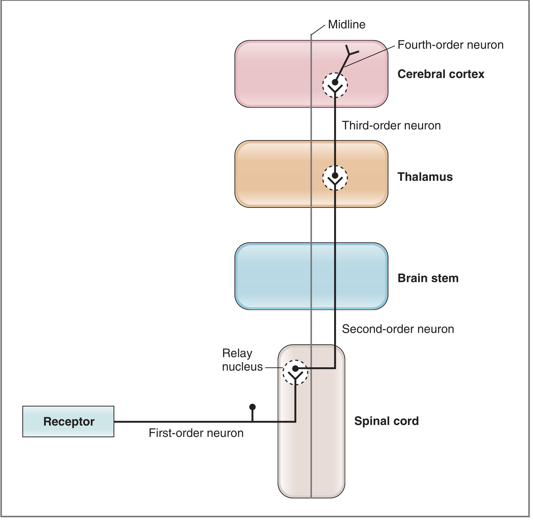
1st Order Neuron (Primary Afferent)
- Cell body location: Dorsal root ganglion (DRG) of the spinal cord (or trigeminal ganglion for facial sensation)
- Extent: From the sensory receptor in the periphery (skin, muscle, joint) → enters the spinal cord via the posterior root → synapses in a relay nucleus in the spinal cord (dorsal horn) or brainstem
- In the somatosensory and olfactory systems, the receptor IS the first-order neuron (no separate synapse needed between receptor and primary afferent)
- Exceptions exist: in the visual, auditory, and taste systems, specialized epithelial receptor cells first synapse onto the first-order neuron
2nd Order Neuron
- Cell body location: Relay nucleus in the spinal cord or brainstem
- Extent: From the relay nucleus → axon crosses the midline (decussates) → ascends all the way up to the thalamus
This differs by pathway:
| Pathway | Modality | Where 2nd order cell body sits | Where it decussates |
|---|---|---|---|
| Spinothalamic (anterolateral) | Pain, temperature, crude touch | Dorsal horn of spinal cord (Rexed laminae I, IV-V) | Crosses in the anterior white commissure of the spinal cord at the same level, then ascends contralaterally |
| Dorsal column - Medial Lemniscus (DCML) | Fine touch, vibration, proprioception | Nucleus gracilis / nucleus cuneatus in the caudal medulla | Crosses as internal arcuate fibers in the medulla, forming the medial lemniscus |
| Trigeminothalamic | Facial sensation (CN V) | Trigeminal nuclei in the brainstem | Crosses in the brainstem |
Key distinction: In the spinothalamic tract, the 1st order neuron synapses and the 2nd order crosses immediately at the spinal cord level. In the DCML pathway, the 1st order ascends ipsilaterally all the way to the medulla before synapsing on the 2nd order neuron, which then crosses.
3rd Order Neuron
- Cell body location: Relay nuclei in the thalamus - specifically the Ventral Posterior Lateral (VPL) nucleus for body sensations, and the Ventral Posterior Medial (VPM) nucleus for facial sensations (from the trigeminal nerve)
- Extent: From the thalamus → axons project through the posterior limb of the internal capsule → ascend to the primary somatosensory cortex (postcentral gyrus, parietal lobe)
- Many 2nd-order neurons converge on a single 3rd-order neuron in the thalamus
4th Order Neuron
- Cell body location: Primary somatosensory cortex (postcentral gyrus, parietal lobe - Brodmann areas 3, 1, 2)
- Extent: These are the cortical neurons where conscious sensory perception occurs. They process, integrate, and distribute the sensory information further within the cortex (to association areas)
- The receptive field of a 4th-order neuron is the largest of all the orders - it represents a wide area of the body surface
Summary Table
| Neuron Order | Cell Body / Location | Projects To | Key Feature |
|---|---|---|---|
| 1st | Dorsal root ganglion / trigeminal ganglion | Spinal cord dorsal horn OR brainstem relay nuclei | Primary afferent; receptor may BE the neuron |
| 2nd | Spinal cord dorsal horn (spinothalamic) OR nucleus gracilis/cuneatus in medulla (DCML) | Thalamus (VPL/VPM) | Crosses midline (decussates) |
| 3rd | Thalamus (VPL for body, VPM for face) | Primary somatosensory cortex | Relay in thalamus; passes through internal capsule |
| 4th | Primary somatosensory cortex (postcentral gyrus) | Association cortices | Site of conscious perception |
Important Clinical Note - Where Lesions Produce Contralateral Deficits
- A lesion above the decussation causes contralateral sensory loss
- Spinothalamic: decussation is in the spinal cord - so a lesion above the cord affects the opposite side
- DCML: decussation is in the medulla - a lateral medullary stroke can therefore produce ipsilateral face loss (trigeminal, which crosses in brainstem) + contralateral body loss (DCML, which already crossed at medulla)
Sources: - Costanzo Physiology 7th Ed., pp. 80-82 | - Gray's Anatomy for Students, pp. 1326-1327
Intensional n resting tremors
Intentional (intention) tremor occurs during voluntary movement and worsens as the limb approaches a target - it's a sign of cerebellar dysfunction. Resting tremor occurs when the limb is fully supported at rest and disappears with movement - classically seen in Parkinson's disease (basal ganglia dysfunction).
Searching sources...
Reading File
Finding Sources
Finding Sources
Reading File
Reading File
Reading File
Finding Sources
Reading File
Finding Sources
I now have comprehensive material from authoritative neurology textbooks. Let me compile the full answer.
Intention Tremor vs. Resting Tremor
Tremor is defined as rhythmic oscillation of a body part produced by alternating or synchronous contractions of reciprocally innervated antagonist muscles. The two most clinically important and commonly confused types are intention tremor and resting tremor.
Resting Tremor
Definition
A resting tremor occurs when the affected body part is completely relaxed and supported against gravity. It dampens or disappears entirely with voluntary movement and re-emerges when the limb returns to rest.
Characteristics
- Frequency: 5-7 Hz (in Parkinson's disease)
- Distribution: Typically distal - fingers, hands, wrists; can involve lower extremities, chin, lips, jaw
- Pattern: Asymmetrical at onset; often described as "pill-rolling" (rolling motion between thumb and fingers)
- Behavior: Increases with anxiety, stress, and cognitive distraction; suppressed by voluntary movement; disappears during sleep
- Patients may conceal it by holding one hand with the other, sitting on the affected hand, or crossing their legs
Underlying Pathology - Basal Ganglia Dysfunction
- The classic cause is Parkinson's disease - loss of dopaminergic neurons in the substantia nigra pars compacta
- Dopamine deficiency disrupts the balance between the direct and indirect pathways of the basal ganglia
- This leads to abnormal oscillatory activity in thalamo-cortical circuits at rest
- Other causes: Holmes tremor, palatal tremor, drug-induced parkinsonism
Associated Features (Parkinson's Disease)
- Bradykinesia (slowness of movement)
- Cogwheel or lead-pipe rigidity
- Postural instability
- Shuffling gait, stooped posture, masked facies
Intention (Ataxic) Tremor
Definition
An intention tremor occurs during voluntary goal-directed movement and progressively worsens as the limb approaches its target. It is absent at rest. The terminal worsening near the target is sometimes called "terminal tremor."
Characteristics
- Frequency: 2-4 Hz (lower than resting tremor)
- Movement: Irregular, oscillating movements in multiple planes that increase as the target is approached
- Test to elicit: Finger-nose-finger test, heel-shin test - the limb overshoots or oscillates wildly just before reaching the target
- Absent at complete rest; appears only when movement is initiated
Underlying Pathology - Cerebellar Dysfunction
- Caused by lesions of the cerebellum or its connections (superior cerebellar peduncle, dentate nucleus)
- The cerebellum normally fine-tunes ongoing movements by comparing intended vs. actual motor output and making real-time corrections
- When this error-correction circuit is damaged, limb movements become uncoordinated and oscillate, especially near the endpoint of movement
- Also called ataxic tremor because it is a feature of cerebellar appendicular ataxia
Associated Cerebellar Signs (DANISH mnemonic)
| Sign | Description |
|---|---|
| Dysdiadochokinesia | Inability to perform rapid alternating movements |
| Ataxic gait | Wide-based, staggering gait |
| Nystagmus | Involuntary eye movements |
| Intention tremor | Worsens approaching target |
| Slurring of speech | Scanning/cerebellar dysarthria |
| Hypotonia | Reduced muscle tone |
Common Causes
- Multiple sclerosis (demyelination of cerebellar pathways)
- Cerebellar stroke or infarct
- Cerebellar tumors
- Alcohol toxicity (acute and chronic)
- Friedreich's ataxia
- Holmes tremor (combined rest + postural + intention tremor from superior cerebellar peduncle lesions)
Side-by-Side Comparison
| Feature | Resting Tremor | Intention Tremor |
|---|---|---|
| When present | At rest, limb fully supported | During voluntary movement |
| Effect of movement | Disappears with movement | Worsens approaching target |
| Frequency | 5-7 Hz | 2-4 Hz |
| Distribution | Distal (fingers, wrists) | Limbs (finger-nose test) |
| Lesion site | Basal ganglia (substantia nigra) | Cerebellum or its connections |
| Classic disease | Parkinson's disease | Cerebellar disease (MS, stroke) |
| Mechanism | Dopamine deficiency → thalamo-cortical dysrhythmia | Loss of cerebellar error correction |
| Other features | Bradykinesia, rigidity | Dysdiadochokinesia, ataxia, nystagmus |
| During sleep | Disappears | Disappears |
Memory Hook
"Rest = Parkinson's (basal ganglia) | Intention = Cerebellum"
- Resting tremor: the hand shakes when you're NOT trying to use it
- Intention tremor: the hand shakes ONLY when you TRY to reach something
Sources: - Neuroanatomy through Clinical Cases, 3rd Ed., pp. 788-789 | - Bradley and Daroff's Neurology in Clinical Practice, p. 433 | - Costanzo Physiology 7th Ed.
This is a shared conversation. Sign in to Orris to start your own chat.