
Hydronephrosis
hydronephrosis ultrasound kidney dilated pelvis
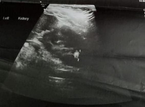
This diagnostic ultrasound image depicts the left kidney in a longitudinal view, labeled 'Left Kidney'. The scan reveals significant hydronephrosis characterized by a dilated renal pelvis measuring 13 mm, appearing as an anechoic (dark) fluid-filled space within the central collecting system. Within the dilated pelvis, there is a large, highly echogenic (bright) focus approximately 2 cm in diameter, labeled with points '1' and '2'. This finding is consistent with a large renal pelvic calculus (urolithiasis). The surrounding renal parenchyma shows a heterogeneous echotexture. This image is clinically significant for demonstrating obstructive uropathy caused by a large stone, leading to pelvic dilatation. It serves as an educational tool for identifying renal stones and secondary signs of obstruction like hydronephrosis in pediatric or adult urology.
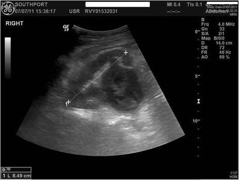
This diagnostic abdominal ultrasound image displays the right kidney in a longitudinal view, demonstrating moderate to severe hydronephrosis. The renal pelvis and collecting system are grossly dilated, appearing as a large, central anechoic (fluid-filled) region. Notably, the dilated space contains thick, hyperechoic (bright) linear internal septations and heterogeneous echogenic debris, which are hallmark sonographic signs of pyonephrosis (infected hydronephrosis). Electronic calipers measure the kidney's longitudinal axis at approximately 8.49 cm. The renal parenchyma appears thinned, suggesting chronic pressure from the dilated collecting system. The surrounding retroperitoneal tissues show a normal speckled echotexture. This image serves as a critical educational example of complicated urinary tract obstruction where fluid stasis has progressed to suppuration, requiring urgent clinical intervention like drainage or nephrostomy.
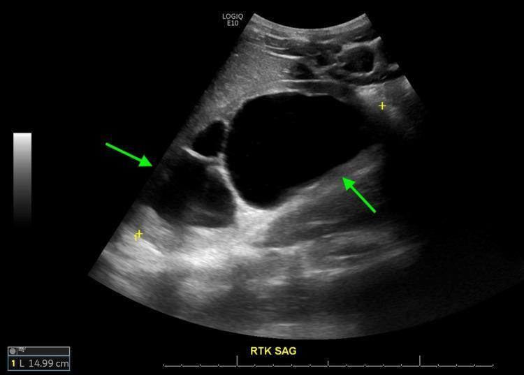
A sagittal view B-mode ultrasound of the left kidney (RTK SAG label in image) demonstrating Grade IV hydronephrosis. The renal pelvis is severely dilated and appears as a large, central anechoic (dark) space. Green arrows point to specific pathological features: the left arrow indicates significant cortical thinning of the renal parenchyma, while the right arrow highlights hyperechoic (bright) debris or proteinaceous material layered within the dependent portion of the dilated renal pelvis. The kidney measures approximately 14.99 cm as indicated by the calipers. These sonographic findings are consistent with chronic obstructive uropathy, such as that caused by posterior urethral valves (PUV). The visible reduction in cortical thickness suggests long-standing pressure and potential chronic kidney disease. This image serves as a diagnostic tool for assessing the severity of urinary tract obstruction and renal parenchymal damage.
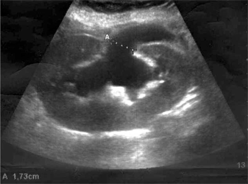
This diagnostic ultrasound image demonstrates a human kidney with signs of severe hydronephrosis. The primary finding is a large, central, anechoic (black) area representing a dilated renal pelvis, from which several elongated anechoic regions radiate outwards, corresponding to dilated renal calyces. This branching configuration creates a typical 'bear paw' or cauliflower-like pattern of pelvicalyceal dilatation. The surrounding renal parenchyma appears relatively hypoechoic and thinned due to the internal pressure of the fluid accumulation. An electronic caliper measurement marked 'A' indicates a transverse diameter of 1.73 cm in the upper portion of the dilated collecting system. The image is clinically significant for illustrating obstructive uropathy, specifically showing how fluid distention distorts the normal renal architecture. It serves as an educational example of graft sonography used to monitor renal transplant complications like acute hydroureteronephrosis.
hydronephrosis
Hydronephrosis
Definition
Causes / Etiology
Congenital
- Atresia of the urethra
- Valve formations in the ureter or urethra
- Aberrant renal artery compressing the ureter
- Abnormal kidney position with torsion or kinking of the ureter
- Idiopathic pelviureteric junction (PUJ) stenosis - the most common cause of unilateral hydronephrosis
Acquired
| Category | Examples |
|---|---|
| Calculi (stones) | Renal pelvis, ureter |
| Benign prostatic hyperplasia | Commonest cause in older males |
| Tumors | Bladder carcinoma, carcinoma of the prostate, ureteral tumor, retroperitoneal lymphoma, cervical or uterine carcinoma |
| Inflammatory | Prostatitis, ureteritis, urethritis, retroperitoneal fibrosis |
| Neurogenic | Bladder paralysis following spinal cord damage |
| Pregnancy | Mild, physiological |
| Ureterocele | |
| Schistosomiasis | |
| Phimosis / urethral stricture | Causes bilateral disease |
Key rule: Bilateral hydronephrosis occurs only when obstruction is below the level of the ureters. Obstruction at or above the ureter level produces unilateral disease. - Robbins & Kumar Basic Pathology, p. 530
Pathogenesis
- The papillae are most vulnerable (greatest pressure increase)
- Initial functional defect: impaired tubular concentrating ability
- Later: glomerular filtration begins to fall
- Obstruction also triggers interstitial inflammatory reaction → interstitial fibrosis
Morphology (Gross and Microscopic)
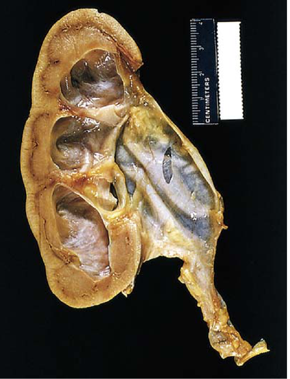
- With subtotal or intermittent obstruction: kidney may be massively enlarged (up to 20 cm); almost entirely replaced by distended pelvicalyceal system; parenchyma compressed and atrophied; obliteration of papillae and flattening of pyramids
- With sudden, complete obstruction: glomerular filtration compromised early; renal function may cease while dilation is still comparatively mild
- Depending on obstruction level, one or both ureters may also be dilated (hydroureter)
- Early: tubular dilation and atrophy
- Later: loss of glomeruli; replacement of parenchyma by fibrous tissue
- Superimposed pyelonephritis is common
Clinical Features
| Scenario | Features |
|---|---|
| Unilateral hydronephrosis | May remain silent for long periods; dull ache or sense of weight in the loin; if rapid onset - severe colicky pain |
| Bilateral hydronephrosis | Anuria and renal failure; symptoms also due to the underlying cause |
| Incomplete bilateral obstruction | Paradoxically causes polyuria (due to tubular concentrating defects) rather than oliguria - can obscure the diagnosis |
| Distal obstruction (below bladder) | Dominant symptoms of bladder distention |
Investigations
1. Ultrasound (First-line)
| Grade | Features |
|---|---|
| Mild | Dilation of pelvis and calyces; parenchymal architecture retained |
| Moderate | Medullary pyramids start to flatten; increasing pelvicalyceal dilation |
| Severe | Pelvis and calyces appear ballooned; corticomedullary differentiation lost; parenchyma thin |
- Renal vasculature can mimic hydronephrosis (use color Doppler to confirm flow)
- Parapelvic cysts can be confused with hydronephrosis (cysts are round/well-circumscribed; hydronephrosis is an irregular branching structure)
- False negatives occur with acute/partial obstruction, volume depletion, and retroperitoneal fibrosis
2. Intravenous Urography (IVU / IVP)
- Earliest changes: decreased concavity then flattening of minor calyces (intrarenal pelvis) or change in renal pelvis first (extrarenal pelvis)
- Progression: dilation of major calyces → clubbing (convexity) of minor calyces → grossly distended pelvis with an acute angle at the ureteropelvic junction
- Delayed films (6 hours) may be needed; if pelvis/calyces still not seen → retrograde urography
3. MR Urography (MRU)
- Plausible alternative to diuretic renogram; far superior anatomic resolution; no radiation
- Provides: pinpoint localization of anatomic abnormalities, differential function, and drainage assessment
- Gadolinium contrast used; furosemide (1 mg/kg, max 20 mg) administered
- Particularly useful in children when avoiding radiation is paramount - Campbell-Walsh Urology
4. Isotope Renography (MAG3 Diuretic Renogram)
- Detects dilatation and obstruction of the renal collecting system
- Provides differential renal function - useful before surgical intervention
5. Whitaker Test
- Measures intrapelvic pressure by percutaneous puncture of the kidney
- Used in specialized units; helps differentiate obstructed from non-obstructed systems
Complications
- Superimposed pyelonephritis (very common)
- Pyonephrosis - infected hydronephrosis; pus in collecting system (look for internal echoes on ultrasound in febrile patient with flank pain)
- Renal failure (bilateral obstruction)
- Irreversible parenchymal damage (prolonged obstruction)
Treatment Principles
- Treat the underlying cause (e.g., stone removal, resection of obstructing tumor, BPH management)
- Pyeloplasty - surgical correction for PUJ obstruction; long-term renal function post-pyeloplasty is generally preserved per a 2025 systematic review (PMID 40205125)
- Nephrostomy - percutaneous drainage for infected/obstructed kidney (pyonephrosis) as urgent intervention
- Ureteric stenting (JJ stent) - temporary or definitive decompression
- Removal of obstruction usually permits full return of function within a few weeks; however, longstanding obstruction produces irreversible changes
Antenatal / Fetal Hydronephrosis
- Detected on prenatal ultrasound; most common fetal urinary tract anomaly
- Causes include PUJ obstruction, vesicoureteral reflux, posterior urethral valves, ureterocele
- Severity graded by anteroposterior renal pelvis diameter on ultrasound
- Most mild cases resolve spontaneously; severe or bilateral cases require postnatal workup and may need early surgical intervention