
Modified radical mastoidectomy
modified radical mastoidectomy surgical steps diagram
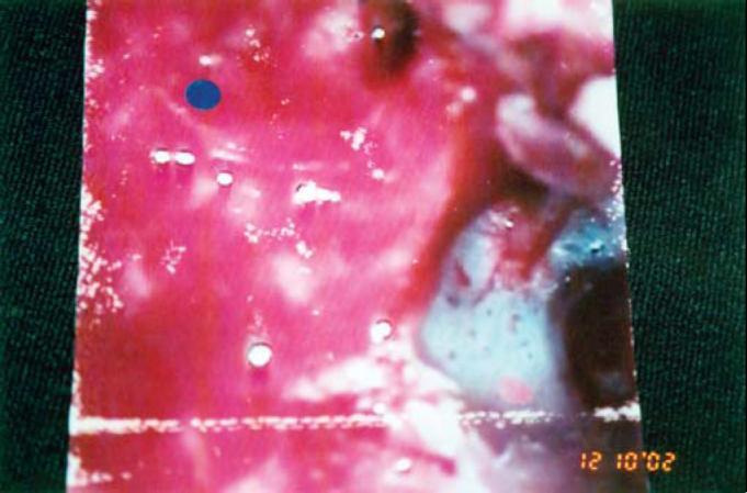
A clinical transoperative photograph depicting a modified radical mastoidectomy in a patient with cholesteatoma of the external auditory canal (CEAC). The surgical field shows the mastoid cavity with evident erosion of the posterior wall of the external auditory canal (EAC). Key anatomical features include an intact tympanic membrane (TM) and a preserved thin bone band near the tympanic ring. The photograph demonstrates the moist, erythematous surface of the middle ear and mastoid structures, with visible white bony landmarks and reddish soft tissue typical of surgical exploration for cholesteatoma management. The image is framed by green surgical draping, and a digital timestamp is visible in the lower right corner. This visual serves to illustrate the intraoperative findings and surgical extent required to manage extensive mastoid involvement while preserving the ossicle chain and facial nerve integrity.
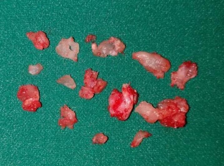
This clinical photograph shows multiple fragments of excised surgical tissue from a canal wall down modified radical mastoidectomy. The specimens, obtained in a piecemeal fashion, are displayed on a green sterile surgical drape. The tissue fragments vary in size and morphology, appearing as irregular, lobulated masses. They exhibit a heterogeneous color profile consisting of fleshy pink and vibrant red areas indicative of vascularization or granulation tissue, interspersed with paler, translucent, and firmer white regions suggestive of fibro-osseous components or calcified stroma. The specimen represents a benign fibro-osseous lesion involving the middle ear and mastoid cavity. This visual serves as an educational example of macroscopic specimen evaluation in otolaryngology and surgical pathology, highlighting the characteristic appearance of tissue removed during ear canal and mastoid surgery for benign neoplasms or chronic inflammatory processes.
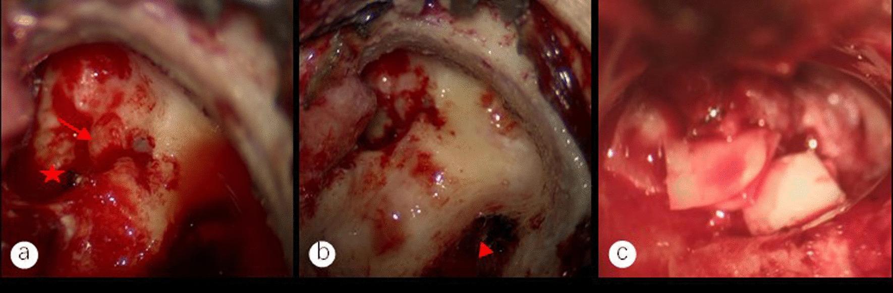
This composite of three intraoperative endoscopic clinical photographs demonstrates key steps in a modified mastoidectomy and tympanoplasty for middle ear cholesteatoma. Panel (a) shows the exposed facial nerve canal (red arrow) and the stapedial base (red star) after the removal of cholesteatoma epithelium. Hemorrhagic granulation tissue is visible near the stapes, which is managed via bipolar cautery to prevent recurrence. Panel (b) depicts the surgical exposure of the sigmoid sinus vein wall (red triangle) following mastoid bone removal, showing a preserved basilar membrane after the removal of overlying disease. Panel (c) illustrates the reconstruction phase, showing the placement of an auricular cartilage graft to restore the ossicular chain connection to the vestibular window (Type III tympanoplasty). These images highlight the surgical management of cholesteatoma involving critical neurovascular structures like the facial nerve and sigmoid sinus, emphasizing preservation of anatomical integrity while ensuring complete lesion eradication in an otolaryngology context.
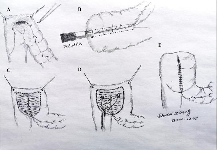
This medical schematic diagram illustrates the sequential surgical steps for creating a colonic urinary reservoir (neobladder) following a radical cystectomy. The procedure is depicted in five stages: Step A shows the initial antimesenteric incision in a folded segment of the sigmoid colon or rectum using electrocautery and stay sutures. Step B demonstrates the introduction of a laparoscopic Endo GIA stapler into the lumen for detubularization. Step C reveals the internal surface of the opened bowel segment with visible staple lines along the medial edges. Step D illustrates the implantation of bilateral ureteric stents into the reservoir, establishing an anti-reflux mechanism. Finally, Step E shows the completed pouch after the anterior seromuscular wall has been closed with continuous or interrupted sutures, transforming the tubular intestine into a wide, reservoir-like structure. This illustration serves as an educational tool for urological surgeons to understand the intraluminal stapling technique and detubularization logic required for urinary diversion.
Modified Radical Mastoidectomy (MRM)
Definition and Concept
- Cummings Otolaryngology Head and Neck Surgery, block 31
- Shambaugh Surgery of the Ear, block 5 & 7
Comparison of Mastoid Procedures
| Feature | Canal Wall Up | Modified Radical (MRM) | Radical Mastoidectomy |
|---|---|---|---|
| Posterior canal wall | Preserved | Removed | Removed |
| Tympanic membrane | Reconstructed | Preserved/reconstructed remnant | Sacrificed |
| Ossicles | Preserved/reconstructed | Remnants preserved | Removed |
| Eustachian tube | Patent | Patent | Obliterated |
| Middle ear access | Limited | Full | All mucosa removed |
| Cavity cleaning | Not required | Required (lifelong) | Required |
| Hearing outcome | Best potential | Moderate | Poor |
Indications
- Cholesteatoma extending into the attic, antrum, or mastoid - especially when extensive canal wall destruction is already present
- Occasional otologic surgeon confronted with cholesteatoma (less technically demanding than the staged canal-wall-up technique)
- Patients unwilling or unable to undergo the two-stage canal-wall-up approach
- Recurrent cholesteatoma after previous failed canal-wall-up surgery
- The only functioning ear (safer single-stage approach)
- Poor eustachian tube function (high risk of retraction pocket reformation)
- Circumstances where second-look surgery is impractical
-
CT shows enlarged, smooth-walled antrum (large cholesteatoma cavity)
-
Persistent otorrhea despite treatment
-
Evidence of complications (facial palsy, labyrinthine fistula, intracranial spread)
-
Shambaugh Surgery of the Ear, p. 541
Contraindications to Open Cavity (MRM)
- Chronic otitis media without cholesteatoma
- Acute coalescent mastoiditis
- Persistent secretory or allergic otitis media
- Tuberculous otitis media (treat medically first)
- Relative: wide exposure of sigmoid sinus, dura, or facial nerve due to aggressive disease
Preoperative Assessment
- Microscopic examination and cleaning; remove pus, mucus, cholesteatoma debris
- Polyps removed carefully (may attach to facial nerve, labyrinthine fistula matrix, or stapes)
- HRCT temporal bone (non-contrast): evaluates erosion of semicircular canals, cochlea, fallopian canal, dural plates, sigmoid sinus; scutum erosion on CT confirms attic cholesteatoma
- MRI with gadolinium: adjunct for suspected intracranial extension, meningoencephalocele, sigmoid sinus thrombosis
- Preoperative ear preparation: 1.5% acetic acid irrigations + antibiotic drops for weeks; oral fluoroquinolones (10-14 days) for extensive mucosal infection
Surgical Technique
Anesthesia and Preparation
- General anesthesia
- Povidone-iodine preparation of ear canal and auricle
- Injection of 1% lidocaine with 1:100,000 epinephrine into postauricular region and canal (hemostasis)
Incision
- Vascular strip incisions in the ear canal
- Postauricular incision ~1 cm behind the postauricular crease
- Plane developed between subcutaneous tissue and the temporalis muscle/mastoid periosteum
- Harvest of temporalis fascia (for later tympanoplasty graft) - set aside to dry
- Horizontal incision superior to the temporal line through temporalis; vertical incision creates musculoperiosteal flap
Middle Ear Entry
- Canal wall flaps elevated and rotated anteriorly
- Middle ear entered first: disease removed from mesotympanum using malleus handle and incus as landmarks
- Cholesteatoma, polyps, and granulation tissue removed from all regions except posterosuperior quadrant initially
- Atrophic tympanic membrane removed; middle ear prepared for grafting
- Once landmarks identified, posterosuperior quadrant inspected - if disease extends to attic, dissection stops and Gelfoam + epinephrine is packed into middle ear
Bone Work (Mastoidectomy)
- Simple mastoidectomy begun with large cutting bur; canal wall left up initially in all but the most contracted cavities
- All mastoid air cells removed with exposure of:
- Middle and posterior fossa dural plates
- Sigmoid sinus
- Digastric ridge
- Bony canal wall
- As labyrinth is approached: lateral capsule of cholesteatoma opened, matrix left on bony labyrinth - inspected under high magnification for blue line (labyrinthine fistula)
- Facial nerve (vertical segment) identified; facial recess opened using digastric ridge and lateral semicircular canal as landmarks
Taking Down the Canal Wall
- If incus involved with cholesteatoma: incudostapedial joint identified through facial recess, cut, and incus removed
- Posterior canal wall taken down with rongeur
- Facial ridge lowered until thin bone layer remains over vertical facial nerve segment
- Chorda tympani nerve is sacrificed
- Disease removed from oval window region and horizontal facial nerve segment
- Malleus (or remnant) removed by cutting tensor tympani tendon at cochleariform process - provides access to anterior epitympanum
- Anterior epitympanum drilled flush with anterior canal wall
- Inferior canal wall drilled until confluent with mastoid tip (no bony overhang = avoids "sink trap")
Key Anatomical Landmarks and Goals
- Anterior buttress (posterior canal wall meets tegmen) - totally removed for smooth continuity between mastoid tegmen and tegmen tympani
- Posterior buttress (posterior canal wall meets EAC floor lateral to facial nerve) - also removed
- Facial ridge lowered to fallopian canal - improves sinus tympani and hypotympanum exposure
- Sinus tympani - most difficult area; if disease present and stapes absent, further dissection required
- Final result: one large continuous cavity with floor of mastoid cavity flush with floor of bony EAC
Tympanoplasty (the "Modification")
- The critical step distinguishing MRM from radical mastoidectomy
- Tympanic membrane reconstructed using harvested temporalis fascia/areolar tissue
- This seals off the middle ear mucosa and eliminates the expected intermittent discharge from exposed mucosa
- Ossicular reconstruction performed if sufficient remnants remain (hearing secondary consideration)
Meatoplasty
- Mandatory for all open cavity procedures
- Enlarges the ear canal meatus to allow adequate postoperative inspection and cleaning of the cavity
- A poorly performed MRM is characterized by: incomplete removal of posterior canal wall, high facial ridge, inadequate meatoplasty
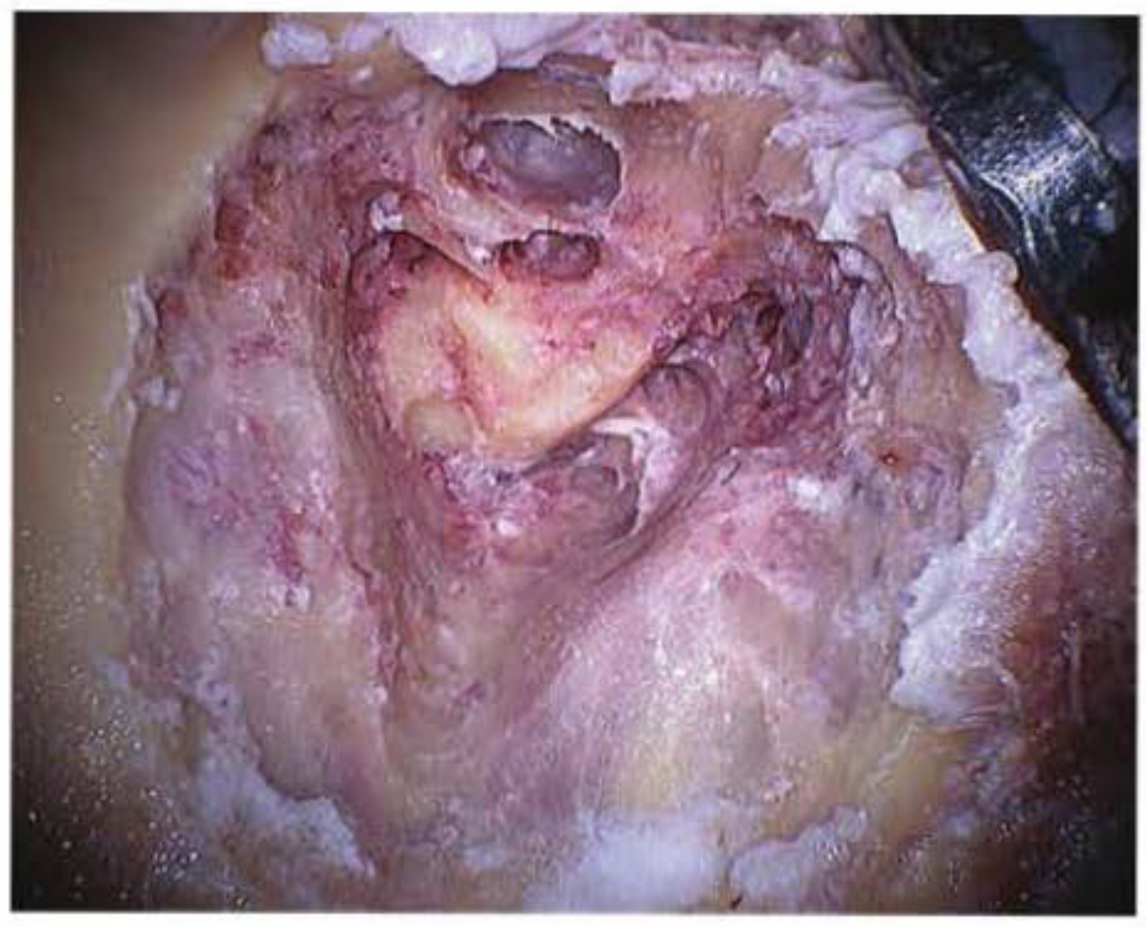
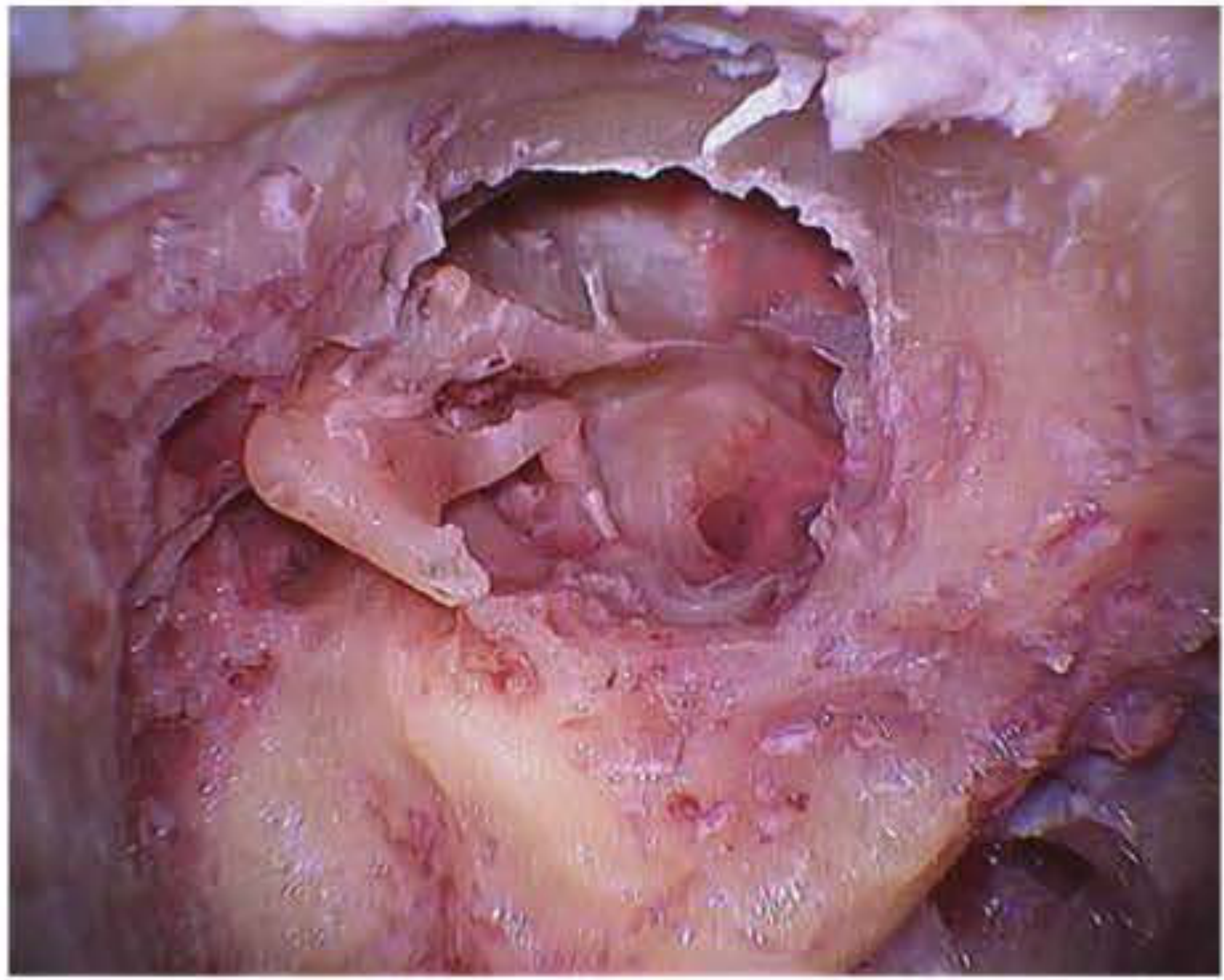
Intraoperative Images
Complications
| Category | Complication |
|---|---|
| Intraoperative | Facial nerve injury, sensorineural hearing loss (labyrinthine fistula), CSF leak (dural tear), sigmoid sinus injury |
| Early postoperative | Wound infection, hematoma, vertigo |
| Late | Residual/recurrent cholesteatoma (esp. sinus tympani), chronic cavity discharge, cavity problems requiring revision, caloric vestibular reaction to water |
Postoperative Cavity Care
- Lifelong periodic cleaning of the mastoid cavity (office visits)
- Avoidance of water entry (caloric vertigo effect with water exposure)
- This is the major disadvantage compared to canal-wall-up technique
- Patients counseled preoperatively about this requirement
MRM vs. Canal-Wall-Up (CWU) - Key Comparison
- Single-stage procedure in most cases
- No need for planned second-look surgery
- Lower recurrence risk (better visualization)
- Less demanding technically
- Lifelong cavity cleaning
- No water in ear (activity restriction)
- Caloric vertigo risk with water
- Larger surgical cavity
- Poorer hearing rehabilitation potential
- Shambaugh Surgery of the Ear, p. 541-544