
sphingolipidosis
sphingolipidosis storage disease histology pathology
sphingolipidosis lysosomal storage disease
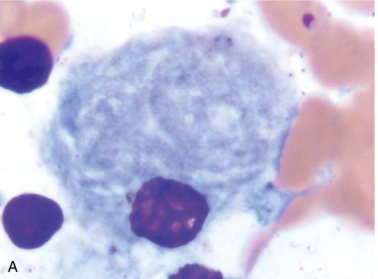
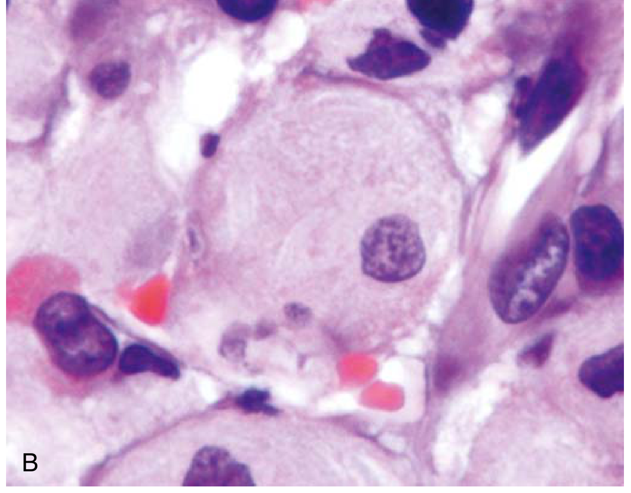
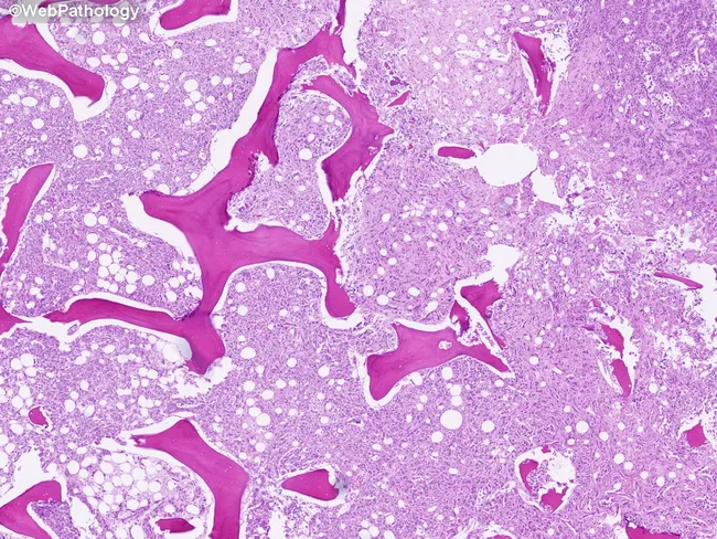
Gaucher disease foam cells bone marrow histology
Sphingolipidoses
- Lippincott Illustrated Reviews: Biochemistry, 8e, p. 594-596
- Robbins & Kumar Basic Pathology, p. 118-120
Biochemical Background
| Class | Head Group | Example |
|---|---|---|
| Sphingomyelin | Phosphocholine | Myelin sheaths |
| Cerebrosides | Single sugar (Glc or Gal) | Galactocerebroside |
| Sulfatides | Galactose + sulfate | White matter |
| Globosides | Oligosaccharide chain | RBC membranes |
| Gangliosides | Oligosaccharide + NANA | Neuronal membranes |
Complete Sphingolipid Degradation Pathway with Diseases
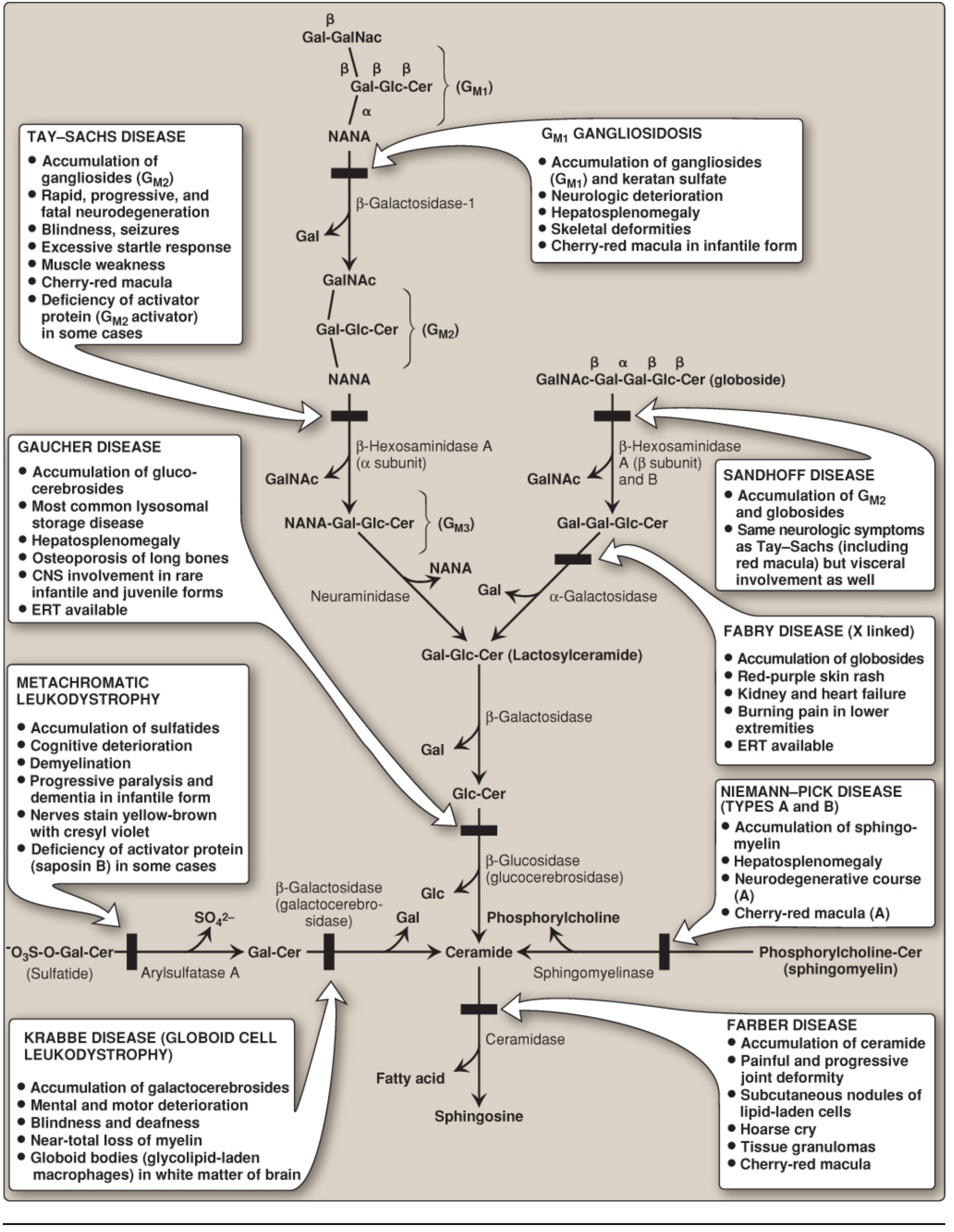
General Properties (Shared by All Sphingolipidoses)
- Single enzyme defect - only one specific sphingolipid accumulates per disease
- Progressive - clinical course is relentlessly progressive
- Autosomal recessive inheritance - except Fabry disease, which is X-linked
- Phenotypic variability - different clinical subtypes exist within a single disease (e.g., Gaucher types 1, 2, 3)
- Population genetics - Gaucher, Tay-Sachs, and Niemann-Pick diseases are enriched in Ashkenazi Jewish populations; Tay-Sachs also has higher frequency in Irish-American, French-Canadian, and Louisiana Cajun populations
- Diagnosis - enzyme activity measured in peripheral leukocytes or cultured fibroblasts; DNA testing; prenatal diagnosis via amniocentesis or chorionic villus sampling
Individual Diseases - Summary Table
| Disease | Deficient Enzyme | Accumulating Lipid | Key Features |
|---|---|---|---|
| Gaucher | Glucocerebrosidase (β-glucosidase) | Glucocerebroside | Hepatosplenomegaly, bone pain, Gaucher cells ("crumpled tissue paper") |
| Niemann-Pick A/B | Acid sphingomyelinase | Sphingomyelin | Hepatosplenomegaly, cherry-red macula (type A), neurodegeneration (type A) |
| Niemann-Pick C | NPC1/NPC2 (lipid transporter) | Cholesterol + GM1/GM2 | Vertical gaze palsy, ataxia, dystonia - distinct from A/B |
| Tay-Sachs | β-Hexosaminidase A (α subunit) | GM2 ganglioside | Cherry-red macula, neurodegeneration, NO visceral involvement |
| Sandhoff | β-Hexosaminidase A+B (β subunit) | GM2 + globoside | Same as Tay-Sachs + visceral involvement |
| Fabry | α-Galactosidase A | Globoside/Gb3 | X-linked; burning pain in extremities, angiokeratomas, renal/cardiac failure |
| Krabbe | β-Galactocerebrosidase | Galactocerebroside | Globoid bodies in white matter, demyelination, motor/mental deterioration |
| Metachromatic Leukodystrophy | Arylsulfatase A | Sulfatides | Peripheral neuropathy, demyelination, nerves stain yellow-brown with cresyl violet |
| Farber Disease | Ceramidase | Ceramide | Painful joint deformity, subcutaneous nodules, hoarse cry, cherry-red macula |
| GM1 Gangliosidosis | β-Galactosidase-1 | GM1 + keratan sulfate | Neurodegeneration, hepatosplenomegaly, skeletal deformities, cherry-red macula |
Key Diseases in Detail
1. Gaucher Disease - Most Common Sphingolipidosis
| Type | Features | CNS? |
|---|---|---|
| Type 1 (99% of cases) | Hepatosplenomegaly (spleen can be massive), bone involvement (osteopenia, lytic lesions, osteonecrosis), cytopenias | No CNS |
| Type 2 (acute neuronopathic) | Presents in infancy; severe neurologic deterioration, convulsions | Yes - early, severe |
| Type 3 (chronic neuronopathic) | Later onset; neurologic signs milder | Yes - late, milder |
- Enzyme replacement therapy (ERT) - recombinant glucocerebrosidase infusions (lifelong)
- Substrate reduction therapy - miglustat (oral inhibitor of glucosylceramide synthase)
- Bone marrow transplantation (macrophages are hematopoietic in origin)
2. Niemann-Pick Disease
- Type A: Infantile onset, severe neurodegeneration, hepatosplenomegaly, cherry-red macula - usually fatal by age 3
- Type B: Milder, organomegaly without CNS involvement
3. Tay-Sachs Disease
- Deficiency of β-hexosaminidase A (α subunit mutation) - GM2 accumulates exclusively in neurons
- Infants present by 6 months: poor feeding, lethargy, floppiness, then progressive neurodegeneration
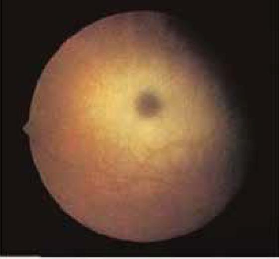
- Cherry-red spot on fundoscopy - the fovea appears red because it lacks the opacified ganglion cells that surround it
- Exaggerated startle response, blindness, seizures
- Death usually by age 3-5
- NO visceral involvement (distinguishes from Sandhoff)
4. Fabry Disease - Only X-linked Sphingolipidosis
- α-Galactosidase A deficiency; X-linked recessive (females can be carriers with variable expression)
- Globotriaosylceramide (Gb3) accumulates in vascular endothelial cells throughout the body (brain, heart, kidneys, skin)
- Triad: burning/painful neuropathy in extremities, angiokeratomas (red-purple skin rash), progressive renal and cardiac failure
- ERT available (agalsidase alfa or beta)
5. Krabbe Disease (Globoid Cell Leukodystrophy)
- β-Galactocerebrosidase deficiency - galactocerebroside accumulates in white matter
- Pathology: globoid bodies - glycolipid-laden macrophages in white matter
- Near-total loss of myelin; mental and motor deterioration, blindness, deafness
6. Metachromatic Leukodystrophy (MLD)
- Arylsulfatase A deficiency (or saposin B activator protein deficiency)
- Sulfatides accumulate in CNS and peripheral nerves
- Nerves stain yellow-brown (metachromatic) with cresyl violet dye
- Progressive demyelination - peripheral neuropathy → cognitive decline → paralysis, dementia
7. Farber Disease
- Ceramidase deficiency - ceramide accumulates
- Classic triad: painful progressive joint deformity, subcutaneous lipogranulomas, hoarse cry (laryngeal involvement)
- Cherry-red macula; tissue granulomas
Diagnosis
| Method | Details |
|---|---|
| Enzyme assay | Leukocytes or cultured fibroblasts - most definitive for classic forms |
| DNA/molecular | Identifies specific mutations; useful for carrier testing |
| Histology | Characteristic cell morphology (Gaucher cells, globoid bodies, metachromasia) |
| Prenatal | Cultured amniocytes or chorionic villi for enzyme activity |
| Shell-like inclusion bodies | Seen in Tay-Sachs |
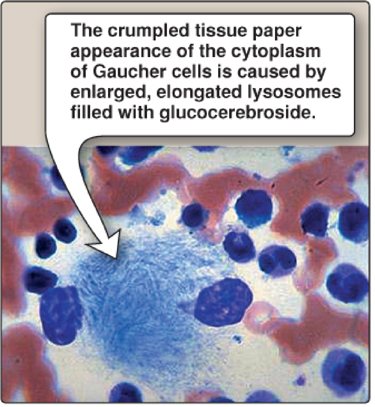
| "Crumpled tissue paper" cytoplasm | Seen in Gaucher |
| Zebra bodies | Seen in Niemann-Pick type A |
Classification within Lysosomal Storage Diseases
Recent Evidence
sphingolipidosis
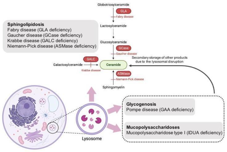
sphingolipidosis lysosomal storage disease treatment
cherry red spot macula fundus Tay-Sachs Niemann-Pick
Sphingolipidoses
Biochemical Basis
| Sphingolipid Class | Head Group | Main Location |
|---|---|---|
| Sphingomyelin | Phosphocholine | Myelin, cell membranes |
| Galactocerebroside | Galactose | White matter myelin |
| Sulfatides | Galactose + sulfate | White matter |
| Globosides | Oligosaccharide chain | RBC membranes |
| Gangliosides (GM1, GM2) | Oligosaccharide + NANA | Neuronal membranes |
Complete Degradation Pathway and Disease Map
General Properties Shared by All Sphingolipidoses
- Single substrate accumulates - only the specific substrate of the deficient enzyme accumulates per disease
- Normal synthesis rate - the lipid continues to be made normally; only catabolism is blocked
- Progressive - relentlessly progressive course
- Autosomal recessive - all sphingolipidoses are AR except Fabry disease, which is X-linked recessive
- Phenotypic and allelic variability - multiple disease subtypes from different mutations in the same gene (e.g. Gaucher types 1, 2, 3)
- Population clustering - Gaucher, Tay-Sachs, and Niemann-Pick types A/B are enriched in Ashkenazi Jewish populations; Tay-Sachs also has increased frequency in Irish-American, French-Canadian, and Louisiana Cajun populations
- Secondary cellular injury - beyond storage, macrophage activation and cytokine release (IL-1, IL-6, TNF) cause additional organ damage
- Impaired autophagy - lysosomal dysfunction also impairs autophagy, leading to accumulation of dysfunctional mitochondria and polyubiquitinated proteins, which triggers free radical generation and apoptosis
Disease-by-Disease Summary Table
| Disease | Deficient Enzyme | Accumulating Lipid | Key Clinical Features |
|---|---|---|---|
| Tay-Sachs | β-Hexosaminidase A (α-subunit) | GM2 ganglioside | Cherry-red macula, neurodegeneration, exaggerated startle, NO visceral disease |
| Sandhoff | β-Hexosaminidase A + B (β-subunit) | GM2 + globoside | Same neurologic features as Tay-Sachs + visceral involvement |
| GM1 Gangliosidosis | β-Galactosidase-1 | GM1 + keratan sulfate | Neurodegeneration, hepatosplenomegaly, skeletal deformities, cherry-red macula |
| Gaucher | Glucocerebrosidase (β-glucosidase) | Glucocerebroside | Hepatosplenomegaly, bone disease, Gaucher cells; types 2 and 3 have CNS involvement |
| Niemann-Pick A/B | Acid sphingomyelinase | Sphingomyelin | Hepatosplenomegaly; type A = neurodegeneration + cherry-red macula; type B = organomegaly only |
| Niemann-Pick C | NPC1/NPC2 (lipid transporter - NOT a hydrolase) | Cholesterol + GM1/GM2 | Vertical supranuclear gaze palsy, ataxia, dystonia, psychomotor regression |
| Fabry | α-Galactosidase A | Globotriaosylceramide (Gb3) | X-linked; burning neuropathy, angiokeratomas, renal/cardiac failure |
| Krabbe | β-Galactocerebrosidase | Galactocerebroside | Demyelination, globoid bodies, mental/motor deterioration, blindness, deafness |
| Metachromatic Leukodystrophy | Arylsulfatase A (or saposin B) | Sulfatides | Peripheral neuropathy, demyelination, nerves stain yellow-brown with cresyl violet |
| Farber Disease | Ceramidase | Ceramide | Painful joint deformity, subcutaneous lipogranulomas, hoarse cry, cherry-red macula |
Individual Diseases in Detail
1. Tay-Sachs Disease
- Onset: 3-6 months - motor weakness, poor feeding, lethargy, floppiness
- Progressive: deafness, blindness, spasticity → rigidity
- Exaggerated startle response (hyperekplexia)
- Death by age 2-3 years from respiratory infections
- No hepatosplenomegaly (distinguishes from Sandhoff)
- Carrier frequency in Ashkenazi Jews: 1 in 30; disease incidence 1:3600
2. Gaucher Disease - Most Common Sphingolipidosis
| Type | Name | CNS | Notes |
|---|---|---|---|
| Type 1 (99%) | Chronic non-neuronopathic | Absent | Hepatosplenomegaly (spleen can become massive), bone disease (osteopenia, lytic lesions, osteonecrosis), cytopenias; compatible with long life |
| Type 2 | Acute infantile neuronopathic | Severe, early | Onset 3-6 months; fatal in 2nd year from pulmonary infections |
| Type 3 | Chronic neuronopathic | Mild, late onset | Intermediate severity |
- ERT - recombinant glucocerebrosidase (imiglucerase, velaglucerase); mannose-6-phosphate modification targets enzyme to macrophage lysosomes
- Substrate reduction therapy - miglustat (oral inhibitor of glucosylceramide synthase)
- Bone marrow transplantation (curative in principle; macrophages are hematopoietic)
- Gene therapy (emerging - HSCs engineered to express GBA)
3. Niemann-Pick Disease
- Type A: Severe ASM deficiency; sphingomyelin accumulates in macrophages (liver, spleen, bone marrow, lungs) AND neurons. Macrophage cytoplasm shows fine vacuolation ("foam cells"). EM shows "zebra bodies" (concentric lamellated myelin figures). Cherry-red macula present. Fatal by age 3.
- Type B: Milder; organomegaly without CNS involvement; compatible with survival into adulthood
4. Fabry Disease
- Burning/painful neuropathy in hands and feet (acroparesthesias) - often the earliest symptom
- Angiokeratomas - red-purple skin rash, usually over the bathing-trunk distribution
- Progressive renal failure + cardiomyopathy/cardiac arrhythmias
5. Krabbe Disease (Globoid Cell Leukodystrophy)
6. Metachromatic Leukodystrophy (MLD)
7. Farber Disease (Lipogranulomatosis)
- Painful progressive joint deformity
- Subcutaneous lipid-laden granulomas (nodules)
- Hoarse cry (laryngeal granuloma)
Diagnosis
| Method | Application |
|---|---|
| Enzyme activity assay | Leukocytes or cultured fibroblasts - definitive for classic forms |
| DNA/molecular testing | Identifies specific mutations; carrier detection; prenatal diagnosis |
| Histology | Gaucher cells ("crumpled tissue paper"), globoid bodies (Krabbe), metachromatic staining (MLD), zebra bodies (Niemann-Pick), onion-skin lysosomes (Tay-Sachs) |
| Prenatal diagnosis | Cultured amniocytes or chorionic villi for enzyme activity |
| Newborn screening | Available for select disorders (e.g. Fabry, Gaucher, Krabbe in some programs) using dried blood spot enzyme assays |
Lysosomal Storage Disease Classification (Context)
Recent Evidence
- Neuroinflammation as a target (PMID 40992103): glial activation (microglia, astrocytes) is a major driver of CNS injury in sphingolipidoses beyond simple substrate accumulation - representing a new therapeutic angle for Tay-Sachs, Krabbe, and MLD
- Gene therapy (PMID 40782413): HSC gene therapy is in trials for several LSDs including Krabbe and MLD, with CNS penetration a key challenge
- Current and emerging therapies (PMID 39826077): substrate reduction therapy, pharmacological chaperones, and mRNA/gene therapies are expanding the treatment landscape beyond ERT
fatty liver
fatty liver hepatic steatosis histology macrovesicular
Light-microscopy histology of liver tissue demonstrates macrovesicular steatosis. Imaging modality: Histology using Hematoxylin and Eosin (H&E) stain on paraffin-embedded sections. Anatomical localization: hepatic parenchyma of the liver, with hepatocytes arranged in plates separated by sinusoids and preserved lobular architecture visible at low magnification. Visual features: numerous intracellular lipid vacuoles produce large clear droplets that displace hepatocyte nuclei to the periphery; cytoplasm otherwise eosinophilic; occasional subtle ballooning is not evident; portal tracts and central veins are discernible but not markedly inflamed. Pathological interpretation: macrovesicular steatosis consistent with fatty liver changes; most compatible with nonalcoholic fatty liver disease (NAFLD) or alcoholic fatty liver disease (AFLD) depending on patient history. In this image, steatosis is the predominant finding with minimal inflammatory infiltrate and no definitive fibrosis at this level of magnification. Clinical significance: fatty change indicates hepatocellular stress from metabolic syndrome or hepatotoxic exposure; may precede steatohepatitis, fibrosis, cirrhosis, and increased risk of hepatocellular carcinoma with ongoing injury. Potential clinical use cases: assessment of steatosis burden in NAFLD/NASH studies, biopsy-guided staging, therapeutic monitoring, and education for trainees. The image serves as an educational reference for recognizing hepatocellular lipid accumulation and distinguishing it from other hepatic pathologies such as steatohepatitis and cholestatic injury.
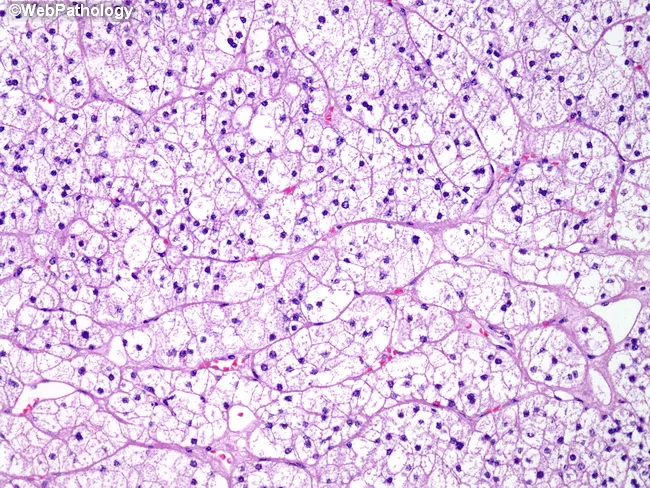
Light microscopic histology image of liver tissue stained with Hematoxylin and Eosin (H&E). The sample shows hepatic parenchyma arranged in broad cords separated by prominent sinusoids. The cytoplasm of many hepatocytes appears markedly pale or clear due to intracellular lipid accumulation, consistent with macrovesicular steatosis. Nuclei are scattered throughout each cell, often displaced toward the cell periphery, reflecting lipid droplet expansion. The overall architecture remains preserved without convincing lobular inflammation, Mallory-Denk hyaline, or confluent necrosis at this magnification. Portal tracts are not prominent in this field, suggesting midzonal or hepatocellular predominance of the fatty change. Capillarization of sinusoids or mild vascular congestion is not conspicuous. The pattern is compatible with fatty liver changes, which can be seen in nonalcoholic fatty liver disease (NAFLD), alcoholic fatty liver disease, or other etiologies of steatosis. Clinically, these findings correlate with metabolic syndrome, obesity, type 2 diabetes, or excess alcohol intake. The image provides educational value for recognizing macrovesicular steatosis and distinguishing it from other hepatocellular pathologies such as steatohepatitis, viral hepatitis, or cholestasis. This histology is essential for diagnosing hepatic steatosis and guiding further metabolic or lifestyle interventions. Correlation with patient history and imaging enhances diagnostic confidence and guides management and follow-up care.
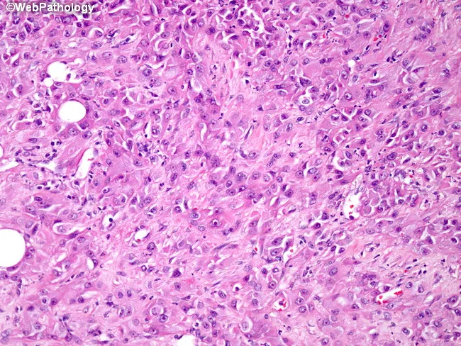
This histopathology slide depicts liver parenchyma prepared for light microscopy and stained with hematoxylin and eosin (H&E). The tissue shows polygonal hepatocytes arranged in an orderly plate-like architecture with cords separated by sinusoids. The cytoplasm is eosinophilic and mildly granular; nuclei are round or oval with inconspicuous nucleoli. Several large, clear vacuolar spaces within hepatocytes are compatible with macrovesicular fatty change (steatosis). The overall architecture appears preserved, but occasional cytoplasmic ballooning and mild cytoplasmic rarefaction may reflect early hepatocellular stress. Ductal structures and portal tracts are not the dominant features in this field and appear limited to scattered portal elements. There is no conspicuous necrosis, active inflammation, or significant fibrosis visible at this magnification, though sampling bias may obscure subtle changes. The image emphasizes hepatocellular morphology, lipid accumulation, and cellular detail that are essential for assessing fatty liver disease, steatohepatitis, or metabolic hepatopathy. Clinically, these findings can correlate with nonalcoholic fatty liver disease (NAFLD), alcoholic hepatopathy, or other causes of hepatic steatosis. Differential considerations include steatosis without inflammation, steatohepatitis, drug-induced liver injury with fatty change, or minimal chronic hepatitis. The slide is suitable for teaching hepatic histology, fatty change assessment, and correlating histologic pattern with clinical liver function abnormalities.
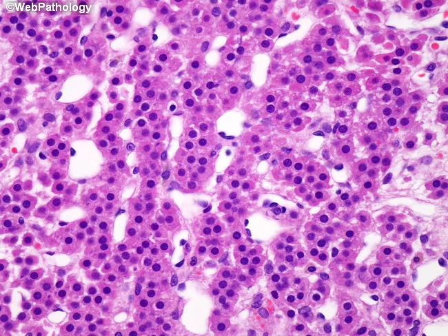
High-magnification brightfield histology of liver tissue stained with hematoxylin and eosin. The hepatic parenchyma displays preserved lobular architecture with cords of polygonal hepatocytes separated by narrow to wide sinusoids. A prominent feature is diffuse intracellular lipid accumulation, manifested as numerous clear cytoplasmic vacuoles that displace occasional nuclei to the cell margin. Both macrovesicular and microvesicular fat droplets are evident, creating a foamy to bubbly cytoplasmic appearance in many hepatocytes. The overall pattern is hepatocellular steatosis; there is no conspicuous bridging necrosis or lobular inflammation visible in this field, though mild cytoplasmic swelling of some hepatocytes can be appreciated. Bile ducts and portal tracts appear unremarkable in this view. The appearance is compatible with fatty liver changes, seen in nonalcoholic fatty liver disease (NAFLD) or alcoholic fatty liver disease, and may reflect metabolic syndrome, insulin resistance, or hepatotoxic exposure. Clinically, such steatosis can be reversible with weight reduction, glucose control, and avoidance of hepatotoxins. Pathologic significance lies in assessing the degree of steatosis and distinguishing simple steatosis from steatohepatitis, which would warrant closer clinical management. This image is suitable for educational reference in hepatology, pathology, and diagnostic imaging.
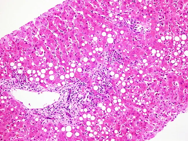
Light microscopy of a liver biopsy stained with Hematoxylin and Eosin demonstrates hepatic parenchymal fatty change with preserved lobular architecture. Numerous large lipid droplets occupy hepatocyte cytoplasm, producing clear vacuoles that give a characteristic macrovesicular steatosis appearance. Scattered hepatocytes exhibit mild ballooning degeneration, reflecting cellular stress. A modest inflammatory infiltrate is present in select portal tracts and within sinusoids, consistent with low-grade inflammation. Kupffer cells appear mildly enlarged, in keeping with portal-sinusoidal immune activation. No bridging necrosis, significant cholestasis, or overt lobular disarray is evident in this field. The pattern is most compatible with fatty liver injury but can be seen with metabolic syndrome, obesity, excessive alcohol intake, or drug/toxin exposure. In the setting of infectious mononucleosis from Epstein-Barr virus, hepatic involvement is common but usually subclinical; histology can show portal and sinusoidal lymphocytic infiltrates with rare atypical lymphocytes, though steatosis is not a defining feature. Therefore, correlation with serology and clinical history is essential for etiologic assignment. This image is a useful teaching reference for recognizing steatosis on liver biopsy, distinguishing fat-laden hepatocytes from inflammatory nodular processes, and framing the differential diagnosis of viral hepatitis versus fatty liver disease in hepatology, pathology, and medical education.
"metabolic dysfunction-associated steatotic liver disease" MASLD
Fatty Liver Disease (Hepatic Steatosis)
Nomenclature (2023 Update)
| Old Term | New Term (2023 Delphi Consensus) |
|---|---|
| NAFLD (nonalcoholic fatty liver disease) | MASLD - Metabolic dysfunction-Associated Steatotic Liver Disease |
| NASH (nonalcoholic steatohepatitis) | MASH - Metabolic dysfunction-Associated SteatoHepatitis |
| Alcoholic fatty liver | MetALD or alcohol-associated liver disease |
Epidemiology
- 20-30% of the general population in North America and Europe has NAFLD/MASLD
- 60-75% of obese or diabetic patients have NAFLD; 20-25% of these have NASH/MASH
- Now the most common cause of chronic liver disease in the United States, affecting up to 30-46% of the population
- Expected to be (or already is) the leading indication for liver transplantation, surpassing viral hepatitis
- NASH is a major cause of cryptogenic cirrhosis (fat disappears as fibrosis advances, leaving only cirrhosis without an apparent etiology)
- Strong driver of the rising incidence of HCC in Western countries - Mulholland & Greenfield's Surgery, 7e; Tietz Textbook of Laboratory Medicine, 7e
Etiology and Risk Factors
Metabolic / Primary Causes (MASLD)
- Obesity (especially central/visceral adiposity)
- Type 2 diabetes mellitus / insulin resistance
- Dyslipidemia (hypertriglyceridemia, low HDL)
- Metabolic syndrome - nearly half of individuals meeting metabolic syndrome criteria have NAFLD
- 90% of NAFLD patients have at least one of these risk factors; 30% have three or more
Secondary Causes
| Category | Examples |
|---|---|
| Alcohol | Alcoholic fatty liver disease (ALD) - threshold >20 g/day |
| Drugs/toxins | Corticosteroids, amiodarone, methotrexate, tamoxifen, valproate, irinotecan (chemotherapy) |
| Nutritional | Total parenteral nutrition, rapid weight loss, protein malnutrition |
| Metabolic disorders | Wilson disease, lipodystrophy, abetalipoproteinemia |
| Endocrine | Hypothyroidism, PCOS, hypopituitarism |
| GI surgery | Jejunoileal bypass, extensive small bowel resection |
Pathogenesis
The "Two-Hit" Model (Classic)
- First hit - insulin resistance → hepatic steatosis (sensitizes liver to further injury)
- Second hit - oxidative stress, inflammation, lipotoxicity → steatohepatitis → fibrosis
Biochemical Mechanisms of Fat Accumulation
- Oxidized by mitochondria (β-oxidation)
- Esterified into TG and exported as VLDL
- Incorporated into phospholipids and cholesteryl esters
| Mechanism | Detail |
|---|---|
| Increased FFA delivery | Visceral adipose lipolysis → portal FFA flood |
| Insulin resistance | Impaired suppression of adipose lipolysis + hepatic de novo lipogenesis (DNL) |
| Increased DNL | Dietary fructose activates ChREBP and SREBP-1 → promotes DNL |
| Impaired β-oxidation | Mitochondrial dysfunction |
| Decreased VLDL export | Reduced TG export |
| Adipokine dysregulation | ↑ Leptin, TNF-α, IL-6, resistin; ↓ Adiponectin → promotes inflammation and fibrosis |
| Bile acid dysregulation | Impaired FXR signaling → ↑ DNL, ↓ β-oxidation |
Progression to Steatohepatitis (MASH/NASH)
- Lipotoxicity → mitochondrial dysfunction, ER stress
- Unfolded protein response activation
- Inflammasome activation → IL-1β, IL-18 secretion
- Gut-derived bacterial endotoxin (LPS) → hepatic stellate cell activation → fibrosis
Histopathology
Simple Steatosis (MASLD without MASH)
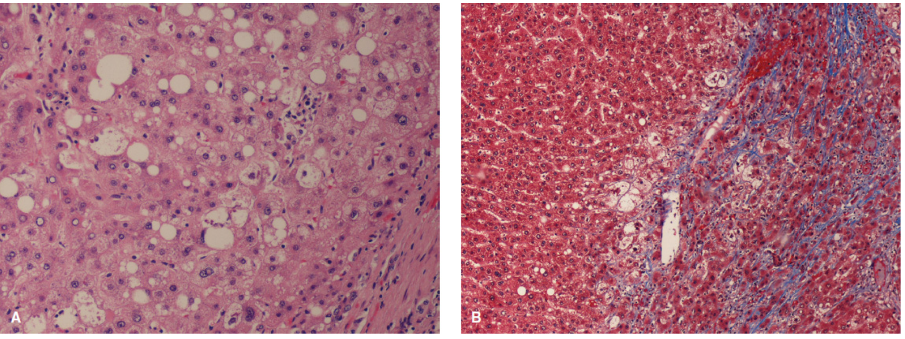
Steatohepatitis (MASH/NASH) - requires ALL of:
- Steatosis (macrovesicular, zone 3 predominant)
- Hepatocyte ballooning degeneration - swollen, rarefied cytoplasm
- Lobular inflammation - mixed inflammatory infiltrate
- Mallory-Denk bodies - eosinophilic cytoplasmic inclusions (aggregates of cytokeratins 8/18)
- Pericellular/perisinusoidal fibrosis (zone 3, "chicken-wire" pattern) on trichrome stain
Grading and Staging (NAS - NAFLD Activity Score)
| Component | Score |
|---|---|
| Steatosis | 0-3 (5-33%, 33-66%, >66%) |
| Lobular inflammation | 0-3 |
| Hepatocyte ballooning | 0-2 |
| Total NAS ≥5 = NASH likely; NAS ≤2 = NASH unlikely | |
| Fibrosis stage | F0 = none → F4 = cirrhosis |
Disease Spectrum and Natural History
Simple Steatosis (MASLD)
↓ (~20% progress over 10+ years)
Steatohepatitis (MASH/NASH)
↓ (~20% progress)
Fibrosis (F1→F4)
↓
Cirrhosis
↓
HCC / Liver failure / Transplant
- Up to 20% of patients with NASH progress to cirrhosis
- >30% macrovesicular steatosis in donor livers correlates with increased graft failure risk in transplantation
Diagnosis
Clinical Presentation
- Usually asymptomatic - discovered incidentally on imaging or elevated LFTs
- May have vague RUQ discomfort or fatigue
- Signs of metabolic syndrome (obesity, hypertension, dyslipidemia, T2DM)
- Advanced disease: signs of portal hypertension, hepatomegaly
Laboratory Tests
| Test | Finding |
|---|---|
| AST, ALT | Mildly elevated (ALT > AST in NAFLD; AST > ALT in alcoholic) |
| GGT | Elevated |
| Lipid panel | Hypertriglyceridemia, low HDL |
| Fasting glucose / HbA1c | Insulin resistance / T2DM |
| CK-18 fragments | Serum marker of hepatocyte apoptosis - indicator of NASH |
| PIINP | Marker of fibrosis |
Non-Invasive Fibrosis Scores
| Score | Components |
|---|---|
| NAFLD Fibrosis Score | Age, BMI, AST/ALT ratio, platelets, albumin, diabetes |
| BARD score | BMI + AST/ALT ratio + diabetes |
| FIB-4 index | Age × AST / (platelets × √ALT) |
| Enhanced Liver Fibrosis (ELF) test | Direct fibrosis markers (TIMP-1, PIIINP, HA) |
Imaging
- Ultrasound - increased echogenicity ("bright liver"); sensitive for >30% steatosis
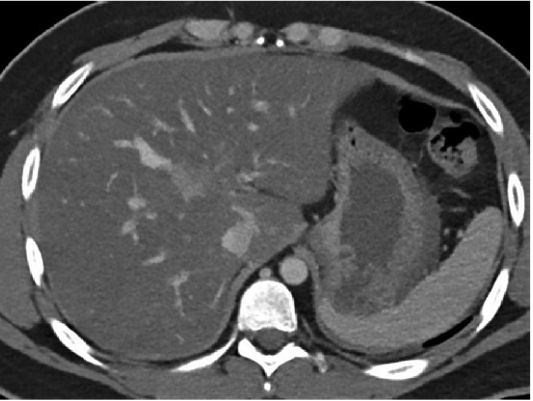
- CT - fatty liver appears hypodense relative to spleen (normal liver HU > spleen); liver:spleen ratio <0.8 indicates steatosis (see CT image above)
- MRI-PDFF (proton density fat fraction) - most accurate non-invasive quantification of hepatic fat
- FibroScan/Transient elastography - liver stiffness measurement for fibrosis staging; CAP (controlled attenuation parameter) for steatosis
Liver Biopsy
- Distinguishing simple steatosis from NASH
- Staging fibrosis
- Excluding other diagnoses
Management
Lifestyle Modification (First-line for all)
- Weight loss 5-10% - associated with significant histologic improvement in steatosis and inflammation
- >10% weight loss - may improve fibrosis
- Aerobic exercise - reduces hepatic fat independent of weight loss
- Mediterranean diet, avoidance of fructose/high-sugar beverages
- Alcohol avoidance - even modest alcohol accelerates fibrosis in NASH
Pharmacological Therapy
| Drug | Mechanism | Evidence |
|---|---|---|
| Resmetirom (Rezdiffra) | Thyroid hormone receptor-β agonist | FDA-approved 2024 for MASH with moderate-to-advanced fibrosis (F2-F3) - first approved drug |
| Pioglitazone (thiazolidinedione) | PPARγ agonist - improves insulin sensitivity | Improves steatosis, inflammation, AND fibrosis in NASH + T2DM |
| GLP-1 agonists (liraglutide, semaglutide) | Weight loss + direct hepatic effects | Significant histologic improvement in NASH; semaglutide in trials |
| Vitamin E (800 IU/day) | Antioxidant | Benefit in non-diabetic NASH; not recommended in diabetics or cirrhosis |
| Metformin | Insulin sensitizer | Does NOT improve histology - not recommended for NASH |
| Obeticholic acid (FXR agonist) | Bile acid pathway | Improves fibrosis; pruritus is common side effect |
| Probiotics | Gut microbiome modulation | Meta-analysis: ↓ transaminases, cholesterol, TNF-α, insulin resistance |
Bariatric Surgery
- For patients with obesity (BMI >40 or >35 with comorbidities) who fail lifestyle modification
- Produces dramatic improvement or resolution of NAFLD/NASH
- Improves fibrosis in most patients
Liver Transplantation
- Indicated for NASH cirrhosis with decompensation or HCC
- NAFLD/NASH now a leading or the leading indication for liver transplantation in the US
- Post-transplant: NAFLD can recur in the graft; metabolic syndrome management is essential
- Donor livers with >30% macrovesicular steatosis have increased graft failure risk
Alcoholic Fatty Liver Disease (ALD)
- Acetaldehyde toxicity
- Generation of reactive oxygen species
- Altered NAD⁺/NADH ratio → impaired fatty acid oxidation → fat accumulation