
Hypertension definition cause pathology clinical future complications for5 marks
Reading File
Now let me search the medical library for comprehensive hypertension content.
Finding Sources
Finding Sources
Finding Sources
Finding Sources
Reading File
Reading File
Let me look for the dedicated hypertension sections in Goldman-Cecil Medicine and the Kidney textbook.
Finding Sources
Finding Sources
Excellent! Harrison's and Katzung's have great hypertension content. Let me read those sections now.
Reading File
Reading File
Let me read the Harrison's block 30 section specifically on hypertension definition and pathophysiology, and the Katzung section on clinical presentation/pathophysiology.
Finding Sources
Reading File
Now let me read the Harrison's dedicated hypertension chapter (block 30) - Definition, Primary, Secondary sections.
Reading File
Reading File
I now have excellent content from Harrison's and Morgan & Mikhail's for a comprehensive 5-mark answer. Let me read the Harrison's definition and classification section directly.
Reading File
I now have comprehensive content from multiple authoritative sources. Let me compile the complete 5-mark answer on hypertension.
Hypertension - Definition, Causes, Pathology, Clinical Features & Complications
(Harrison's Principles of Internal Medicine 22E | Morgan & Mikhail's Clinical Anesthesiology 7e | Katzung's Basic and Clinical Pharmacology 16e)
1. Definition
Hypertension is defined as a sustained elevation of arterial blood pressure above normal limits, assessed on two or more separate occasions.
ACC/AHA 2017 Classification (adults ≥18 years):
| Category | Systolic (mmHg) | Diastolic (mmHg) |
|---|---|---|
| Normal | <120 | <80 |
| Elevated | 120-129 | <80 |
| Stage 1 Hypertension | 130-139 | 80-89 |
| Stage 2 Hypertension | ≥140 | ≥90 |
| Hypertensive urgency | >180/120 | No organ damage |
| Hypertensive emergency | >180/120 | With organ damage |
High BP is a leading risk factor for cardiovascular disease (CVD), including ischemic and hemorrhagic stroke, coronary heart disease (CHD), heart failure, peripheral arterial disease, chronic kidney disease/end-stage kidney disease (ESKD), dementia, and all-cause mortality.
2. Causes / Etiology
A. Primary (Essential) Hypertension - 90-95% of cases
No identifiable single cause. Contributing factors include:
- Genetic predisposition (familial aggregation)
- High dietary sodium intake
- Obesity and insulin resistance / hyperinsulinemia
- Sedentary lifestyle
- Excessive alcohol intake
- Sympathetic nervous system overactivity
- Activation of the renin-angiotensin-aldosterone system (RAAS)
- Abnormal intracellular calcium and sodium handling in vascular smooth muscle cells
B. Secondary Hypertension - 5-10% of cases
| Cause | Mechanism |
|---|---|
| Chronic kidney disease | Impaired sodium excretion, RAAS activation |
| Renal artery stenosis | Reduced renal perfusion → RAAS activation |
| Primary hyperaldosteronism | Excess aldosterone → Na+/water retention |
| Cushing's syndrome | Excess cortisol → vasoconstriction |
| Pheochromocytoma | Excess catecholamines |
| Acromegaly | Excess GH/IGF-1 |
| Pregnancy / pre-eclampsia | Endothelial dysfunction |
| Oral contraceptives (estrogen) | Na+ retention, vasoconstriction |
| Obstructive sleep apnea | Sympathetic activation, hypoxia |
| Coarctation of the aorta | Mechanical obstruction |
3. Pathology / Pathophysiology
BP = Cardiac Output (CO) × Total Peripheral Resistance (TPR)
Hypertension arises from abnormalities in one or both of these factors.
Key Pathophysiological Mechanisms:
-
Renin-Angiotensin-Aldosterone System (RAAS) activation
- Angiotensin II causes potent vasoconstriction and aldosterone release → increased Na+ and water retention → raised CO and TPR.
-
Sympathetic nervous system (SNS) overactivity
- Increases heart rate, myocardial contractility, and peripheral vasoconstriction.
- Exaggerated vasopressor responses seen in hypertensive patients.
-
Endothelial dysfunction
- Reduced nitric oxide (NO) synthesis → impaired vasodilation.
- Increased endothelin (a potent vasoconstrictor).
- Reactive oxygen species contribute to vascular inflammation.
-
Abnormal renal sodium handling
- Impaired natriuresis → volume expansion → elevated CO.
-
Vascular remodeling
- Vascular smooth muscle hypertrophy → irreversibly elevated TPR.
- Increased intracellular Ca²+ → increased vascular tone.
Pathological Changes in Tissues:
- Blood vessels: Hyaline arteriolosclerosis (benign hypertension) - thickening of arteriolar walls with homogenous eosinophilic material; Hyperplastic arteriolosclerosis ("onion-skin" lesion) - in malignant hypertension; Fibrinoid necrosis of arterioles in accelerated/malignant hypertension.
- Heart: Concentric left ventricular hypertrophy (LVH) due to chronic increased afterload; diastolic dysfunction; eventually systolic dysfunction.
- Kidney: Nephrosclerosis - hyalinization of glomeruli, tubular atrophy, interstitial fibrosis. In malignant hypertension, necrotizing glomerulitis + fibrinoid necrosis.
- Brain: Microaneurysms (Charcot-Bouchard aneurysms) in small penetrating arteries.
4. Clinical Features
Hypertension is largely asymptomatic ("the silent killer") until organ damage develops.
Symptoms (when present):
- Headache (typically occipital, worse in the morning) - especially in severe hypertension
- Dizziness / tinnitus
- Epistaxis (nosebleeds)
- Visual disturbances (blurring)
- Palpitations
Signs on Examination:
- Elevated blood pressure on repeated measurements
- Fundoscopy (Keith-Wagener-Barker classification):
- Grade I: Silver wiring (arteriolar narrowing)
- Grade II: AV nipping (arteriovenous nicking)
- Grade III: Flame-shaped hemorrhages, cotton-wool spots (soft exudates)
- Grade IV: Papilledema (malignant hypertension)
- Loud A2 (aortic component of second heart sound)
- Sustained apex beat (LVH)
- S4 gallop (impaired LV relaxation)
- Renal bruits (if renovascular hypertension)
5. Complications
Long-standing uncontrolled hypertension accelerates atherosclerosis and causes hypertensive organ damage across multiple systems:
A. Cardiac Complications
- Left ventricular hypertrophy (LVH) - concentric hypertrophy → diastolic dysfunction
- Coronary artery disease / Angina / MI - accelerated atherosclerosis
- Congestive heart failure - initially diastolic, later systolic
- Aortic dissection - especially with severe/uncontrolled hypertension
B. Cerebrovascular Complications
- Ischemic stroke - thromboembolism from atherosclerotic arteries
- Hemorrhagic stroke - rupture of Charcot-Bouchard microaneurysms
- Hypertensive encephalopathy - breakthrough of cerebral autoregulation
- Vascular dementia - chronic microvascular ischemia
C. Renal Complications
- Nephrosclerosis - gradual loss of renal function
- Proteinuria / microalbuminuria - early marker of hypertensive nephropathy
- Chronic kidney disease (CKD) / End-stage renal disease (ESRD) - hypertension is a major cause
- Malignant hypertension → acute renal failure
D. Ocular Complications (Hypertensive Retinopathy)
- Arteriolar narrowing → hemorrhages, exudates → papilledema (Grade IV)
E. Peripheral Vascular Complications
- Peripheral arterial disease (PAD) - 2.5x increased risk in men, 3.9x in women
- Aortic aneurysm formation
Summary Table of Complications:
| System | Complication |
|---|---|
| Heart | LVH, IHD, MI, CCF, aortic dissection |
| Brain | Stroke (ischemic/hemorrhagic), encephalopathy, dementia |
| Kidney | CKD, ESRD, nephrosclerosis |
| Eyes | Hypertensive retinopathy, papilledema |
| Peripheral vessels | PAD, aortic aneurysm |
Key Points for 5 Marks:
- Definition: sustained BP ≥130/80 (ACC/AHA) or ≥140/90 (traditional)
- 90-95% is essential (primary); 5-10% is secondary
- Pathophysiology centers on RAAS activation, SNS overactivity, endothelial dysfunction, and vascular remodeling
- Largely asymptomatic until organ damage - the "silent killer"
- Major complications: stroke, MI, heart failure, CKD, and retinopathy (target organ damage)
Sources: Harrison's Principles of Internal Medicine 22E (2025), Morgan & Mikhail's Clinical Anesthesiology 7e, Katzung's Basic and Clinical Pharmacology 16e
Entrobiusvermicularis defination etiology pathology clinical features
Reading File
Finding Sources
Finding Sources
Excellent results. Now let me read the most detailed sources in parallel - Robbins Pathology, Red Book (pediatric infectious diseases), Henry's Lab Methods, and Sherris Microbiology.
Reading File
Reading File
Reading File
I have rich content. Let me also fetch the pinworm microscopy image and read the Sherris Microbiology section for additional detail.
Reading File
Loading Image
I now have comprehensive content from multiple authoritative textbooks and a great microscopy image. Here is the full answer:
Enterobius vermicularis (Pinworm / Threadworm)
(Sources: Robbins Cotran Pathologic Basis of Disease | Red Book 2021 | Henry's Clinical Diagnosis by Laboratory Methods | Sherris & Ryan's Medical Microbiology 8e)
1. Definition
Enterobius vermicularis (also called Oxyuris vermicularis) is a human intestinal nematode (roundworm) that causes enterobiasis - the most common helminthic infection in children of all social strata worldwide, including in high-resource countries. It is commonly known as the pinworm or threadworm, named for the female's characteristic sharply pointed tail.
Key facts:
- Belongs to phylum Nematoda, class Secernentea
- Over 1 billion people infected worldwide; an estimated 40 million in the United States alone
- Prevalence 20-30% in some age groups/communities
- E. vermicularis is the common species; E. gregorii has been reported in Europe, Africa, and Asia
- Humans are the only known natural host - pets are not reservoirs
2. Etiology (Morphology & Life Cycle)
Morphology:
| Feature | Description |
|---|---|
| Adult female | 8-13 mm long, cream/white colored, sharply pointed "pin-like" tail |
| Adult male | Smaller (3 mm), ventrally curved tail with copulatory spicule; rarely seen |
| Both sexes | Lateral fin-like ridges (alae) on both sides; prominent esophageal bulb |
| Eggs | Colorless, ovoid, flattened on one side ("bean-shaped"), 20-40 µm × 50-60 µm; thin-shelled |
Life Cycle:
- Ingestion - Infective eggs are swallowed (fecal-oral route) via contaminated hands, food, bedding, clothing, or airborne eggs
- Hatching - Eggs hatch in the small intestine, releasing larvae
- Adult location - Adults establish themselves primarily in the cecum, appendix, and ascending colon
- Mating - Adult males die soon after copulation
- Nocturnal migration - Gravid females migrate at night to the perianal region to deposit eggs on perianal skin folds
- Egg maturation - Eggs become infective within 4-6 hours under optimal conditions
- Auto-infection cycle - Scratching due to pruritus → eggs under fingernails → hand-to-mouth → re-ingestion
- Retroinfection - Newly hatched larvae may occasionally migrate back from anal skin into the rectum
- Incubation period - 2-6 weeks from egg ingestion to gravid female migrating to perianal region
- Female lifespan - Up to 100 days; eggs remain infective in indoor environment for up to 2 weeks
Transmission Routes:
- Fecal-oral (main route) - contaminated hands, food
- Person-to-person - handling contaminated clothing, bedding
- Autoinfection - scratching perianal area → hand-to-mouth
- Environmental surfaces - curtains, carpeting
- Airborne - inhalation of eggs (minor route)
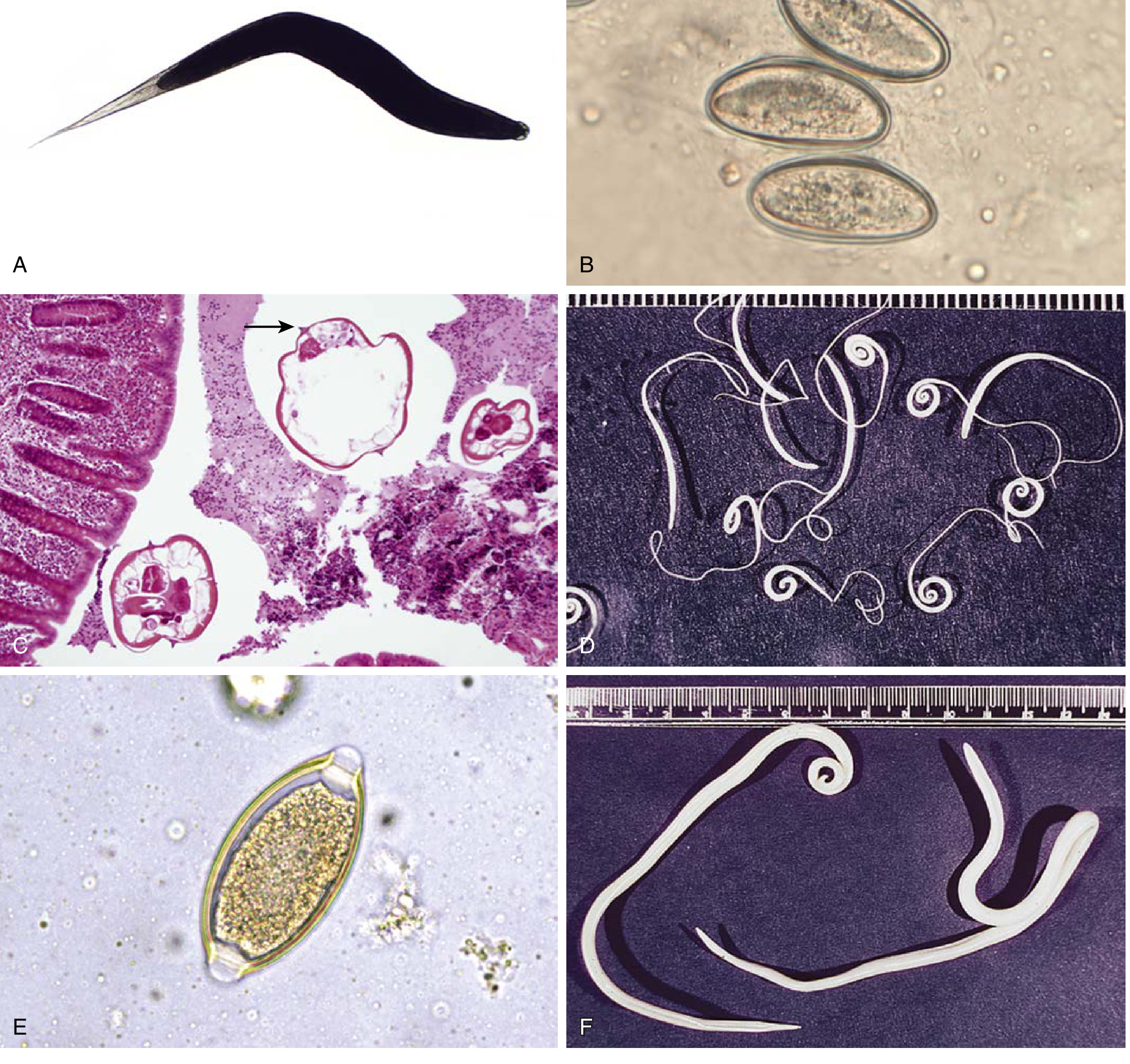
Figure: A - Adult female pinworm with pointed tail. B - Eggs of E. vermicularis (flattened/bean-shaped). C - Cross-sections of adult E. vermicularis in appendix, with lateral alae (arrow) visible on H&E staining.
3. Pathology
- Pinworms do not invade host tissues and live their entire life within the intestinal lumen - this is why they rarely cause serious illness
- No tissue invasion → peripheral eosinophilia is generally NOT seen (and elevated eosinophilia should not be attributed to pinworm)
- No elevated IgE - due to low invasiveness
- The female deposits eggs on the perirectal/perianal mucosa, causing local irritation and intense pruritus
- Scratching causes excoriation of the perianal skin, which can lead to secondary bacterial superinfection
- Adult worms are found in the lumen of the appendix and cecum - visible on histology as cross-sections with characteristic lateral alae (identification feature on H&E stain)
- Appendix involvement: Worms are found in the lumen of the appendix; in some cases associated with signs of acute appendicitis, but also found in histologically normal appendices (incidental finding)
- Aberrant migration - Adult worms occasionally migrate to ectopic sites:
- Vagina, fallopian tubes, uterus, peritoneal cavity → can cause granulomatous reaction, salpingitis, pelvic peritonitis
- Urethra → urethritis
- Eosinophilic enterocolitis has been rarely reported
- Eggs remain viable and infective outside the body for extended periods
4. Clinical Features
Symptoms:
| Feature | Details |
|---|---|
| Perianal pruritus (pruritus ani) | Most common symptom; intense, worse at night (nocturnal); caused by migrating females depositing eggs |
| Pruritus vulvae | Rare; from worm migration to female genitalia |
| Sleep disturbance | Due to nocturnal pruritus; irritability, restlessness |
| Perineal/perianal excoriation | From scratching; risk of secondary bacterial infection |
| Asymptomatic | Many/most infections cause no symptoms |
Associated Features (disputed causal link):
- Teeth grinding at night (bruxism)
- Enuresis (bedwetting) - should be ruled out in evaluation of enuresis
- Weight loss / poor appetite
- Irritability in children
Complications from Aberrant Migration:
- Vaginitis / vulvovaginitis - common in young girls; worms migrate from perianal area to vagina
- Urethritis
- Salpingitis (fallopian tube inflammation)
- Pelvic peritonitis
- Appendicitis (worms occlude lumen; seen especially in Bailey & Love Surgery)
- Eosinophilic granulomas at ectopic sites
Epidemiological Pattern:
- Primarily affects preschool and school-aged children
- Clusters within families and institutions (schools, daycare)
- Also infects primary caregivers of infected children
5. Diagnosis
| Method | Details |
|---|---|
| Cellophane/Scotch tape test | Gold standard - tape applied to perianal skin at night or on waking; pressed on slide; eggs seen under microscope |
| Pinworm paddle test | Clear plastic adhesive paddle pressed on perianal folds; eggs visible on microscopy |
| Timing | Best done before morning bathing; repeat on 3 different days increases sensitivity from ~50% (single test) to ~90% |
| Stool examination | NOT helpful - eggs/worms rarely found in stool |
| Direct visualization | Adult female worms (white, 8-13 mm) may be visible in the perianal region at night |
6. Treatment
| Drug | Notes |
|---|---|
| Pyrantel pamoate | Over-the-counter; first-line; single dose, repeat after 2 weeks |
| Mebendazole | Prescription; effective |
| Albendazole | Effective; not FDA-approved specifically for pinworms in the US |
- Treat all household members simultaneously (due to high transmission within families)
- Repeat dose at 2 weeks to eliminate worms acquired after first treatment
- Wash bedding, clothing; trim fingernails; maintain hand hygiene
Key Points to Remember:
- E. vermicularis = most common helminthic infection in children in developed countries
- Lives in cecum/colon; females migrate nocturnally to perianal skin to lay eggs
- Eggs become infective in 4-6 hours; remain viable for 2 weeks indoors
- No tissue invasion → NO eosinophilia, NO elevated IgE
- Classic symptom = nocturnal perianal pruritus causing sleep disturbance
- Diagnosis = Scotch tape test (NOT stool examination)
- Treatment = pyrantel pamoate / mebendazole / albendazole (repeat after 2 weeks; treat entire household)
Sources: Robbins Cotran Pathologic Basis of Disease | Red Book 2021 (Committee on Infectious Diseases) | Henry's Clinical Diagnosis and Management by Laboratory Methods | Sherris & Ryan's Medical Microbiology 8e | Yamada's Textbook of Gastroenterology
This is a shared conversation. Sign in to Orris to start your own chat.