
Airway assessment, preparation and management of difficult airway. Md Anesthesia exam short note
"difficult airway" AND management
ASA 2022 practice guidelines management difficult airway airway assessment preparation
https://www.asahq.org/~/media/sites/asahq/files/public/resou…
Airway assessment, preparation and management of difficult airway
Definition
1. Airway assessment
A. History
- Previous difficult mask ventilation or intubation, airway-alert card, anesthetic records
- Obstructive sleep apnea, snoring, CPAP use
- Stridor, hoarseness, dysphagia, dyspnea when supine
- Facial trauma, burns, airway tumor/infection, neck irradiation or surgery
- Rheumatoid arthritis, ankylosing spondylitis, acromegaly
- Pregnancy, obesity, diabetes, craniofacial syndromes
- Aspiration risk and urgency of surgery
B. General examination
- Obesity, short neck, beard, edentulous state
- Facial deformity, trauma, micrognathia, macroglossia
- Neck mass, goiter, scars, restricted neck movement
- Signs of upper-airway obstruction
C. Bedside airway tests
| Test | Abnormal finding suggesting difficulty |
|---|---|
| Mouth opening | Interincisor distance less than 3 cm |
| Mallampati classification | Class III or IV |
| Thyromental distance | Less than 6 to 6.5 cm or less than 3 fingerbreadths |
| Sternomental distance | Less than 12.5 cm |
| Upper-lip bite test | Inability to bite upper lip with lower incisors, Class III |
| Neck movement | Reduced atlanto-occipital extension, less than 35 degrees |
| Jaw protrusion | Poor mandibular protrusion |
| Nasal patency | Important if nasal intubation is planned |
| Front-of-neck access | Identify and mark cricothyroid membrane, especially in obesity or neck pathology |
D. Predicting difficult facemask ventilation
- M: Mask seal problem, beard, facial deformity
- O: Obesity or upper-airway obstruction
- A: Age more than 55 years
- N: No teeth
- S: Stiff lungs or cervical spine
E. Predicting difficult laryngoscopy/intubation
- L: Look externally for facial/neck abnormalities
- E: Evaluate 3-3-2 rule
- M: Mallampati score
- O: Obstruction
- N: Neck mobility
2. Preparation for a suspected difficult airway
A. Planning and communication
- Call for experienced help early, including a second anesthesiologist and surgeon/ENT support when appropriate.
- Explain risks and obtain informed consent.
- Conduct a team briefing: primary plan, backup plan, rescue plan, and extubation plan.
- Decide whether surgery can be deferred, performed under regional anesthesia, or done with awake intubation.
- Ensure continuous monitoring: ECG, non-invasive BP, pulse oximetry and capnography.
B. Oxygenation
- Preoxygenate with 100% oxygen using a tight-fitting mask for 3-5 minutes or eight vital-capacity breaths.
- Use head-elevated or ramped position in obesity and pregnancy.
- Consider CPAP/NIV for poor respiratory reserve.
- Continue oxygen during airway attempts, preferably with nasal oxygenation/high-flow nasal oxygen.
- Confirm any successful tracheal intubation or SGA ventilation with continuous waveform capnography.
C. Prepare equipment: difficult-airway cart
- Suction, working bag-mask device, oral/nasal airways
- Various masks, two-handed ventilation capability
- Laryngoscopes with different blades
- Video laryngoscope
- Bougie, stylet, tube exchanger, appropriately sized endotracheal tubes
- Second-generation SGA and intubating SGA
- Flexible bronchoscope and compatible tubes
- Emergency front-of-neck access kit: scalpel, bougie, cuffed 6.0-mm tube
- Equipment for surgical tracheostomy where indicated
D. Drugs and positioning
- Check drugs, dose calculations, IV access, vasopressors and reversal agents.
- Prefer a ramped, head-up position where feasible.
- Topicalize airway and provide carefully titrated sedation if awake intubation is planned.
- Avoid deep sedation that causes airway obstruction or loss of spontaneous ventilation.
3. Management of difficult airway
Fundamental principles
- Oxygenation takes priority over intubation.
- Limit repeated attempts to prevent trauma, edema, bleeding, and hypoxemia.
- Call for help early.
- Use capnography to confirm ventilation and tube placement.
- Move through a preplanned algorithm rather than persisting with a failing technique.
A. When to choose awake intubation
- Anticipated difficult intubation plus difficult mask or SGA ventilation
- Increased risk of aspiration
- Intolerance of apnea, for example severe hypoxemia or low reserve
- Expected difficulty with emergency invasive airway access
- Significant upper-airway obstruction or distorted airway anatomy
B. Difficult intubation but ventilation is adequate
- Stop and optimize conditions: head position, suction, external laryngeal manipulation, use of stylet/bougie.
- Use video laryngoscopy early.
- Change technique, device, blade, operator, or route rather than repeating the same failed attempt.
- Use flexible bronchoscopy or intubation via an SGA where suitable.
- Consider waking the patient, postponing surgery, or using regional/local anesthesia if safe.
- If proceeding, have a clear rescue plan and limit attempts.
C. Failed intubation with inadequate ventilation
- Declare failure and call for help.
- Give 100% oxygen.
- Optimize facemask ventilation: two-person technique, oral/nasal airway, jaw thrust, repositioning.
- Insert a second-generation SGA to restore oxygenation.
- Do not continue repeated traumatic laryngoscopy attempts.
D. Cannot intubate, cannot oxygenate: CICO
- Scalpel-bougie cricothyrotomy
- Identify cricothyroid membrane.
- Make a horizontal stab incision with scalpel.
- Insert bougie into trachea.
- Railroad a cuffed 6.0-mm tracheal tube.
- Inflate cuff, ventilate, and confirm with waveform capnography.
E. Extubation of the difficult airway
- Extubate only when fully awake, adequately oxygenated, warm, and with full reversal of neuromuscular block.
- Ensure reintubation equipment and skilled help are present.
- Consider extubation over an airway-exchange catheter in selected high-risk cases.
- Extubate in a head-up position and provide post-extubation oxygen/CPAP if appropriate.
- Document the difficulty clearly, inform the patient, and provide an airway-alert record.
Short conclusion
10 page short note
"difficult airway" AND management
2022 ASA practice guidelines management difficult airway awake intubation extubation
Airway Assessment, Preparation and Management of Difficult Airway
MD Anaesthesia Exam Note
Introduction
- Difficult facemask ventilation
- Difficult supraglottic airway device (SAD) placement or ventilation
- Difficult laryngoscopy
- Difficult tracheal intubation
- Difficult emergency front-of-neck airway (FONA) access
Oxygenation is more important than intubation.
1. Definitions
Difficult facemask ventilation
Difficult laryngoscopy
Difficult tracheal intubation
Failed intubation
Cannot intubate, cannot oxygenate (CICO)
2. Objectives of Airway Assessment
- Difficult mask ventilation
- Difficult insertion or ventilation through an SAD
- Difficult laryngoscopy or intubation
- Aspiration risk
- Rapid desaturation during apnoea
- Difficult cricothyrotomy or tracheostomy
- Routine induction and intubation
- Video laryngoscopy as the first technique
- Awake tracheal intubation
- Regional or local anaesthesia
- Elective invasive airway access
- Postponement/cancellation of surgery
3. Preoperative Airway Assessment
A. History
1. Previous airway problems
- Previous difficult intubation or difficult mask ventilation
- Previous awake intubation, fiberoptic intubation, tracheostomy or cricothyrotomy
- Airway alert card, medical records, imaging or discharge summary
- Dental injury during prior anaesthesia
2. Symptoms of airway obstruction
- Snoring and obstructive sleep apnoea
- Use of CPAP at home
- Stridor
- Voice change or hoarseness
- Dysphagia or odynophagia
- Dyspnoea, especially in supine position
- Inability to lie flat
- Noisy breathing
3. Relevant disorders
- Obesity
- Diabetes mellitus
- Rheumatoid arthritis
- Ankylosing spondylitis
- Acromegaly
- Down syndrome and other craniofacial syndromes
- Mucopolysaccharidoses
- Cervical spine disease or trauma
- Head and neck malignancy
- Goitre
- Previous neck surgery or radiotherapy
- Facial trauma, burns or infection
- Deep neck infection, Ludwig angina, epiglottitis
- Pregnancy
4. Aspiration risk
- Full stomach
- Intestinal obstruction
- Gastro-oesophageal reflux
- Hiatus hernia
- Pregnancy
- Diabetes with gastroparesis
- Emergency surgery
5. Likelihood of rapid desaturation
- Morbid obesity
- Pregnancy
- Severe lung disease
- Paediatric age group
- Sepsis
- Reduced functional residual capacity
B. General Examination
- Obesity and body habitus
- Large breasts or chest deformity affecting laryngoscopy position
- Short, thick neck
- Facial asymmetry
- Micrognathia or retrognathia
- Macroglossia
- Prominent upper incisors
- Facial trauma or burns
- Beard, which impairs facemask seal
- Neck masses, enlarged thyroid or scars
- Restricted neck movements
- Signs of respiratory distress or stridor
C. Bedside Airway Examination
1. Mouth opening
- Normal: more than 3.5 cm
- Suggestive of difficulty: less than 3 cm or less than two fingerbreadths
2. Dentition
- Prominent upper incisors
- Loose teeth
- Large incisors
- Dentures
- Edentulous state
- Poor dental condition
3. Mallampati classification
| Class | Structures visible |
|---|---|
| I | Soft palate, fauces, uvula and pillars |
| II | Soft palate, fauces and uvula |
| III | Soft palate and base of uvula |
| IV | Only hard palate visible |
4. Thyromental distance
- Normal: more than 6.5 cm
- Difficult airway likely: less than 6 cm or less than three fingerbreadths
5. Sternomental distance
- Less than 12.5 cm suggests difficulty.
6. Neck movement
- Cervical spondylosis
- Rheumatoid arthritis
- Ankylosing spondylitis
- Halo traction
- Cervical collar
- Previous cervical spine surgery
- Trauma
7. Mandibular protrusion and upper lip bite test
| Class | Finding |
|---|---|
| I | Lower incisors can bite upper lip above vermilion line |
| II | Lower incisors bite below vermilion line |
| III | Lower incisors cannot bite upper lip |
8. Nasal examination
- Patency of each nostril
- Deviated nasal septum
- Polyps
- Epistaxis tendency
- Previous nasal trauma or surgery
- Anticoagulant use
9. Front-of-neck assessment
- Hyoid bone
- Thyroid cartilage
- Cricothyroid membrane
- Cricoid cartilage
- Tracheal rings
- Sternal notch
4. Airway Assessment Mnemonics
A. LEMON: Prediction of difficult laryngoscopy
-
L: Look externally
Facial trauma, beard, obesity, large tongue, short neck, micrognathia, neck mass. -
E: Evaluate 3-3-2 rule
- 3 fingerbreadths mouth opening
- 3 fingerbreadths from mentum to hyoid
- 2 fingerbreadths from hyoid to thyroid notch
-
M: Mallampati score
Class III or IV suggests difficulty. -
O: Obstruction
Stridor, tumor, infection, foreign body, hematoma, epiglottitis. -
N: Neck mobility
Reduced atlanto-occipital extension predicts difficult laryngoscopy.
B. MOANS: Prediction of difficult mask ventilation
-
M: Mask seal problem
Beard, facial deformity, scars, trauma. -
O: Obesity or obstruction
Obesity, large tongue, OSA, tumor. -
A: Age
Older than 55 years. -
N: No teeth
Difficult mask seal. -
S: Stiff lungs or cervical spine
Bronchospasm, restrictive lung disease, pulmonary edema, cervical immobility.
C. RODS: Prediction of difficult SAD placement
- R: Restricted mouth opening
- O: Obstruction
- D: Distorted airway
- S: Stiff lungs or cervical spine
D. SHORT: Factors associated with difficult FONA
- S: Surgery or scar
- H: Hematoma
- O: Obesity
- R: Radiation
- T: Tumor
5. Preparation for Difficult Airway
A. Formulate a strategy
- Plan A: Primary technique for securing the airway
- Plan B: Rescue oxygenation technique
- Plan C: Final noninvasive rescue strategy
- Plan D: Emergency FONA
- Extubation and postoperative airway plan
- Who performs the first attempt
- Who provides assistance
- Which device will be used first
- Number of permitted attempts
- When to call senior help
- When to proceed to FONA
B. Communication and help
- Inform the patient when feasible.
- Obtain informed consent for awake intubation or invasive airway access if appropriate.
- Call an experienced anaesthesiologist early.
- Alert ENT or surgical team when surgical airway may be required.
- Assign clear roles: airway operator, drug administrator, assistant, timekeeper and person calling for help.
- Use closed-loop communication.
C. Monitoring
- Pulse oximetry
- ECG
- Non-invasive blood pressure
- Capnography after ventilation is established
- Neuromuscular monitoring where relaxant is used
D. Positioning
Sniffing position
Ramped position
E. Preoxygenation and apnoeic oxygenation
- 100% oxygen with a well-sealed facemask for 3 minutes of tidal breathing, or
- Eight vital-capacity breaths over 60 seconds
- Head-up or ramped position
- CPAP or non-invasive ventilation
- High-flow nasal oxygen where available
- Nasal oxygen continued during laryngoscopy
F. Equipment preparation
Basic equipment
- Suction, checked and functioning
- Facemasks in multiple sizes
- Oral and nasopharyngeal airways
- Self-inflating bag and breathing circuit
- Capnography
Intubation equipment
- Direct laryngoscopes and blades of different sizes
- Video laryngoscope
- Appropriately sized endotracheal tubes
- Stylets and bougies
- Tube exchanger
- Magill forceps
- Flexible intubating scope
- Optical stylet if available
Rescue oxygenation equipment
- Second-generation SAD
- Intubating SAD
- Equipment for fiberoptic intubation through SAD
FONA equipment
- Scalpel
- Bougie
- Cuffed tracheal tube, usually internal diameter 6.0 mm
- Alternative commercial cricothyrotomy kit
- Surgical tracheostomy set where available
6. Choice Between Awake and Asleep Intubation
Indications for Awake Tracheal Intubation
- Anticipated difficult intubation with anticipated difficult mask or SAD ventilation
- High aspiration risk
- Intolerance of apnoea, such as severe hypoxemia or low respiratory reserve
- Expected difficulty in emergency invasive airway access
- Significant upper-airway obstruction
- Unstable cervical spine
- Severe facial or airway trauma
- Severe hemodynamic instability
Advantages of Awake Intubation
- Maintains spontaneous ventilation
- Maintains pharyngeal muscle tone and airway patency
- Preserves protective airway reflexes
- Reduces aspiration risk
- Allows patient cooperation
- Avoids the catastrophe of inducing anaesthesia in a patient who cannot be oxygenated or intubated
Techniques of Awake Intubation
- Flexible bronchoscopic intubation
- Awake video laryngoscopic intubation
- Intubating SAD-assisted intubation
- Optical stylet-assisted intubation
- Retrograde intubation in selected cases
- Awake tracheostomy in severe obstruction
7. Conduct of Awake Intubation
A. Preparation
- Explain procedure and obtain cooperation.
- Establish intravenous access.
- Apply full monitoring.
- Give supplemental nasal oxygen.
- Keep suction ready.
- Prepare backup plan including FONA.
- Use antisialagogue, if not contraindicated, to reduce secretions.
- Avoid excessive sedation.
B. Sedation
- Dexmedetomidine infusion
- Remifentanil infusion
- Small titrated doses of midazolam
- Small doses of ketamine in selected patients
C. Topical Anaesthesia
- Nebulization
- Spray-as-you-go through bronchoscope
- Gargles or viscous preparation
- Nasal pledgets and vasoconstrictor for nasal route
- Glossopharyngeal, superior laryngeal and transtracheal blocks when appropriate
D. Confirmation
- Confirm tracheal location with continuous waveform capnography.
- Secure tube before inducing general anaesthesia.
8. Management of Unanticipated Difficult Airway
General Principles
- Recognize difficulty early.
- Call for help.
- Maintain oxygenation continuously.
- Optimize each attempt.
- Change something with every new attempt.
- Limit attempts to avoid trauma, bleeding and edema.
- Avoid fixation on intubation when oxygenation is failing.
- Transition early to rescue oxygenation or FONA.
Plan A: Attempt Tracheal Intubation
Before first attempt
- Optimize position
- Ensure adequate preoxygenation
- Check suction
- Use external laryngeal manipulation when required
- Use a bougie/stylet as appropriate
- Consider video laryngoscope early
- Select the most experienced available operator
If laryngoscopy is difficult
- Improve head and neck position
- Use ramping
- Optimize blade type and size
- Use video laryngoscopy
- Use bougie or stylet
- Apply external laryngeal manipulation
- Suction secretions/blood
- Change operator
- Consider flexible scope or SAD-guided intubation
Plan B: Restore Oxygenation with SAD
- Rapid restoration of oxygenation and ventilation
- May act as a conduit for fiberoptic or guided tracheal intubation
- Buys time for reassessment and decision-making
- Consider intubation through the SAD.
- Consider waking the patient.
- Consider postponing surgery.
- Consider regional or local anaesthesia when suitable.
Plan C: Facemask Ventilation and Wake the Patient
- Optimize two-person facemask technique.
- Insert oral/nasal airway.
- Use jaw thrust.
- Reposition head and neck.
- Reduce depth of anaesthesia if appropriate.
- Reverse neuromuscular block if necessary and feasible.
- Wake the patient unless there is an overriding emergency indication to continue.
Plan D: CICO and Emergency FONA
Recognition of CICO
- Intubation is unsuccessful, and
- Facemask ventilation is unsuccessful, and
- SAD oxygenation is unsuccessful or impossible, and
- Oxygen saturation is falling or cannot be maintained.
“Cannot intubate, cannot oxygenate. Proceed to front-of-neck access.”
Emergency FONA
Equipment
- No. 10 scalpel blade
- Bougie
- Cuffed 6.0-mm tracheal tube
- Capnography
Steps
- Position neck extended if possible.
- Identify cricothyroid membrane by palpation.
- Make a transverse skin incision if landmarks are difficult.
- Make a horizontal stab incision through the cricothyroid membrane.
- Turn scalpel blade caudally to maintain opening.
- Introduce bougie into trachea.
- Railroad a cuffed 6.0-mm tracheal tube over the bougie.
- Inflate cuff and ventilate.
- Confirm with waveform capnography.
- Secure the tube and arrange definitive airway management.
9. Role of Neuromuscular Blockade
- Relieve laryngospasm
- Improve mouth opening
- Improve facemask ventilation
- Improve laryngoscopy
- Permit SAD insertion
10. Extubation of the Difficult Airway
- Upper airway obstruction
- Laryngospasm
- Bronchospasm
- Hypoventilation
- Aspiration
- Airway edema
- Failure to clear secretions
- Inability to reintubate
A. Identify High-Risk Extubation
- Difficult airway at induction
- Multiple or traumatic intubation attempts
- Airway edema
- Head and neck surgery
- Cervical spine surgery
- Maxillofacial surgery
- Airway bleeding
- Prolonged prone surgery
- Obesity or OSA
- Reduced respiratory reserve
- Residual neuromuscular block
- Inadequate consciousness
- Significant secretions
B. Extubation Strategy
- Ensure surgery is complete and hemostasis achieved.
- Reverse neuromuscular block objectively.
- Ensure patient is awake and obeying commands when appropriate.
- Ensure adequate spontaneous ventilation and oxygenation.
- Suction pharynx thoroughly.
- Place patient head-up where beneficial.
- Ensure experienced help and reintubation equipment are present.
- Provide supplemental oxygen after extubation.
Extubation over an airway-exchange catheter
Delayed extubation
11. Postoperative Documentation and Communication
- Record details of airway assessment.
- Document all techniques, devices and number of attempts.
- Record Cormack-Lehane grade and successful device.
- Mention mask ventilation grade and SAD use.
- Document complications such as hypoxemia, trauma or dental injury.
- Inform the patient and relatives.
- Provide a written difficult-airway alert.
- Inform the surgeon and recovery/ICU team.
- Update electronic medical records.
- Advise the patient to disclose the event before future anaesthesia.
12. Summary of ASA-Based Difficult Airway Approach
-
Assess likelihood and clinical impact of:
- Difficult cooperation
- Difficult facemask ventilation
- Difficult SAD placement
- Difficult laryngoscopy
- Difficult intubation
- Difficult invasive airway access
-
Provide supplemental oxygen throughout airway management.
-
Decide between:
- Awake versus post-induction airway management
- Noninvasive versus invasive technique
- Preservation versus loss of spontaneous ventilation
- Video laryngoscopy as an initial approach
-
Develop primary and backup plans.
-
Confirm ventilation and tracheal tube placement with exhaled carbon dioxide.
Conclusion
Plan the airway, prepare the rescue, oxygenate continuously, limit attempts, and never delay FONA in CICO.
Make a difficult-airway algorithm flowchart
A clear educational difficult-airway management algorithm flowchart for anaesthesia trainees. Include these exact steps and decision branches: Start: Airway assessment and preparation - call for help, monitor, position, preoxygenate, continuous oxygen, difficult-airway cart, prepare front-of-neck access. Decision: Anticipated difficult airway? If yes, decision: awake intubation indicated? Criteria: difficult ventilation expected, aspiration risk, cannot tolerate apnea, difficult emergency front-of-neck access. If yes: Awake tracheal intubation - topicalize, oxygenate, maintain spontaneous ventilation, flexible scope or video laryngoscope - confirm waveform capnography - proceed to anaesthesia. If no: Induce anaesthesia with Plan A. Plan A: first tracheal intubation attempt, optimize position, video laryngoscopy, bougie/stylet, limit attempts, confirm capnography. Decision: successful intubation? yes proceed. No: Decision: oxygenation adequate with facemask or second-generation supraglottic airway? yes: Plan B, restore oxygenation, reassess, intubate through supraglottic airway or wake patient/postpone/regional anaesthesia. no: CICO, declare cannot intubate cannot oxygenate, call help, immediate Plan D emergency front-of-neck access using scalpel-bougie cricothyrotomy, ventilate and confirm waveform capnography, then definitive airway. Include a final horizontal footer: Oxygenation first. Change technique with each attempt. Limit attempts. Never delay emergency front-of-neck access in CICO. Use green for successful/oxygenation pathways, amber for reassessment/rescue, red for CICO. All text must be highly readable and professionally typeset.
ASA 2022 difficult airway algorithm awake intubation CICO emergency invasive airway access
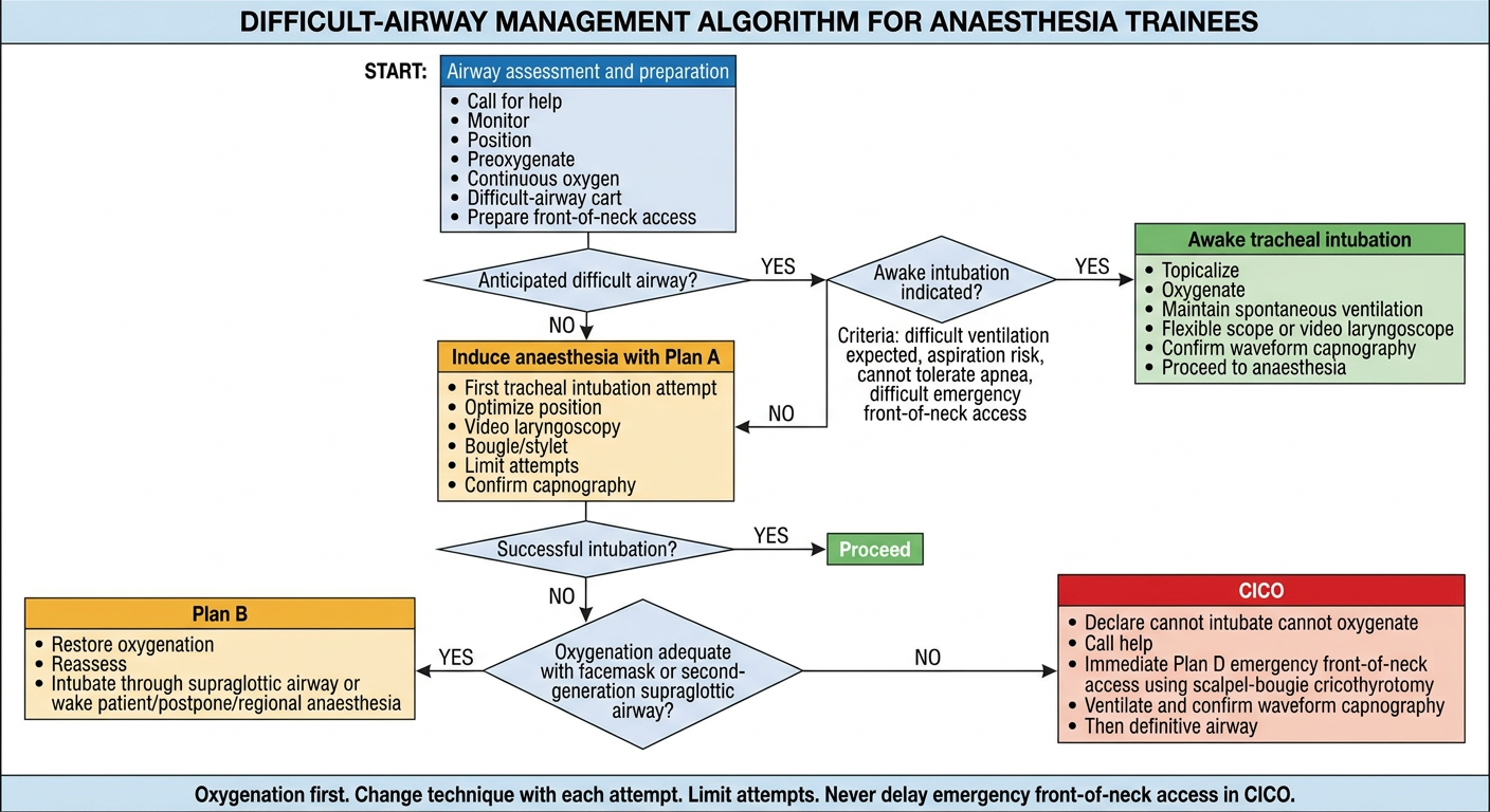
Difficult-Airway Algorithm: Exam Flowchart
                         AIRWAY ASSESSMENT
                                  │
                                  ▼
      Predict difficulty with mask ventilation, SAD, intubation,
              aspiration, apnoea tolerance and FONA
                                  │
                                  ▼
       PREPARE: call for help + monitoring + difficult-airway cart
       suction + ramped position + preoxygenation + nasal oxygen
                    identify/mark cricothyroid membrane
                                  │
                                  ▼
             Is a difficult airway anticipated?
                    ┌─────────────┴─────────────┐
                    │                           │
                   YES                          NO
                    │                           │
                    ▼                           ▼
      Is AWAKE intubation indicated?       Induce anaesthesia
                    │                      with pre-planned Plan A
      ┌─────────────┼─────────────┐                 │
      │             │             │                 ▼
 Difficult        High       Cannot tolerate     First intubation
 ventilation   aspiration        apnoea           attempt: optimize
 expected        risk         or difficult       position, VL/DL,
                             FONA anticipated     bougie/stylet
      │                                             │
      ▼                                             ▼
 AWAKE TRACHEAL INTUBATION                  Is intubation successful?
 Topical anaesthesia, oxygen,                       │
 spontaneous ventilation,                    ┌──────┴──────┐
 flexible scope / videolaryngoscope          │             │
      │                                      YES            NO
      ▼                                       │             │
 Confirm waveform capnography                 ▼             ▼
      │                                 Proceed with    Can oxygenation
      ▼                                  anaesthesia    be maintained by
 Induce anaesthesia                                        mask or SAD?
                                                            │
                                                     ┌──────┴──────┐
                                                     │             │
                                                    YES            NO
                                                     │             │
                                                     ▼             ▼
                                           PLAN B: Restore     CICO
                                           oxygenation with    Declare:
                                           second-generation   "Cannot intubate,
                                           SAD or facemask     cannot oxygenate"
                                                     │             │
                                                     ▼             ▼
                                            Reassess options:   PLAN D:
                                            • Intubate through  Immediate emergency
                                              SAD with scope    FONA
                                            • Alternative       Scalpel-bougie
                                              intubation method cricothyrotomy
                                            • Wake patient      │
                                            • Postpone /        ▼
                                              regional technique Ventilate and confirm
                                                                 waveform capnography
Essential rules
- Oxygenate continuously throughout.
- Confirm ventilation, SAD placement and tracheal intubation using continuous waveform capnography.
- Use the most suitable first technique, often video laryngoscopy in predicted difficulty.
- Limit attempts and alter device, operator or technique with each subsequent attempt.
- If ventilation is adequate but intubation fails, consider waking the patient rather than persisting.
- In CICO, do not delay emergency front-of-neck access.