
Astrocytoma
astrocytoma brain tumor histology MRI
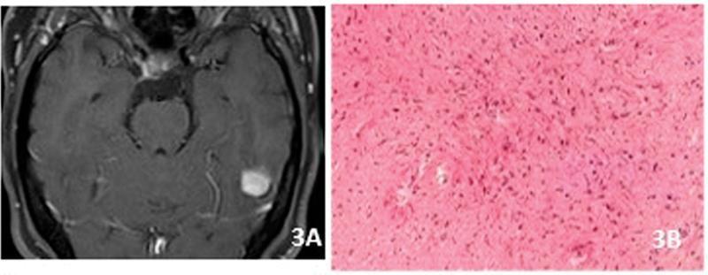
This composite educational figure illustrates the neuroimaging and histopathological features of a pilocytic astrocytoma (WHO Grade 1). Panel A displays an axial T1-weighted post-gadolinium MRI of the brain, showing a discrete, well-circumscribed, intensely enhancing nodular lesion located in the temporal lobe. Notably, there is an absence of perilesional vasogenic edema or significant mass effect on the adjacent parenchyma. Panel B provides a corresponding high-magnification light microscopy view of the tumor using Hematoxylin and Eosin (H&E) staining. The histology demonstrates a classic biphasic pattern characterized by 'piloid' cells—astrocytes with elongated, hair-like bipolar processes. Diagnostic hallmarks visible include Rosenthal fibers, which appear as brightly eosinophilic, worm-like proteinaceous aggregates, and a background of fibrillary matrix. These visual findings correlate with the slow-growing, benign nature of this pediatric-type diffuse low-grade glioma, frequently associated with MAP kinase pathway alterations.
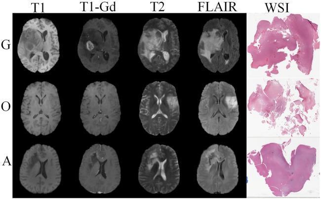
This medical comparison chart displays axial brain MRI sequences and corresponding histopathological whole slide images (WSI) for three glioma subtypes: Glioblastoma (G), Oligodendroglioma (O), and Astrocytoma (A). The MRI modalities include T1-weighted, T1-gadolinium contrasted (T1-Gd), T2-weighted, and Fluid-Attenuated Inversion Recovery (FLAIR). The Glioblastoma row shows a large, heterogeneous mass with significant peripheral enhancement on T1-Gd and extensive hyperintense peritumoral edema on T2 and FLAIR images. The Oligodendroglioma and Astrocytoma rows demonstrate smaller, less infiltrative masses with varying signal intensities. The final column displays Hematoxylin and Eosin (H&E) stained WSI biopsy sections, illustrating the histological differences in tissue architecture and cellularity between the subtypes, such as the more fragmented appearance of the oligodendroglioma sample compared to the more cohesive astrocytoma section. This visual resource is intended for neuroradiology and pathology education, emphasizing the integration of multi-modal imaging and histology for definitive brain tumor classification.
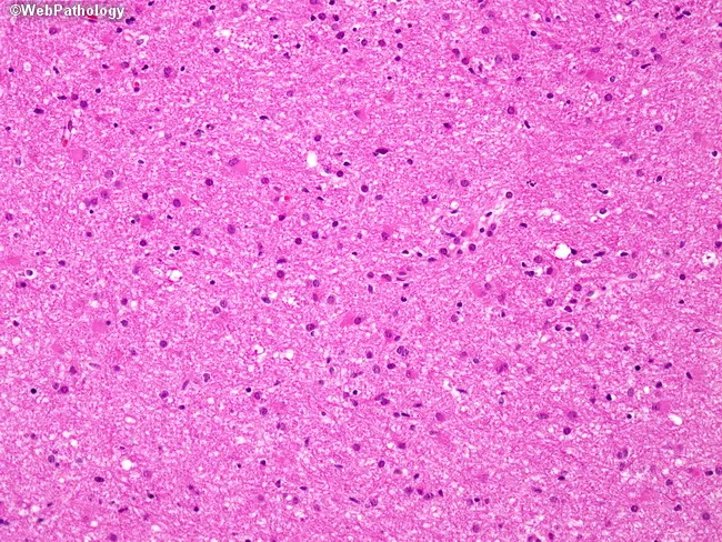
Imaging modality: light microscopy of a hematoxylin and eosin stained brain tissue section from a cerebral hemisphere tumor. Primary subject: diffuse astrocytoma, WHO Grade II (infiltrating astrocytoma). Specimen: brain parenchyma/tumor tissue. Imaging perspective: microscopic histology field. Anatomical context: central nervous system, brain (cerebral hemispheres), intra-axial glial neoplasm displaying diffuse infiltration. Visual features: moderately increased cellularity with mildly enlarged, variably hyperchromatic nuclei; mild nuclear pleomorphism; delicate, interconnected astrocytic processes creating a fibrillary background; abundant eosinophilic cytoplasm in some astrocyte-like cells; absence of overt necrosis or microvascular proliferation in this field; mitotic activity not evident at this magnification. Pathology: low-grade, infiltrative glioma consistent with WHO Grade II astrocytoma; preserves overall brain architecture; growth pattern extraparenchymal infiltration challenging complete resection; lacks features defining higher grades (no brisk mitoses, microvascular proliferation, or necrosis). Diagnostic significance: confirms low-grade astrocytic neoplasm; implications for prognosis and management; potential progression to higher-grade gliomas (III–IV) with time or treatment resistance; imaging correlation with MRI typically demonstrates non-destructive, infiltrative lesion; informs surgical planning, adjuvant radiotherapy/chemotherapy decisions; differential considerations include oligodendroglioma and pilocytic astrocytoma in specific contexts. Clinical relevance: presents with seizures/ focal deficits in adults (4th–6th decades), guiding neuro-oncologic workup and longitudinal surveillance. This image exemplifies low-grade glial tumor histology.
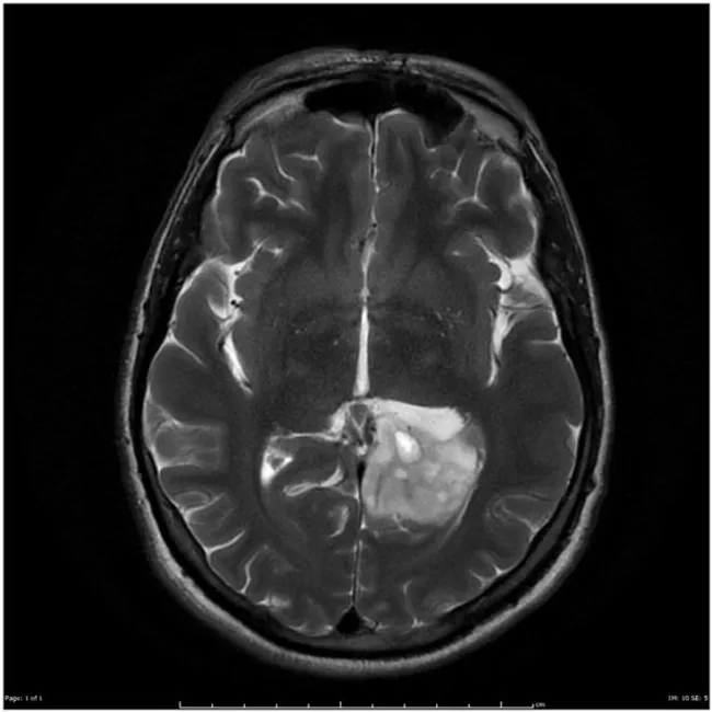
Magnetic Resonance Imaging (MRI) of the brain, axial plane, with gadolinium contrast, including post‑contrast T1-weighted sequences and accompanying T2/FLAIR imaging. A left occipito‑temporal intra-axial mass is evident, showing irregular, ill‑defined enhancement after contrast with surrounding vasogenic edema and mild mass effect on adjacent cortex and sulci. The radiographic phenotype is most compatible with an infiltrative glial neoplasm of intermediate to high grade, with differential diagnosis including anaplastic astrocytoma versus glioblastoma. Perfusion or spectroscopy data (not shown) would further aid grading and treatment planning. The patient underwent surgical resection. Histology demonstrated astrocytic tumor cells with increased cellularity, nuclear pleomorphism, and brisk mitotic activity, without definitive necrosis, consistent with anaplastic astrocytoma (WHO Grade III). Location in the left occipito‑temporal region correlates with potential language and visual processing effects and explains seizure propensity. Imaging features—irregular enhancement, infiltrative margins, edema, and no definite necrosis—support high‑grade designation, though overlap with Grade IV lesions exists. Clinically, these tumors are managed with maximal safe resection followed by radiotherapy with concomitant and adjuvant temozolomide; MGMT status may influence prognosis. This case exemplifies imaging‑pathology correlation in adult supratentorial gliomas and informs differential diagnosis, surgical planning, and oncologic management discussions for neuroradiology, neuro-oncology, and neurosurgical trainees. Case courtesy of Radiopaedia, Dr. Frank Gaillard.
"astrocytoma"[MeSH Terms] AND "classification"[MeSH Terms]
Astrocytoma
1. Classification (WHO 2021)
| WHO Grade | Type | Key Molecular Feature |
|---|---|---|
| 1 | Pilocytic astrocytoma | BRAF fusion (KIAA1549-BRAF); IDH wild-type |
| 2 | Astrocytoma, IDH-mutant | IDH1/IDH2 mutation + TP53 + ATRX |
| 3 | Astrocytoma, IDH-mutant (anaplastic) | + mitoses; higher cellularity |
| 4 | Astrocytoma, IDH-mutant, Grade 4 | + microvascular proliferation/necrosis OR CDKN2A/B homozygous deletion |
- IDH-mutant astrocytomas (grades 2-4) are classified under "Adult-type diffuse gliomas" along with oligodendroglioma and IDH-wild-type glioblastoma
- Pilocytic astrocytoma (grade 1) is a circumscribed astrocytic tumor, classified separately
- Grade 1 is not used for diffuse gliomas by convention, as all diffuse gliomas are considered malignant
- Homozygous deletion of CDKN2A/CDKN2B automatically assigns Grade 4, even if histology suggests lower grade
2. Epidemiology
- Pilocytic astrocytoma: Children and young adults; most common cerebellar tumor in children
- IDH-mutant diffuse astrocytoma: Peak incidence in the 4th-6th decades
- Spinal astrocytoma: Peak in 3rd-4th decade; slight male predominance (55%); most common intramedullary tumor in children (up to 90%)
- Most common spinal site: thoracic cord (~70%), followed by cervical cord
3. Pathogenesis
IDH-Mutant Astrocytomas
- Driver mutation: IDH1 (most common) or IDH2 - leads to production of 2-hydroxyglutarate, an oncometabolite that inhibits epigenetic regulators
- Co-mutations: TP53 (tumor suppressor) and ATRX (chromatin remodeling) - both frequently inactivated
- Arise in deep white matter, infiltrate extensively; poorly defined borders
- Grades 2-3: no necrosis; Grade 4: + microvascular proliferation and/or necrosis
Pilocytic Astrocytoma
- BRAF activation: KIAA1549-BRAF fusion in 80-90% (especially cerebellar tumors); activates the MAPK/ERK pathway
- No IDH1/IDH2 mutations
- Often has a cyst + mural nodule architecture
4. Clinical Features
Presentation
- Seizures: First symptom in ~2/3 of patients with cerebral astrocytoma; 60-75% have recurrent seizures during illness
- Headache: Relatively late, due to raised ICP
- Focal neurologic deficits: Depend on location
- Children (cerebellar): Gait unsteadiness, unilateral ataxia, raised ICP (headache, vomiting)
- Low-grade tumors can be clinically silent for years before symptoms emerge
5. Pathology / Morphology
Grade 2 IDH-Mutant
- Macroscopically: poorly defined, gray, infiltrative mass - expands brain without forming a discrete mass
- Microscopically: mild-moderate increase in glial nuclei, nuclear pleomorphism, GFAP-positive fibrillary background; tumor cells found many cm from the main lesion
- No necrosis or mitoses
Grade 3 (Anaplastic)
- More densely cellular, greater nuclear pleomorphism, mitotic figures present
Grade 4 IDH-Mutant
- Greater cellularity, cytological atypia, high proliferative activity
- Microvascular proliferation and/or necrosis (unlike IDH-wild-type glioblastoma, large central necrosis is usually absent)
Pilocytic Astrocytoma
- Bipolar cells with long, thin "hair-like" (piloid) processes - GFAP positive
- Rosenthal fibers (eosinophilic, worm-like protein aggregates) - classic hallmark
- Eosinophilic granular bodies, microcysts
- Mitoses and necrosis are rare
6. Imaging
MRI Features (Cerebral)
| Sequence | Low-Grade | High-Grade |
|---|---|---|
| T1 | Isointense or hypointense | Heterogeneous, hypointense |
| T2/FLAIR | Hyperintense | Hyperintense + edema |
| Gadolinium | Minimal/variable enhancement | Irregular enhancement (Grade 3-4) |
- Low-grade: homogenous T1 hypointense, T2 hyperintense infiltrating mass; poorly defined borders; little/no contrast enhancement
- High-grade: irregular enhancement, vasogenic edema, mass effect
- Cysts and calcification common, especially in cerebellar tumors
- Pilocytic astrocytoma: intensely enhancing mural nodule in a cyst
Spinal MRI
- Solid component: T1 isointense/hypointense, T2 hyperintense
- Cystic/necrotic component: T1 hypointense, strongly T2 hyperintense
- Low-grade fibrillary astrocytomas: often no enhancement
- Associated syringomyelia may occur; borders of syrinx cavity do not enhance
7. Treatment
Surgery
- First-line: Maximal safe surgical resection for all grades
- Modern brain mapping (language and motor mapping intraoperatively) allows larger, safer resections
- Pilocytic astrocytoma: complete resection is often curative
- In spinal astrocytomas: typically only biopsy is feasible (infiltrative, poorly defined margins)
- Repeated operations prolong survival in some patients
Radiotherapy
- Conformal radiation therapy (~5,300 cGy, focused on tumor) is standard post-operatively for higher-grade tumors
- For low-grade in younger patients: radiation may be deferred (serial imaging surveillance); delaying avoids dementia and hypopituitarism
- A randomized trial showed early RT extended progression-free survival (5.3 vs 3.4 years) but did not improve overall survival (~7 years both groups)
Chemotherapy
- Temozolomide (TMZ): Has replaced PCV as the standard agent in most centers
- PCV (procarbazine + CCNU + vincristine): One randomized trial showed benefit when added to RT (13.8 vs 7.8 years median survival - Buckner et al)
- Tumors with oligodendroglial components respond particularly well to chemotherapy
- MGMT methylation status influences response to TMZ
Novel/Emerging
- BRAF inhibitors (e.g., dabrafenib): For pilocytic/BRAF-altered astrocytomas - targeting the MAPK pathway
- IDH inhibitors: Under investigation for IDH-mutant gliomas
- Oncolytic viruses: Direct intratumoral injection; promising responses in preliminary data; awaiting controlled trials
8. Prognosis
| Grade | Median Survival |
|---|---|
| Grade 2 (IDH-mutant) | >10 years |
| Grade 3 (IDH-mutant) | ~3-5 years |
| Grade 4 (IDH-mutant) | <2 years (better than IDH-wt GBM) |
| Pilocytic (Grade 1) | Near-normal lifespan after complete resection |
- Low-grade astrocytomas can be static for several years, then progress
- Eventual malignant transformation is part of the natural history
- 10-year survival for supratentorial low-grade gliomas: 11-40% with surgery + conformal RT (Shaw et al.)
- KIAA1549-BRAF fusion in cerebellar pilocytic astrocytoma may be associated with better overall survival
- Spinal low-grade: 5-year survival >75%; high-grade spinal glioma carries poor prognosis
9. Key Biomarkers Summary
| Marker | Significance |
|---|---|
| IDH1/IDH2 mutation | Better prognosis vs IDH-wild-type; defines astrocytoma subtype |
| ATRX loss | Supports astrocytoma (not oligodendroglioma) |
| TP53 mutation | Co-mutation in diffuse astrocytomas |
| CDKN2A/B deletion | Auto-assigns Grade 4 regardless of histology |
| BRAF fusion | Pilocytic astrocytoma; target for MAPK inhibitors |
| MGMT methylation | Predicts response to temozolomide |
| 1p/19q codeletion | ABSENT in astrocytoma (present in oligodendroglioma - helps differentiate) |
| GFAP | Positive in all astrocytic tumors (IHC marker) |
- Adams and Victor's Principles of Neurology, 12th Ed, pp. 663-664
- Robbins & Kumar Basic Pathology (Robbins Pathology), pp. 857-860
- Grainger & Allison's Diagnostic Radiology, p. 1265
- Bradley and Daroff's Neurology in Clinical Practice