
Cor pulmonale
"cor pulmonale" AND management
cor pulmonale right heart failure ECG chest X-ray
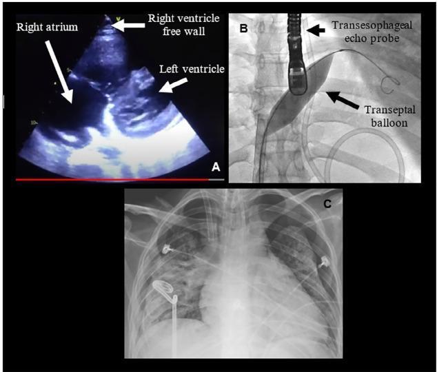
This composite clinical figure illustrates the diagnosis and management of severe pulmonary hypertension and its complications. Panel A shows a transthoracic echocardiogram (TTE) in a patient with acute cor pulmonale. Labeled structures include a dilated right atrium and left ventricle, with an arrow highlighting the right ventricle free wall, demonstrating significant right heart strain. Panel B is a fluoroscopic image capturing an atrioseptostomy procedure. It depicts a transesophageal echo (TEE) probe positioned in the esophagus and a transeptal balloon inflated across the interatrial septum to create a right-to-left shunt. Panel C is an anteroposterior thoracic X-ray demonstrating bilateral pneumothorax with limited lung expansion. Visible medical devices include bilateral thoracic drainage tubes (chest tubes), an endotracheal tube, and ECG leads. The image set provides a comprehensive visual summary of hemodynamic monitoring, interventional rescue maneuvers for right ventricular failure, and subsequent pulmonary complications in a critical care setting.
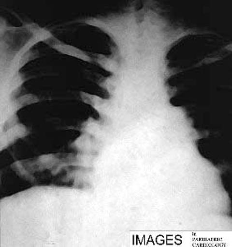
**Imaging Modality:** Frontal Chest X-ray (CXR). **Anatomical Region:** Thoracic cavity, focusing on the cardiomediastinal silhouette and pulmonary vasculature. **Observed Pathology:** Findings are consistent with cor pulmonale secondary to severe pulmonary hypertension. **Characteristic Visual Features:** * **Cardiac Morphology:** Pronounced cardiomegaly with a "boot-shaped" appearance, characterized by an upturned apex and a prominent right heart border, indicating right ventricular and right atrial hypertrophy. * **Vascular Markings:** Significant prominence and dilation of the central pulmonary arteries (hilar engorgement). There is a marked discrepancy between the enlarged central vessels and the "pruning" or attenuation of peripheral pulmonary vascular markings. * **Mediastinum:** Expansion of the transverse cardiac diameter and a bulging pulmonary trunk segment along the left heart border. * **Lung Fields:** Increased lucency in the peripheral lung zones due to decreased perfusion (oligemia). **Diagnostic Cues:** The combination of central pulmonary artery dilation and right-sided heart enlargement without signs of left-sided failure (e.g., absence of Kerley B lines or pleural effusions) is highly suggestive of pulmonary arterial hypertension leading to chronic cor pulmonale.
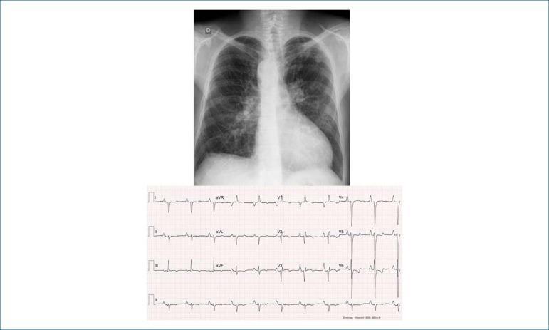
This composite educational image includes a posteroanterior (PA) chest radiograph and a 12-lead electrocardiogram (ECG) demonstrating signs of congenital heart disease and hemodynamic stress. The chest X-ray reveals a moderately enlarged cardiac silhouette with an elongated, rounded apex and an excavated middle arch, suggesting ventricular dominance. A notable anatomical variation is the right-sided aortic arch. The pulmonary vasculature appears prominent, particularly in the hila and lower lung fields, which may indicate systemic-pulmonary collateral circulation. The accompanying ECG shows a sinus rhythm with markers of right-sided heart strain and cavity overload. Key findings include peaked P waves (P-pulmonale) in leads I, II, and left precordial leads, and a significant right-axis deviation of the QRS complex (+180º) with deep S waves from V3 through V6. T-wave inversions in the precordial leads further support right ventricular strain. This combination of findings is characteristic of complex cyanotic congenital heart disease, such as Tetralogy of Fallot or pulmonary atresia with a right-sided aortic arch.
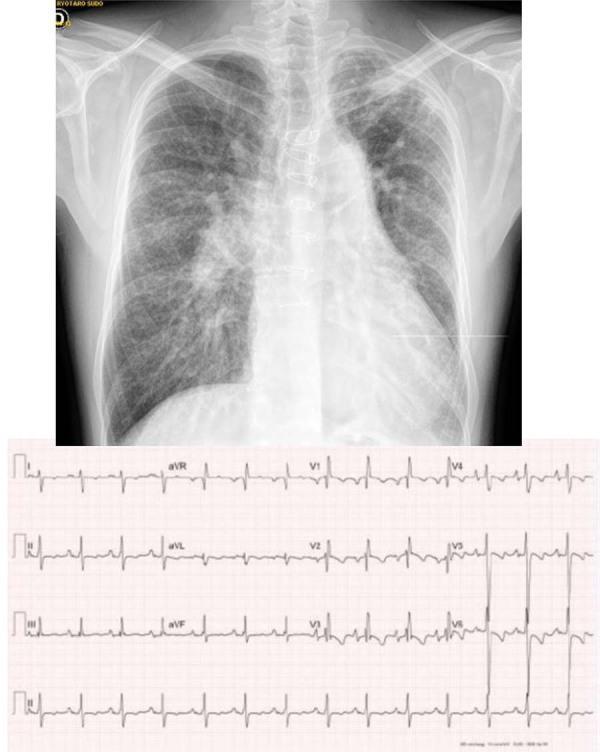
Educational composite containing a posteroanterior chest X-ray and a 12-lead electrocardiogram (ECG) demonstrating complex congenital heart disease. The chest radiograph shows cardiomegaly with a prominent left ventricular contour and a bulging middle arch. A striking pulmonary vascular asymmetry is visible: the right lung shows increased hilar vascularity with peripheral tapering suggestive of pulmonary arterial hypertension, while the left lung exhibits significantly reduced vascular markings with thin, sparse vessels indicating pulmonary hypoperfusion. Below, the 12-lead ECG shows sinus rhythm with right axis deviation (+110 degrees) and signs of right cavity overload, including apiculate P waves (P-pulmonale) and a predominant S-wave pattern in leads V4-V6. Diffuse T-wave inversions are present in the precordial leads, reflecting generalized ventricular repolarization changes. This combination is clinically relevant for evaluating complex cyanotic heart defects, such as double outlet right ventricle with asymmetric pulmonary flow or pulmonary atresia.
Cor Pulmonale
Source: Robbins & Kumar Basic Pathology, p. 363
Pathogenesis Overview
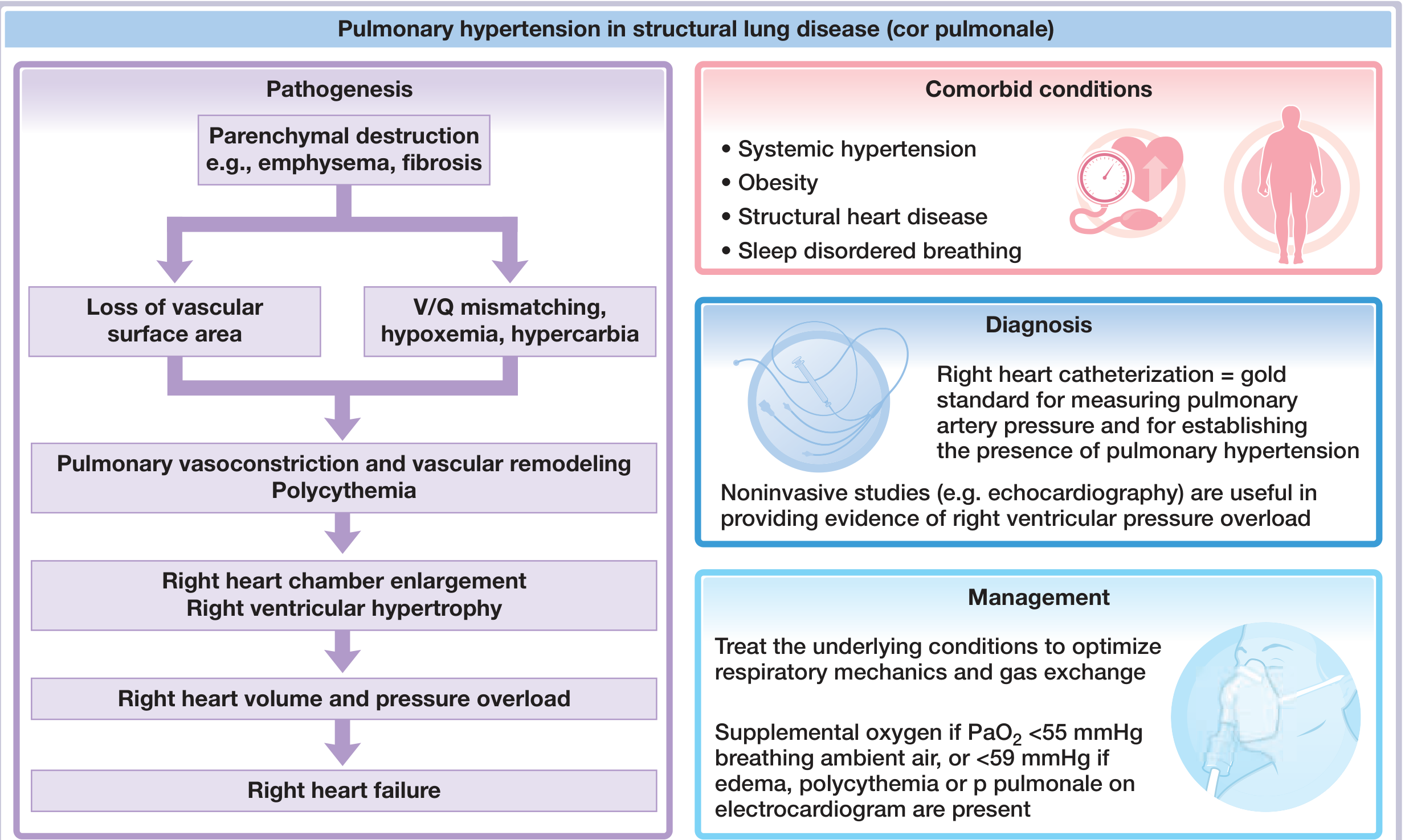
- Parenchymal destruction (emphysema, fibrosis) → loss of pulmonary vascular surface area
- V/Q mismatch → hypoxemia ± hypercarbia
- Hypoxic pulmonary vasoconstriction + vascular remodeling
- Polycythemia (hypoxia-driven erythropoiesis) → increased blood viscosity → raised pulmonary vascular resistance
- Elevated pulmonary artery pressure → RV pressure and volume overload
- RV hypertrophy → eventually right heart failure
Etiology / Causes
| Category | Examples |
|---|---|
| Obstructive lung disease | COPD (most common), asthma |
| Restrictive / parenchymal | Pulmonary fibrosis, sarcoidosis, pneumoconiosis |
| Vascular | Chronic thromboembolic disease, primary PAH |
| Chest wall / neuromuscular | Kyphoscoliosis, obesity hypoventilation, obstructive sleep apnea |
| Acute | Massive pulmonary embolism |
Acute vs. Chronic
| Feature | Acute Cor Pulmonale | Chronic Cor Pulmonale |
|---|---|---|
| Onset | Sudden (e.g., massive PE) | Slow and insidious |
| RV pathology | Dilation only; may even be normal size if sudden death | RV hypertrophy + dilation; RV wall may equal LV thickness |
| Cardiac output | Reduced | Normal or increased until late stages |
| Reversibility | Often reversible | Progressive; failure ensues late |
Robbins & Kumar Basic Pathology, p. 363
Morphology (Gross Pathology)
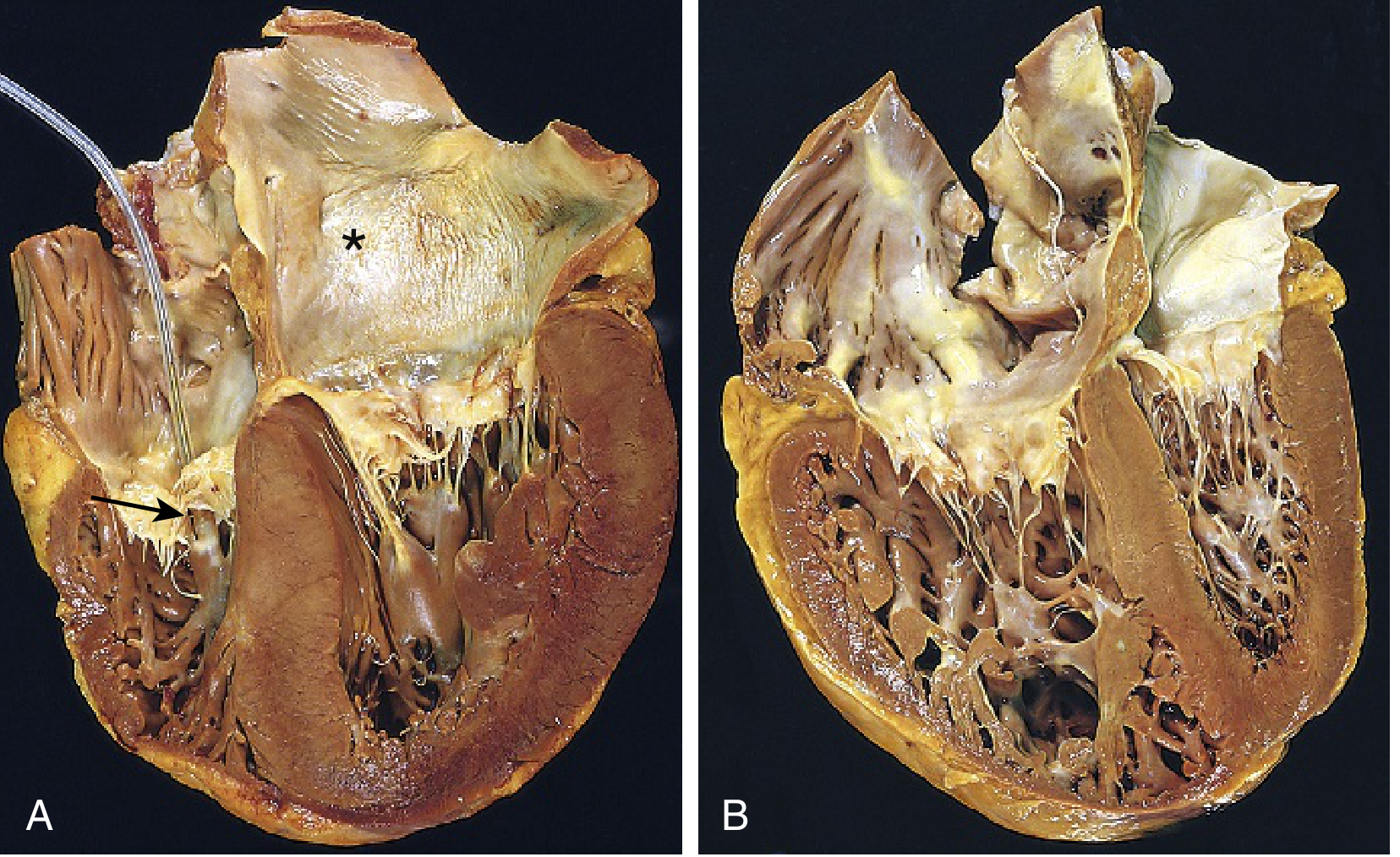
Pathophysiology of Edema
- Reduced renal plasma flow → impaired urinary sodium excretion
- Hypercapnia → kidneys compensate for respiratory acidosis by retaining HCO3- and excreting H+, causing secondary sodium and water retention
- Impaired oxygen tissue delivery → altered renal (and systemic) cellular metabolism → salt and water dysregulation
- Intracellular acidosis from buffering chronic respiratory acidosis → augments organ dysfunction
Fuster and Hurst's The Heart, 15th Edition, p. 1775
Right Ventricular Response
- Early/compensated: RV hypertrophies to handle increased afterload; cardiac output is maintained or elevated due to increased intravascular volume stretching myocardial fibers
- "Pink puffer" (emphysema phenotype): Often has lower RV end-diastolic volumes due to lung hyperinflation reducing venous return
- "Blue bloater" (chronic airways disease with hypoxemia/hypercapnia): More likely to develop overt pulmonary vascular disease and cor pulmonale
- Late/decompensated: RV stiffness, ventriculo-arterial uncoupling, and RV dyssynchrony dominate; ventricular interdependence then impairs LV systolic and diastolic function as well
Fuster and Hurst's The Heart, 15th Edition, p. 1775-1776
Clinical Manifestations
- Symptoms: Dyspnea on exertion (most common), fatigue, syncope or near-syncope on exertion, ankle swelling
- Signs: Elevated JVP, right ventricular heave (parasternal lift), loud P2, tricuspid regurgitation murmur (pansystolic at left sternal border), hepatomegaly, peripheral pitting edema, cyanosis
- Signs of the underlying lung disease dominate the clinical picture (barrel chest, wheeze, crepitations)
Investigations
Chest X-ray (CXR)
- Cardiomegaly with right-sided predominance ("boot-shaped" heart)
- Prominent central pulmonary arteries (hilar engorgement)
- Pruning of peripheral pulmonary vascular markings
- Absence of pulmonary venous congestion (no Kerley B lines), distinguishing it from LV failure
ECG
- P pulmonale - peaked P waves >2.5 mm in lead II (tall, narrow, due to right atrial enlargement)
- Right axis deviation (QRS axis >+90°)
- Right ventricular hypertrophy - dominant R in V1, deep S in V5-V6, R:S ratio >1 in V1
- Right bundle branch block (complete or incomplete)
- S1Q3T3 pattern in acute cor pulmonale (massive PE)
- ST depression and T wave inversion in V1-V4 (RV strain)
Echocardiography
- RV dilation and hypertrophy
- Flattening/paradoxical motion of the interventricular septum ("D-shaped" LV in short axis)
- Tricuspid regurgitation - allows estimation of RV systolic pressure (RVSP) via Bernoulli equation
- Reduced RV ejection fraction; impaired RV function indices (TAPSE, S')
- Right heart catheterization is the gold standard for measuring pulmonary artery pressure (mPAP ≥25 mmHg defines pulmonary hypertension)
Other
- ABG: Hypoxemia (PaO2 low), hypercapnia in advanced disease
- CBC: Polycythemia (secondary erythrocytosis)
- PFTs: Confirm underlying obstructive or restrictive lung disease
- CT PA / V/Q scan: Rule out thromboembolic disease
- NT-proBNP / BNP: Elevated; useful for severity assessment and prognosis
Management
1. Oxygen Therapy
- Indicated if resting PaO2 <55 mmHg on ambient air
- Or PaO2 <59 mmHg if edema, polycythemia, or P pulmonale on ECG are also present
- Target: maintain SpO2 ≥90%, SaO2 ≥90%
- Goal: reverse hypoxic vasoconstriction and reduce pulmonary artery pressure
2. Treat the Underlying Cause
- COPD: Bronchodilators, inhaled corticosteroids, pulmonary rehabilitation, smoking cessation
- OSA: CPAP/BiPAP
- Pulmonary fibrosis: Antifibrotics (pirfenidone, nintedanib)
- Chronic PE: Anticoagulation; pulmonary thromboendarterectomy if eligible
- Acute cor pulmonale (PE): Anticoagulation, thrombolysis, or embolectomy
3. Diuretics
- Reduce fluid overload and peripheral edema
- Use cautiously - aggressive diuresis can reduce RV preload and worsen output
- Spironolactone may be added
4. Pulmonary Vasodilators
- Role is limited and controversial in cor pulmonale due to lung disease (Group 3 PH)
- Unlike Group 1 PAH, pulmonary vasodilators may worsen V/Q mismatch and systemic hypoxemia
- Reserved for patients with disproportionate pulmonary hypertension (i.e., out of proportion to the degree of lung disease) - specialist decision
- Options in PAH (Group 1): phosphodiesterase-5 inhibitors (sildenafil), endothelin receptor antagonists (bosentan), prostacyclins
5. Phlebotomy
- For symptomatic polycythemia (hematocrit >65%)
- Reduces blood viscosity and pulmonary vascular resistance
6. Cardiac Transplantation / Lung Transplantation
- For end-stage disease refractory to medical therapy
- Lung transplant can normalize RV function
Prognosis
Key Distinguishing Points
| Feature | Cor Pulmonale | LV Failure |
|---|---|---|
| Primary pathology | Lung disease | LV disease |
| Cardiac output | Normal/elevated (until late) | Reduced |
| Edema mechanism | Renal Na+ retention, hypercapnia | Reduced CO, RAAS activation |
| CXR | Dilated RV/RA, central PA dilation | Cardiomegaly, pulmonary venous congestion, Kerley B lines |
| S3 gallop | Right-sided (left sternal border, increases with inspiration) | Left-sided (apex) |
| Treatment target | Improve oxygenation and lung mechanics | Diuretics, ACEi, beta-blockers, SGLT2i |
- Robbins & Kumar Basic Pathology, p. 363
- Fuster and Hurst's The Heart, 15th Edition, pp. 1773-1778 (Chapter 59, Lewis J. Rubin)
- Fishman's Pulmonary Diseases and Disorders, 2-Volume Set