
Hypersensitivity rection
Reading File
Now let me search the medical library for hypersensitivity reactions comprehensively.
Finding Sources
Finding Sources
Excellent results. Let me read the most authoritative sources - Robbins Pathology and Cellular Molecular Immunology sections on hypersensitivity.
Reading File
Reading File
Great content retrieved. Let me now read more details on Type I (immediate) and Type IV hypersensitivity from Robbins, plus look for clinical examples and images.
Reading File
Finding Sources
Let me get more detail on each type and fetch the key images.
Reading File
Loading Image
Excellent! Now let me get the Type II, III, and IV details plus key images.
Reading File
Reading File
Loading Image
Now let me get the Type IV section and its mechanism image.
Reading File
Reading File
Loading Image
I now have all the essential content for a comprehensive, well-illustrated answer. Let me compile it.
Hypersensitivity Reactions
Hypersensitivity reactions are pathological immune responses directed against antigens that cause tissue injury. The classic Gell and Coombs (1963) classification divides them into four types based on the underlying immunologic mechanism.
Overview Table
| Type | Name | Mediator | Onset | Prototypic Disorders |
|---|---|---|---|---|
| I | Immediate / Anaphylactic | IgE + mast cells | Minutes | Anaphylaxis, asthma, hay fever, urticaria |
| II | Antibody-mediated (cytotoxic) | IgG / IgM vs. cell surface | Hours | Autoimmune hemolytic anemia, Goodpasture syndrome |
| III | Immune complex-mediated | IgG/IgM complexes deposited in tissues | Hours-days | SLE, serum sickness, post-streptococcal GN |
| IV | Cell-mediated (delayed) | T lymphocytes (CD4+/CD8+) | 48-72 h | Contact dermatitis, TB, type 1 diabetes, MS |
Type I - Immediate (IgE-mediated) Hypersensitivity
Mechanism: A previously sensitized individual is re-exposed to an allergen. The allergen cross-links IgE antibodies already bound to Fc receptors on mast cells, triggering degranulation and release of:
- Preformed mediators - histamine, heparin, tryptase, chymase (released within seconds)
- Newly synthesized mediators - leukotrienes (LTC4, LTD4), prostaglandins, platelet-activating factor
- Cytokines - IL-4, IL-5, IL-13, TNF (drive the late-phase reaction)
Two phases:
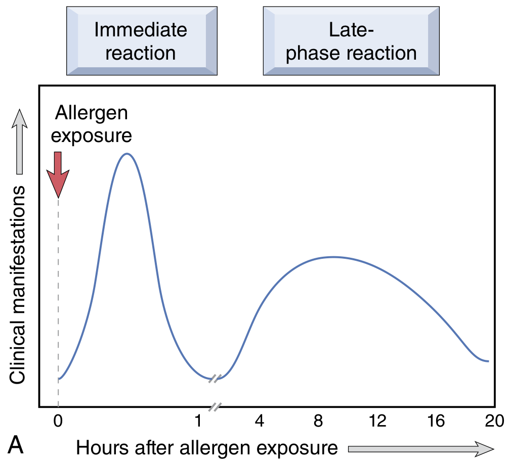
- Immediate phase (within minutes): vasodilation, vascular leakage, smooth muscle spasm, glandular secretion
- Late phase (2-24 hours later): tissue infiltration with eosinophils, neutrophils, basophils, and CD4+ T cells; mucosal epithelial damage. This phase is NOT blocked by antihistamines - requires steroids.
Clinical forms:
| Portal of Entry | Manifestation |
|---|---|
| Systemic (IV/bee sting) | Anaphylaxis - vascular shock, bronchospasm, widespread edema |
| Skin | Urticaria (hives), atopic dermatitis |
| Airways | Allergic rhinitis (hay fever), asthma |
| GI tract | Food allergy, gastroenteritis |
Atopy: Genetic predisposition to develop type I reactions. Atopic individuals have elevated serum IgE and excess IL-4-producing Th2 cells. Atopic triad = atopic dermatitis + allergic rhinitis + asthma.
Hygiene hypothesis: Improved early childhood hygiene in developed countries reduces antigenic stimulation, shifting the immune response toward Th2, thus paradoxically increasing atopic disease.
Type II - Antibody-Mediated (Cytotoxic) Hypersensitivity
Mechanism: IgG or IgM antibodies bind to antigens on cell surfaces or extracellular matrix and cause injury by three mechanisms:

- Opsonization and phagocytosis - Cells coated with IgG or complement (C3b) are recognized by Fc/C3b receptors on macrophages and destroyed. Complement activation can also form the MAC (membrane attack complex) causing direct lysis.
- Complement- and Fc receptor-mediated inflammation - Antibodies deposited on basement membranes activate complement (generating C5a, C3a), recruiting neutrophils and monocytes that release lysosomal enzymes and ROS.
- Antibody-mediated cellular dysfunction - Antibodies block or stimulate receptors without direct cell killing (e.g., anti-TSH receptor antibodies in Graves disease stimulate thyroid hormone production; anti-AChR antibodies in myasthenia gravis block neuromuscular transmission).
Examples:
| Disease | Antigen | Mechanism |
|---|---|---|
| Autoimmune hemolytic anemia | RBC surface antigens | Opsonization/lysis |
| Goodpasture syndrome | Glomerular/alveolar basement membrane (type IV collagen) | Complement-mediated inflammation |
| Graves disease | TSH receptor | Receptor stimulation (no lysis) |
| Myasthenia gravis | Acetylcholine receptor | Receptor blockade |
| Hemolytic transfusion reaction | ABO blood group antigens | Opsonization/complement lysis |
| Erythroblastosis fetalis | Rh antigen | Maternal IgG crosses placenta, destroys fetal RBCs |
Type III - Immune Complex-Mediated Hypersensitivity
Mechanism: Soluble antigen-antibody (IgG/IgM) complexes form in the circulation and deposit in blood vessel walls and tissues. Pathogenicity depends on complex size - intermediate-sized complexes (formed under slight antigen excess) are the most harmful.
Sequence of events:
- Antigen-antibody complex formation in the circulation
- Deposition in vessel walls, glomeruli, joints, and skin (organs filtering blood at high pressure)
- Inflammation and injury - complement activation releases C3a, C5a (chemotaxins); neutrophils and monocytes are recruited and release lysosomal enzymes and ROS; fibrinoid necrosis of vessel walls results
Morphology: Acute vasculitis with neutrophilic infiltration and fibrinoid necrosis. On immunofluorescence: granular deposits of Ig and complement along glomerular basement membrane (contrast with Type II, which shows linear deposits).
Key disease examples:
| Disease | Antigen |
|---|---|
| Systemic lupus erythematosus | Nuclear antigens (DNA, histones) |
| Poststreptococcal glomerulonephritis | Streptococcal cell wall antigens |
| Serum sickness | Foreign serum proteins |
| Arthus reaction (local) | Foreign proteins (experimental model) |
| Polyarteritis nodosa | Hepatitis B surface antigen (some cases) |
Serum sickness is the classic model - occurring 10 days after antigen injection with fever, urticaria, joint pain, lymphadenopathy, and proteinuria. Complement levels fall (consumed by complexes) and can be used to monitor disease activity.
Type IV - Cell-Mediated (Delayed-Type) Hypersensitivity
Mechanism: Unlike types I-III, this type is antibody-independent; it is mediated by sensitized T lymphocytes. It is called "delayed" because the reaction takes 48-72 hours to develop (compared to minutes-hours for antibody-mediated types).
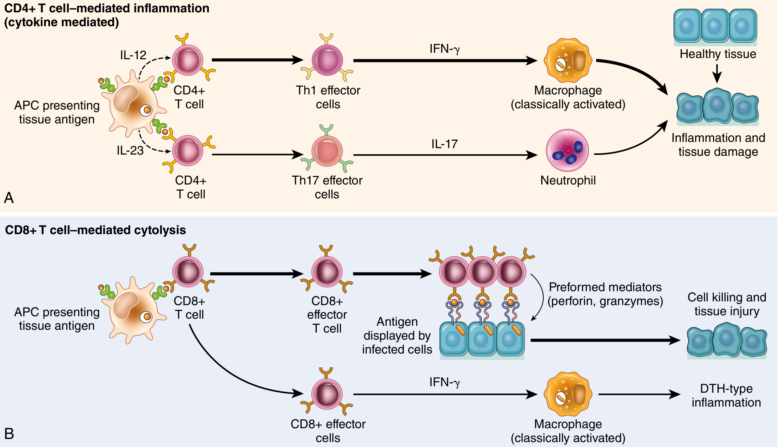
Two main effector arms:
A. CD4+ T cell-mediated inflammation (classical DTH)
- Antigen presented by APC + IL-12 → Th1 differentiation → IFN-γ secretion → macrophage activation → pro-inflammatory mediators, ROS, tissue injury
- IL-23 from APC → Th17 differentiation → IL-17 → neutrophil recruitment
- Prototype: tuberculin skin test reaction; contact dermatitis
B. CD8+ T cell-mediated cytotoxicity
- CTLs kill target cells via perforin/granzymes
- Can also secrete IFN-γ causing DTH-type inflammation
- Important in viral infections, type 1 diabetes
Granulomatous inflammation is a specialized form of type IV hypersensitivity - occurs when a persistent antigen (e.g., M. tuberculosis) cannot be eliminated, leading to chronic macrophage activation and formation of granulomas (aggregates of activated macrophages = epithelioid cells, often with Langhans giant cells).
Clinical examples:
| Disease | Antigen / Trigger | Mechanism |
|---|---|---|
| Contact dermatitis | Urushiol (poison ivy), nickel, chemicals | Th1/Th17 inflammation |
| Tuberculosis | Mycobacterium tuberculosis | Granulomatous inflammation |
| Type 1 diabetes mellitus | Pancreatic islet β-cell antigens | CTL-mediated β-cell destruction |
| Multiple sclerosis | Myelin basic protein | Th1/Th17 → macrophage-mediated demyelination |
| Rheumatoid arthritis | Citrullinated self proteins, collagen? | Th17 cytokines; also immune complexes |
| Inflammatory bowel disease | Enteric bacteria / self antigens | Th1 and Th17 cytokines |
Key Distinctions at a Glance
| Feature | Type I | Type II | Type III | Type IV |
|---|---|---|---|---|
| Immunoglobulin | IgE | IgG/IgM | IgG/IgM | None |
| Antigen location | Soluble (environmental) | Cell surface / ECM | Soluble (circulating) | Cell-associated / tissue |
| Key effector | Mast cell, eosinophil | Complement, phagocyte, NK | Neutrophil, complement | T cells, macrophages, CTLs |
| Onset | Minutes | Hours | Hours-days | 48-72 h |
| Complement activated | No | Yes (classical) | Yes (classical) | No |
| Transfer with serum | Yes | Yes | Yes | No (needs cells) |
Sources: Robbins, Cotran & Kumar - Pathologic Basis of Disease, pp. 198-208 | Cellular and Molecular Immunology, pp. 1240-1244
Apurba sastry
I'll check if Apurba Sastry's textbook is available in the medical library.
Searching sources...
Finding Sources
Searching the Web
Apurba Sastry microbiology textbook topics covered
Unfortunately, Apurba Sastry's "Essentials of Medical Microbiology" is not available in the medical library database I have access to. The library contains titles from publishers like Elsevier (Robbins, Goldman-Cecil, Janeway's, etc.) but does not currently include this book.
Here's what I can tell you about it:
Apurba Sastry - Essentials of Medical Microbiology
Author: Dr. Apurba Sankar Sastry (and Dr. Sandhya Bhat as co-author in some editions)
Publisher: Jaypee Brothers Medical Publishers
Primary use: MBBS (India) - 2nd year microbiology; also popular for USMLE/PG entrance prep
Book Structure
| Part | Content |
|---|---|
| Part I | General Microbiology, Immunology, Hospital Infection Control |
| Part II | Systemic Microbiology (system-based - as per CBME curriculum) |
| - Bloodstream & cardiovascular infections | |
| - GI and hepatobiliary infections | |
| - Respiratory infections | |
| - CNS infections, UTI, STIs, skin/soft tissue infections | |
| Virology | HIV, Hepatitis B & C, COVID-19 chapter, Herpesviruses |
| Mycology | Pathogenic fungi |
| Parasitology | Based on Paniker's framework |
| Clinical Microbiology | Lab diagnosis, sterilization, antibiotics |
| Annexures | Opportunistic infections, transplant infections, national health programmes |
Why it's popular
- Follows the NMC/CBME system-based curriculum (India)
- Clear diagrams and tables
- Well-suited for theory exams AND viva
- Covers hypersensitivity, immunology, and vaccines in good depth in Part I
What I can do for you
Since the book isn't in my library, I can still help you with any topic from this book using other authoritative sources (Robbins, Janeway's Immunobiology, Goldman-Cecil, etc.) that ARE in the library.
Just tell me:
- Which topic or chapter from Apurba Sastry you want to study
- I'll pull the relevant content from equivalent authoritative textbooks and give you a comprehensive answer
Hypersensitivity rection apurba sastry
I'll search for hypersensitivity content specifically as covered in Apurba Sastry's style - focusing on microbiology/immunology angle with practical exam-oriented details.
Searching sources...
Finding Sources
Searching the Web
Apurba Sastry hypersensitivity reaction microbiology immunology types notes
Jawetz Medical Microbiology is the closest international equivalent to Apurba Sastry. Let me read that section plus the Slideshare content which appears to be directly from Apurba Sastry's book.
Reading File
Reading Web Page
https://www.slideshare.net/slideshow/hypersensitivity-pptxhy…
I now have the exact Apurba Sastry content. Let me get the remaining details on Types II, III, IV from Jawetz (closest equivalent) and more Sastry-specific content.
Reading File
Reading Web Page
https://www.scribd.com/document/992409126/Hypersensitivity-R…
I now have everything needed - the exact Apurba Sastry classification table from the SlideShare, and full mechanistic content from Jawetz (which mirrors Sastry's microbiology approach). Let me compile the complete Apurba Sastry-style answer.
Hypersensitivity Reactions
As per Apurba S. Sastry - Essentials of Medical Microbiology
Definition
Hypersensitivity is an abnormal, exaggerated immune response to a specific antigen (allergen) that leads to tissue damage or death in a previously sensitized (presensitized) individual.
- Requires prior sensitization - reaction typically occurs on the second encounter with the antigen
- Classified by Gell and Coombs (1963) into 4 types
- Types I, II, III = Immediate (antibody/humoral mediated)
- Type IV = Delayed (cell-mediated)
Gell and Coombs Classification - Master Table (Apurba Sastry)
| Feature | Type I | Type II | Type III | Type IV |
|---|---|---|---|---|
| Name | Anaphylactic / Allergic | Cytotoxic / Cytolytic | Immune Complex | Delayed-Type Hypersensitivity (DTH) |
| Immunity | Humoral | Humoral | Humoral | Cell-mediated |
| Immediate / Delayed | Immediate | Immediate | Immediate | Delayed |
| Duration of onset | 2 min - 30 min | 5-8 hours | 2-8 hours | 24-72 hours |
| Antigen | Soluble | Cell surface bound | Soluble | Soluble or bound |
| Mediator | IgE | IgG | Ag-Ab complex | T |
| Effector mechanism | Mast cell degranulation | 1. ADCC 2. Complement-mediated cytolysis | Complement activation + inflammatory response | Macrophage activation → phagocytosis / cytotoxicity |
| Desensitization | Easy, but short lasting | Easy, but short lasting | Easy, but short lasting | Difficult, but sustained |
| Typical manifestations | Anaphylaxis, Asthma, Atopic dermatitis | Transfusion reactions, Rh incompatibility, Hemolytic anemia | Arthus reaction, Serum sickness, Glomerulonephritis, Rheumatoid arthritis | Tuberculin test, Granuloma (TB, leprosy), Contact dermatitis |
TYPE I - Immediate (Anaphylactic) Hypersensitivity
Hallmark
Production of IgE by sensitized B cells following allergen contact → mast cell degranulation
Mechanism (Two-Step)
Step 1 - Sensitization:
Allergen → stimulates B cells → produce IgE antibodies → IgE binds via Fc portion to high-affinity FcεRI receptors on mast cells and basophils
Step 2 - Elicitation (on re-exposure):
Same allergen cross-links cell-bound IgE → mast cell degranulation → release of mediators
Mediators
| Mediator | Type | Action |
|---|---|---|
| Histamine | Primary (preformed) | Vasodilation, ↑capillary permeability, bronchospasm |
| Tryptase, chymase | Primary (preformed) | Tissue damage markers |
| Prostaglandins | Secondary (newly formed) | Edema, bronchoconstriction |
| Leukotrienes C4, D4 | Secondary (newly formed) | Vasodilation, ↑vascular permeability |
| Leukotriene B4 | Secondary | Chemoattractant for leukocytes |
| TNF-α, IL-4 | Secondary (cytokines) | Late-phase inflammation |
Outcomes
- Localized response → called Atopy
- Systemic response → called Anaphylaxis
Common Allergens
| Type | Examples |
|---|---|
| Food | Nuts, egg, peas, seafood, beans, milk |
| Plants & pollens | Rye grass, ragweed, timothy grass |
| Proteins | Foreign serum, vaccines |
| Drugs | Penicillin, aspirin |
| Others | Latex, insect venom |
Clinical Examples
- Anaphylactic shock (bee sting, penicillin injection)
- Bronchial asthma
- Allergic rhinitis (hay fever)
- Atopic dermatitis (eczema)
- Urticaria (hives)
- Food allergy
Atopy
- Genetic predisposition to Type I reactions
- Elevated serum IgE; excess Th2 cells
- Strong familial predisposition
- Atopic triad: Atopic dermatitis + Allergic rhinitis + Asthma
Treatment of Anaphylaxis
- Epinephrine (adrenaline) - first line, reverses bronchospasm and hypotension
- Antihistamines - block H1 receptors (useful for rhinitis; NOT effective for late-phase)
- Corticosteroids - for late-phase reaction; needed for asthma
- Airway maintenance, artificial ventilation, cardiac support
Desensitization
- Repeated small doses of allergen → shift from IgE to IgG (blocking antibody) production
- Easy, but short-lasting
TYPE II - Cytotoxic (Antibody-Mediated) Hypersensitivity
Hallmark
IgG antibodies against cell surface or matrix antigens → cell destruction
Mechanisms
- Complement-mediated cytolysis - IgG/IgM + antigen on cell surface → activates classical complement → MAC → cell lysis
- ADCC (Antibody-Dependent Cell-Mediated Cytotoxicity) - NK cells, macrophages bind IgG-coated target via Fc receptors → cell destruction without phagocytosis
- Opsonization - IgG/C3b coat cells → recognized by phagocyte Fc/C3b receptors → phagocytosis
- Antibody-mediated cellular dysfunction - antibody blocks or stimulates receptor without killing the cell (e.g., Graves disease)
Clinical Examples
| Disease | Antigen | Mechanism |
|---|---|---|
| ABO transfusion reaction | ABO blood group antigens on RBCs | Complement lysis |
| Rh incompatibility / Erythroblastosis fetalis | Rh antigen on fetal RBCs | Maternal IgG crosses placenta → fetal RBC destruction |
| Autoimmune hemolytic anemia | RBC surface antigens | Opsonization + complement lysis |
| Goodpasture syndrome | Glomerular + alveolar basement membrane | Complement activation, inflammation |
| Graves disease | TSH receptor | Ab stimulates receptor → hyperthyroidism (no cell lysis) |
| Myasthenia gravis | Acetylcholine receptor | Ab blocks receptor → muscle weakness |
| Drug-induced hemolysis | Drug (e.g., penicillin) bound to RBC surface | IgG to drug-RBC complex → hemolysis |
TYPE III - Immune Complex Hypersensitivity
Hallmark
Ag-Ab (IgG) complexes form in circulation → deposit in tissues → complement activation → inflammation
Mechanism (Three Stages)
- Complex formation - Antigen + IgG → immune complexes in blood (most dangerous = intermediate-sized complexes formed under antigen excess)
- Deposition - complexes lodge in kidneys (glomeruli), joints, blood vessels, skin
- Inflammation - complement activation → C3a, C5a (chemotaxins) → neutrophil recruitment → release of lysosomal enzymes + ROS → fibrinoid necrosis of vessel walls
Morphology
- Fibrinoid necrosis of blood vessels
- Immunofluorescence: granular (lumpy-bumpy) deposits of Ig + C3 along glomerular basement membrane (differs from Type II which shows linear deposits)
- Serum complement (C3) levels fall (consumed) - useful to monitor disease activity
Two Forms
| Form | Description |
|---|---|
| Local (Arthus reaction) | Intradermal antigen in immunized individual → local IgG + complement → local vasculitis; occurs within 12 hours |
| Systemic (Serum Sickness) | Large antigen load → systemic complexes → fever, urticaria, joint pain, lymphadenopathy, proteinuria; occurs 7-10 days after antigen |
Clinical Examples
- Serum sickness (horse antiserum, anti-thymocyte globulin)
- Post-streptococcal glomerulonephritis (Group A β-hemolytic Streptococcus, nephritogenic strains)
- Systemic Lupus Erythematosus (SLE) - nuclear Ag-Ab complexes
- Rheumatoid arthritis - IgM anti-IgG (rheumatoid factor) complexes in joints
- Arthus reaction (experimental local model)
- Polyarteritis nodosa (Hepatitis B)
TYPE IV - Delayed-Type Hypersensitivity (DTH)
Hallmark
T cell-mediated, no antibody involved. Reaction begins 2-3 days after antigen contact. Also called Cell-Mediated Hypersensitivity
Mechanism
- Sensitization: Antigen presented by APC → naive T cell activation → T
DTHcells and memory T cells formed - Elicitation (on re-exposure):
- Antigen re-exposure → T
DTH(CD4+ Th1) cells recognize antigen on APC - Secrete cytokines: IFN-γ, IL-2, TNF → macrophage activation
- Activated macrophages → release lysosomal enzymes, ROS, NO → tissue destruction
- CD8+ CTLs → directly kill target cells via perforin/granzymes
Sub-types
| Sub-type | Onset | Example |
|---|---|---|
| Contact hypersensitivity | 48-72 h | Nickel, formaldehyde, poison ivy, topical drugs |
| Tuberculin-type | 48-72 h | Mantoux test / PPD skin test in TB |
| Granulomatous | Weeks-months | TB, leprosy, histoplasmosis, schistosomiasis |
Granulomatous Hypersensitivity
- Occurs when antigen cannot be eliminated (persistent)
- Chronic macrophage activation → epithelioid cells + Langhans giant cells → granuloma formation
- Seen in: Tuberculosis, Leprosy, Sarcoidosis, Fungal infections
Clinical Examples
- Tuberculin (Mantoux) test - PPD injected intradermally → induration at 48-72h = positive (prior sensitization to TB)
- Contact dermatitis - poison ivy (urushiol), nickel, rubber, formaldehyde
- Type 1 Diabetes mellitus - CTL destruction of β-islet cells
- Multiple sclerosis - T cell attack on myelin
- Granuloma formation in TB and leprosy
Desensitization
- Difficult, but sustained (unlike Types I-III)
Quick Comparison - Apurba Sastry Style
| Type I | Type II | Type III | Type IV | |
|---|---|---|---|---|
| Ig class | IgE | IgG | IgG | None |
| Complement | No | Yes | Yes | No |
| Transfer | Serum | Serum | Serum | Cells only |
| Key test | Skin prick test, serum IgE | Coombs test | C3 levels ↓, IF granular | Patch test, Mantoux test |
| Treatment | Epinephrine, antihistamines, steroids | Treat underlying cause | Steroids | Steroids |
Exam High-Yield Points (Apurba Sastry Focus)
- Only Type IV is transferred by cells (not serum) - used to prove cell-mediated immunity
- Arthus reaction = local Type III (NOT Type I despite being a skin reaction)
- Serum sickness = systemic Type III
- Tuberculin test = Type IV (NOT Type I) - remember 48-72 hour delay
- Granuloma = Type IV (granulomatous hypersensitivity)
- Graves disease = Type II (Ab stimulates receptor) - no tissue damage
- Myasthenia gravis = Type II (Ab blocks receptor)
- IgE is the hallmark antibody of Type I; acts via mast cell FcεRI receptor
- Desensitization is easiest for Types I-III (short-lasting); hardest for Type IV (but sustained)
- Complement levels fall in active Type III disease (consumed by complexes)
Sources: Essentials of Medical Microbiology - Apurba S. Sastry (Jaypee); Jawetz, Melnick & Adelberg's Medical Microbiology 28e, pp. 151-154
This is a shared conversation. Sign in to Orris to start your own chat.