Lab diagnosis of candidiasis
Candida laboratory diagnosis microscopy germ tube pseudohyphae
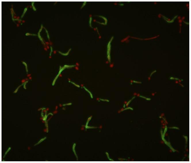
This diagnostic image is a fluorescence microscopy micrograph illustrating the morphological features of Candida albicans. The visual field demonstrates dual-color immunofluorescence staining using mAb K20 to highlight specific fungal cell wall components. Elongated, filamentous structures known as germ tubes exhibit diffuse green fluorescence along their cell walls, indicating the localization of the target antigen during hyphal transition. In contrast, smaller, oval-to-round budding yeast cells (blastospores) show concentrated red fluorescence, typically localized at points of active cellular division or budding sites. The spatial arrangement shows budding cells frequently attached to the lateral or terminal aspects of the germ tubes, representing the proliferative lifecycle of the pathogen. This image is medically significant for studying fungal pathogenesis, cell wall molecular composition, and the diagnostic identification of C. albicans through characteristic germ tube formation and specific monoclonal antibody binding patterns.
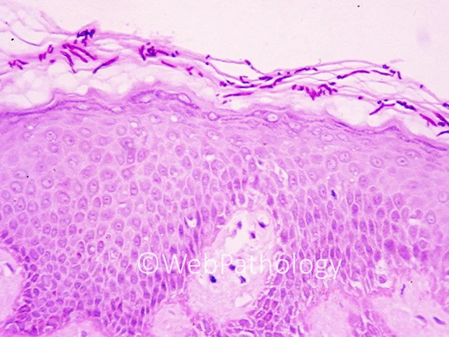
This is a skin histopathology specimen stained with Periodic Acid-Schiff (PAS), imaged under light microscopy. The biopsy shows epidermal and superficial dermal compartments of cutaneous tissue with characteristic fungal elements. PAS positivity highlights both yeast forms and pseudohyphae within the stratum corneum and epidermis, consistent with cutaneous candidiasis. Yeasts appear as small to oval budding cells; pseudohyphae are elongated, branching, and may extend through the keratinocyte layers. The epidermis may demonstrate mild acanthosis with preserved overall architecture; scattered neutrophilic microabscesses can be present in the epidermis or papillary dermis as part of the inflammatory response. These histologic features, in combination with PAS staining, solidify the diagnosis of Candida species infection of the skin. Clinical significance: recognition of intraepidermal yeast and pseudohyphae with PAS staining supports targeted antifungal therapy (topical azoles or oral agents in extensive disease) and prompts assessment for predisposing factors such as moisture, obesity, diabetes, immunosuppression, or antibiotic exposure. Differential considerations include dermatophyte infections (dermatophyte hyphae), Malassezia spp., and other yeasts; however, the presence of pseudohyphae favors Candida. This image is valuable for dermatopathology education, fungal biology research, and diagnostic pathology training, illustrating canonical histologic hallmarks of cutaneous candidiasis and the utility of PAS for fungal visualization.
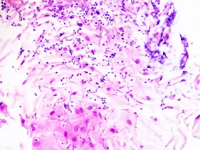
Imaging modality and technique: Light microscopy of an esophageal mucosal biopsy stained with Hematoxylin and Eosin (H&E). Anatomical location: esophageal squamous mucosa of the upper gastrointestinal tract, biopsy specimen from the distal esophagus. Visual features: a mucosal fragment exhibiting desquamated squamous cells and mixed inflammatory debris with an inflammatory infiltrate in the lamina propria. Fungal elements are seen as slender pseudohyphae with budding yeast forms occupying the mucosal surface and occasionally invading superficial epithelium. The organisms appear basophilic/purple on H&E; they are classically highlighted by fungal special stains such as PAS or GMS, which would accentuate wall thickening and budding yeasts. Pathological findings: Candida species with pseudohyphae and budding yeast within inflammatory debris; mucosal inflammation consistent with esophagitis; no extensive necrosis; focal epithelial desquamation; compatible with infectious esophagitis. Diagnostic significance: histologic confirmation of Candida esophagitis; provides definitive etiologic diagnosis in patients with odynophagia, dysphagia, fever, or immunosuppression; informs antifungal therapy and duration; may prompt evaluation for underlying immunodeficiency. Potential clinical use cases: confirmation of suspected fungal esophagitis in HIV/AIDS, organ transplant recipients, chemotherapy, or prolonged steroid use; differentiation from pill esophagitis; this description supports educational and research use in teaching fungal mucosal invasion and antifungal response assessment.
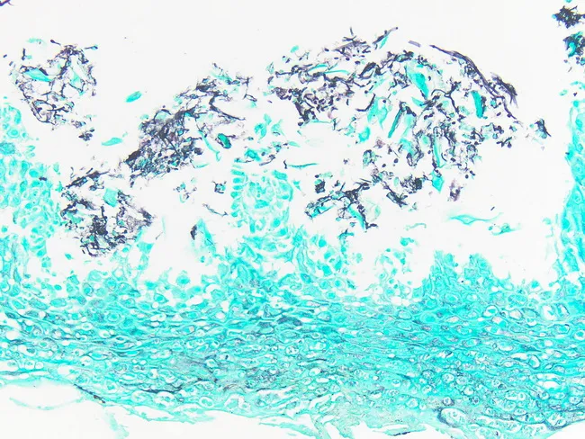
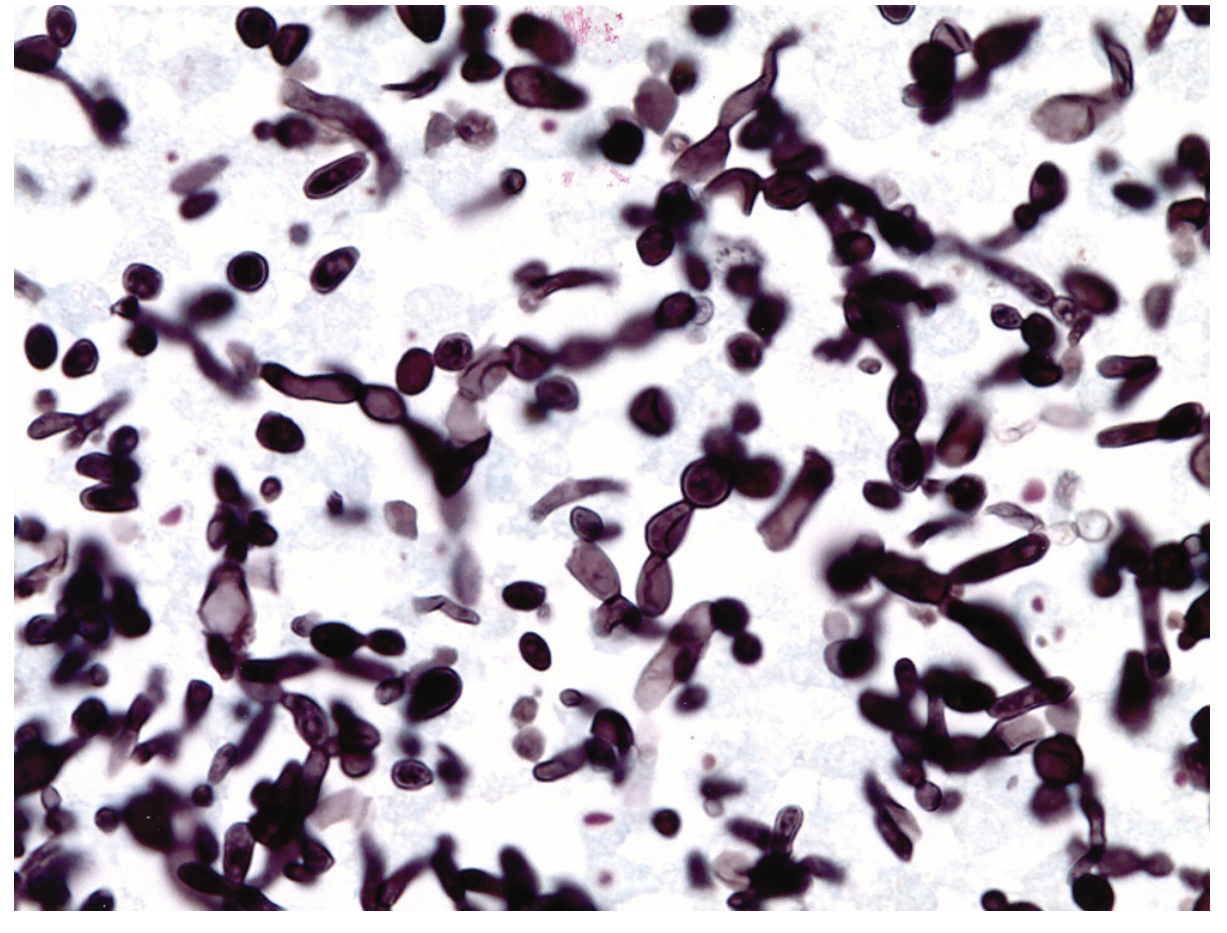
Imaging modality: histopathology with light microscopy of a Gomori methenamine silver (GMS) stained esophageal mucosal biopsy. Primary anatomic site: esophagus, specifically the stratified squamous epithelium of the distal to mid-esophagus with involvement of the lamina propria. The specimen demonstrates surface and intraepithelial fungal elements highlighted by GMS as dark, threadlike structures consisting of yeast forms and slender pseudohyphae, with occasional budding yeasts. The mucosal architecture shows preserved epithelium with focal inflammatory infiltrates and minimal necrosis in this field. Notable features include abundant fungal pseudohyphae and yeast forms within the epithelium and superficial lamina propria, confirming candida infection; the GMS stain provides high-contrast visualization against a light background. Pathological diagnosis: Candida esophagitis with fungal hyphae-like elements and budding yeast consistent with Candida spp., most commonly Candida albicans. Diagnostic significance: histologic confirmation supports clinical odynophagia and dysphagia, particularly in immunocompromised patients, HIV/AIDS, post-therapy states, or post-antibiotic exposure. Differential considerations include other fungal infections (Histoplasma, Cryptococcus) and non-infectious mimics; differentiation relies on morphologic patterns and special stains. Clinical relevance: informs selection and duration of antifungal therapy (e.g., fluconazole, echinocandins) and monitoring response; serves as a teaching example for gastroenterology, pathology, and infectious disease training. This image is educational for diagnostics and differential diagnosis.
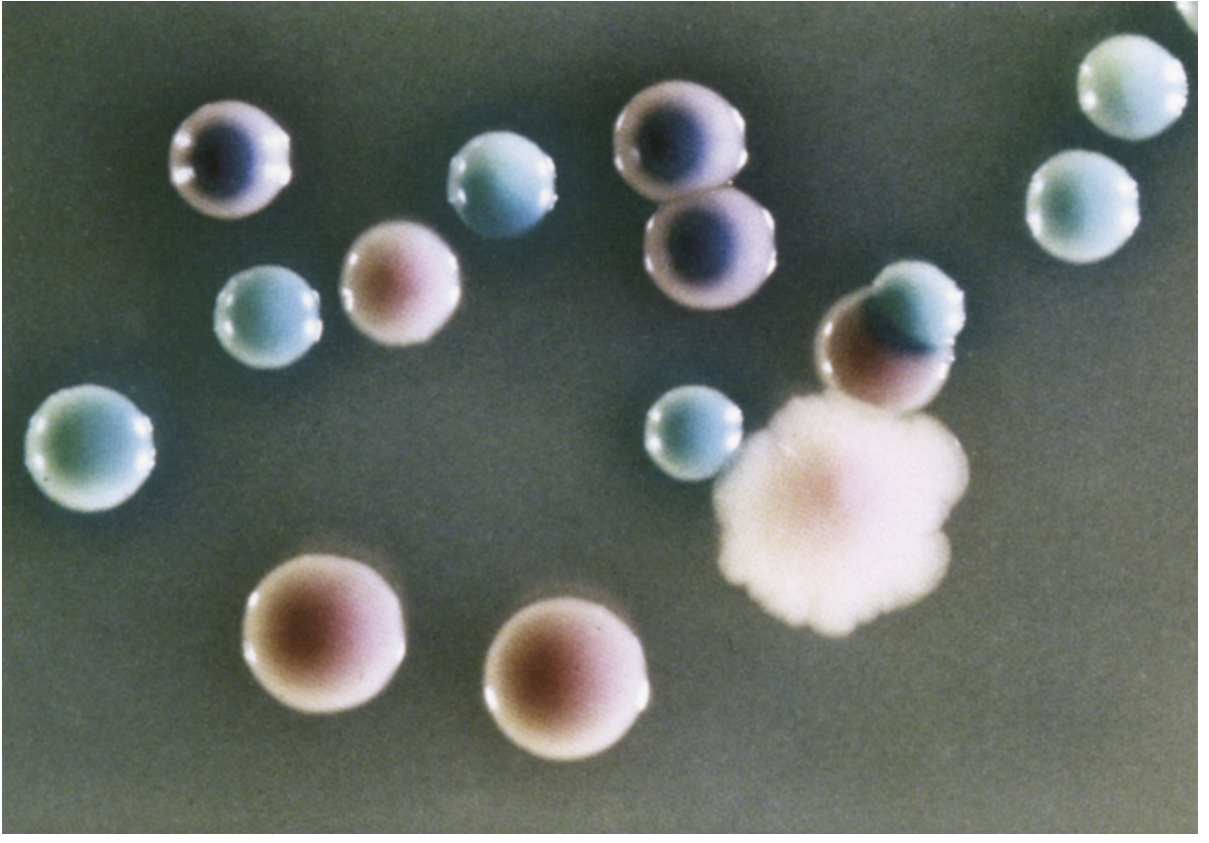
Candida chromagar colony identification C. albicans C. tropicalis
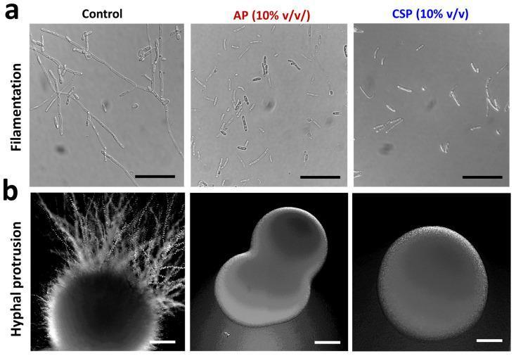
Microscopic and macro-colony imaging demonstrating the inhibitory effects of L. plantarum postbiotics (AP and CSP) on Candida albicans morphogenesis. Row (a) displays phase-contrast microscopic images of C. albicans filamentation in liquid RPMI-1640 media (scale bar 20 µm). The control shows extensive, elongated, and branching hyphal filaments, whereas the AP and CSP (10% v/v) treated groups exhibit a significant reduction in filament length, primarily existing as isolated yeast cells or truncated germ tubes. Row (b) presents the macro-morphology of C. albicans colonies on potato dextrose agar (PDA) after 7 days (scale bar 200 µm). The control colony shows prominent, radiating hyphal protrusions extending into the agar, creating a characteristic 'fuzzy' border associated with virulence. In contrast, colonies treated with AP or CSP display smooth, well-defined circumscribed margins with a total absence of peripheral hyphal extensions. This comparison illustrates the transition from a virulent hyphal/filamentous phenotype to a commensal-like yeast phenotype, highlighting the anti-hyphal and anti-pathogenic potential of the tested postbiotics in both liquid and solid growth environments.
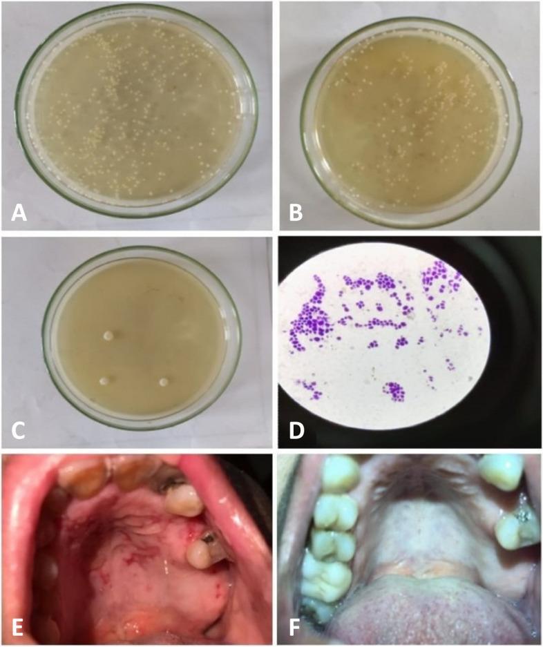
A multi-panel clinical and laboratory figure demonstrating the progression and treatment of oral candidiasis. Panels A, B, and C show Petri dishes containing agar medium with Candida albicans colony-forming units (CFUs). Plate A (baseline) shows confluent growth of small, cream-colored colonies. Plate B (one week post-treatment) shows a moderate reduction in colony density. Plate C (end of treatment) exhibits a marked decrease, with only a few isolated large colonies visible. Panel D is a high-power light micrograph of a Gram-stained smear showing characteristic purple-stained budding yeast cells and pseudohyphae. Panel E is a clinical photograph of the hard palate of a diabetic patient, revealing diffuse, erythematous candidiasis characterized by a bright red, inflamed mucosa with irregular borders. Panel F shows the same patient’s palate after treatment with miconazole-loaded chitosan nanoparticles, demonstrating clinical resolution with a return to a healthy, pink, smooth mucosal appearance and no visible inflammation.
Laboratory Diagnosis of Candidiasis
1. Specimen Collection
| Infection Type | Specimen |
|---|---|
| Mucosal (oral thrush, vaginitis) | Swabs, scrapings |
| Cutaneous | Skin scrapings, nail clippings |
| Urinary | Midstream urine |
| Systemic/invasive | Blood, BAL, CSF, tissue biopsy, peritoneal fluid |
| Hepatosplenic | Liver/spleen biopsy |
2. Direct Microscopy
- Scrapings from mucosal or cutaneous lesions are treated with 10-20% KOH, optionally with calcofluor white (a fluorescent brightener that binds chitin in fungal cell walls)
- Examined under fluorescence microscope
- Shows budding yeast cells (blastoconidia) + pseudohyphae (elongated chains of yeast cells with constrictions at junctions)
- C. albicans, C. dubliniensis, and C. tropicalis can also form true hyphae
3. Culture
- Sabouraud dextrose agar (SDA) - the standard mycologic medium; grows cream/white, smooth, pasty colonies in 24-72 hours
- Blood agar - also supports growth
- Lim broth / BIGGY agar - selective/differential media for Candida
Chromogenic Media - CHROMagar Candida
| Species | Colony color on CHROMagar |
|---|---|
| C. albicans | Green |
| C. tropicalis | Blue/blue-gray |
| C. krusei | Large, rough, pale pink |
| C. glabrata | Smooth, pink/mauve |
| Other species | White to pink |
4. Species Identification Tests
Germ Tube Test (Reynolds-Braude phenomenon)
- Gold standard rapid ID for C. albicans
- Inoculate a few colonies into human or rabbit serum at 37°C for 2-3 hours
- C. albicans produces germ tubes - true hyphae-like extensions without a constriction at the base
- Positive in ~95% of C. albicans isolates
- C. dubliniensis also produces germ tubes (distinguish by growth at 45°C - C. albicans grows, C. dubliniensis does not)
Chlamydospore Production (Cornmeal Agar / Dalmau Plate)
- Culture on cornmeal agar with Tween 80 for 24-48 hours at 25°C
- C. albicans produces characteristic thick-walled chlamydospores (terminal or intercalary) - virtually pathognomonic
- Also reveals morphologic patterns: blastoconidia, pseudohyphae arrangement
Carbohydrate Assimilation/Fermentation (API 20C AUX)
- Tests the ability to utilize specific sugars as carbon sources
- Distinguishes between species based on metabolic profiles
- Classic but time-consuming (48-72 hours)
MALDI-TOF MS (Matrix-Assisted Laser Desorption Ionization - Time of Flight Mass Spectrometry)
- Now the preferred rapid identification method in most clinical labs
- Generates a protein fingerprint of the organism within minutes of culture
- Accurate to species level; cost-effective once established
- Has transformed routine Candida species identification in modern labs
5. Histopathology
| Stain | Appearance |
|---|---|
| GMS (Gomori Methenamine Silver) | Black/dark fungal elements against green background - best contrast |
| PAS (Periodic Acid-Schiff) | Magenta/red yeast and pseudohyphae |
| H&E | Basophilic/purple fungal elements - less sensitive |
6. Blood Cultures (Invasive/Systemic Candidiasis)
- Automated continuous-monitoring blood culture systems (BACTEC, BacT/ALERT) are the standard for candidemia
- Candida typically detects within 24-72 hours of incubation
- Sensitivity is limited (~50%) for deep-seated/non-candidemic invasive infection - hence need for supplementary non-culture methods
- Multiple sets should be drawn; the lysis-centrifugation method (Isolator tube) can improve yield for some species
7. Non-Culture / Serologic / Biomarker Tests
| Test | Target | Notes |
|---|---|---|
| (1→3)-β-D-Glucan (BDG) assay | Cell wall polysaccharide component (fungal pan-marker) | Positive in invasive candidiasis, also aspergillosis, PCP; not specific to Candida; used as screening/surveillance in high-risk patients |
| Mannan antigen / Anti-mannan antibody | Candida cell wall mannoprotein | Higher specificity; combined use improves sensitivity; useful in ICU patients |
| Candida PCR | Fungal DNA in blood/tissue | Rapid, sensitive; not yet universally standardized but increasingly used |
| T2Candida Panel (T2 biosystems) | PCR + T2 magnetic resonance from whole blood | Detects 5 Candida species directly in blood within ~3-5 hours; does not require culture |
8. Antifungal Susceptibility Testing
- Performed by broth microdilution (CLSI M27 method - gold standard) or Etest
- Particularly important for: C. glabrata (reduced azole susceptibility), C. krusei (intrinsically resistant to fluconazole), C. auris (multi-drug resistant emerging pathogen)
- Species identification alone guides empiric therapy choices before susceptibility results are available
Summary Table
| Method | Use | Key Finding |
|---|---|---|
| KOH + calcofluor | Superficial infections | Budding yeasts, pseudohyphae |
| Gram stain | Smears | Gram-positive budding yeasts |
| Culture (SDA) | All types | Cream/white colonies |
| CHROMagar | Species differentiation | Color-coded colony ID |
| Germ tube test | ID C. albicans rapidly | Positive germ tube in serum |
| Chlamydospore (cornmeal agar) | ID C. albicans | Thick-walled terminal spores |
| MALDI-TOF MS | Species ID from culture | Minutes-fast, highly accurate |
| GMS/PAS histostain | Tissue biopsy | Budding yeasts + pseudohyphae |
| Blood culture (automated) | Candidemia | Growth in 24-72 hrs |
| β-D-Glucan assay | Invasive disease | Pan-fungal marker |
| Mannan/anti-mannan | Invasive candidiasis | Candida-specific |
| PCR / T2Candida | Rapid invasive detection | Directly from blood |
- Medical Microbiology 9e, Murray, Rosenthal & Pfaller - Chapter 65 (Candidiasis) and Chapter 60 (Lab Diagnosis of Fungal Infections)
- Tietz Textbook of Laboratory Medicine, 7th Edition - Chapter 87 (Laboratory Diagnosis of Fungal Infections)
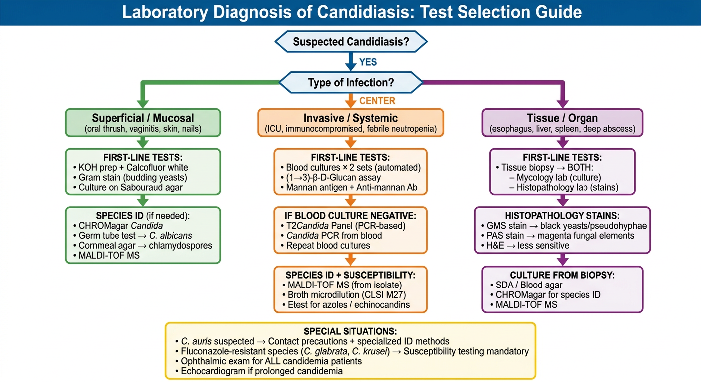
Create a clinical decision flowchart for choosing candidiasis tests
A clean, professional clinical decision flowchart titled "Laboratory Diagnosis of Candidiasis: Test Selection Guide". The flowchart starts at the top with a diamond decision box: "Suspected Candidiasis?" with YES arrow going down. Next diamond: "Type of Infection?" with three branches: - LEFT branch: "Superficial / Mucosal" (oral thrush, vaginitis, skin, nails) - CENTER branch: "Invasive / Systemic" (ICU, immunocompromised, febrile neutropenia) - RIGHT branch: "Tissue / Organ" (esophagus, liver, spleen, deep abscess) LEFT branch (Superficial) leads to a green box: "FIRST-LINE TESTS: • KOH prep + Calcofluor white • Gram stain (budding yeasts) • Culture on Sabouraud agar" Arrow down to: "SPECIES ID (if needed): • CHROMagar Candida • Germ tube test → C. albicans • Cornmeal agar → chlamydospores • MALDI-TOF MS" CENTER branch (Invasive) leads to an orange box: "FIRST-LINE TESTS: • Blood cultures × 2 sets (automated) • (1→3)-β-D-Glucan assay • Mannan antigen + Anti-mannan Ab" Arrow down to: "IF BLOOD CULTURE NEGATIVE: • T2Candida Panel (PCR-based) • Candida PCR from blood • Repeat blood cultures" Arrow down to: "SPECIES ID + SUSCEPTIBILITY: • MALDI-TOF MS (from isolate) • Broth microdilution (CLSI M27) • Etest for azoles / echinocandins" RIGHT branch (Tissue/Organ) leads to a purple box: "FIRST-LINE TESTS: • Tissue biopsy → BOTH: – Mycology lab (culture) – Histopathology lab (stains)" Arrow down to: "HISTOPATHOLOGY STAINS: • GMS stain → black yeasts/pseudohyphae • PAS stain → magenta fungal elements • H&E → less sensitive" Arrow down to: "CULTURE FROM BIOPSY: • SDA / Blood agar • CHROMagar for species ID • MALDI-TOF MS" At the bottom, a shared yellow box across all branches: "SPECIAL SITUATIONS: • C. auris suspected → Contact precautions + specialized ID methods • Fluconazole-resistant species (C. glabrata, C. krusei) → Susceptibility testing mandatory • Ophthalmic exam for ALL candidemia patients • Echocardiogram if prolonged candidemia" Clean white background, color-coded branches (green, orange, purple), bold headers, arrows connecting each step, professional medical style, suitable for hospital reference.