
Treatment of acute pulmonary edema
"acute pulmonary edema" AND treatment
chest x-ray acute pulmonary edema bilateral bat wing infiltrates
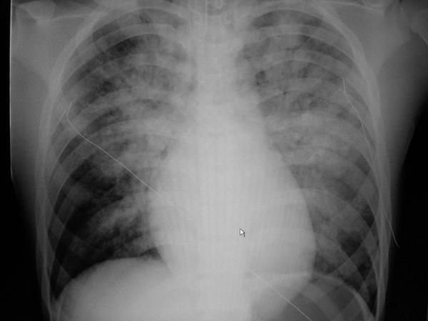
This diagnostic image is an anteroposterior (AP) chest X-ray demonstrating acute bilateral pulmonary edema. The radiograph reveals diffuse, hazy opacities and ill-defined infiltrates throughout both lung fields, exhibiting a characteristic perihilar 'bat-wing' distribution with prominent involvement of the upper and central lung zones. The lung parenchyma shows signs of alveolar consolidation and fluid accumulation. Cardiomegaly is evident, with an enlarged cardiac silhouette and indistinct borders. A thin, radiopaque monitoring line or catheter is visible crossing the right thoracic cavity toward the mediastinum. The mediastinal structures and bony thorax, including the clavicles and ribs, are otherwise unremarkable. This visual is clinically significant for students learning to identify radiological signs of heart failure and pulmonary congestion, as it highlights the hallmark features of cardiogenic pulmonary edema versus acute respiratory distress syndrome (ARDS).
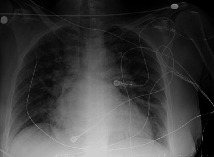
This anterior-posterior (AP) chest X-ray illustrates a case of acute pulmonary edema and cardiomegaly. The primary findings include diffuse, bilateral alveolar opacities and patchy infiltrates that exhibit a predominantly perihilar 'bat-wing' distribution. These densities obscure normal vascular markings and contribute to a mottled, ground-glass appearance throughout the mid-to-lower lung zones. The cardiac silhouette is significantly enlarged, with a widened mediastinum and indistinct heart borders consistent with congestive heart failure. Multiple supportive medical devices are present: a right-sided central venous catheter with its tip at the level of the superior vena cava, several circular radiopaque ECG electrode markers with associated lead wires traversing the anterior chest, and a vertical enteric tube passing through the lower mediastinum. The costophrenic angles are partially blunted by overlying infiltrates or potential pleural effusion. This diagnostic image serves as a classic representation of cardiogenic pulmonary edema for clinical educational purposes.
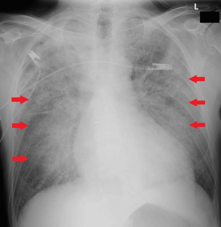
This diagnostic image is an anteroposterior (AP) chest X-ray demonstrating features of acute pulmonary edema. The most striking finding is a 'butterfly shadow' or 'bat-wing' pattern, characterized by bilateral, symmetric, ground-glass opacities predominantly in the perihilar and medullary regions of the lungs. Red arrows point to the peripheral margins of these alveolar infiltrates, where there is relatively more sparing of the cortical lung fields. The cardiac silhouette is enlarged (cardiomegaly), which, in conjunction with the central opacities, is highly suggestive of cardiogenic pulmonary edema. Visible medical support includes radiopaque electrocardiogram (ECG) leads and a central venous line or monitoring device traversing the upper thorax. The bony structures, including the ribs and clavicles, appear intact. This visual presentation is an educational example of pulmonary congestion and alveolar filling commonly seen in acute decompensated heart failure or flash pulmonary edema, particularly in patients with underlying chronic kidney disease.
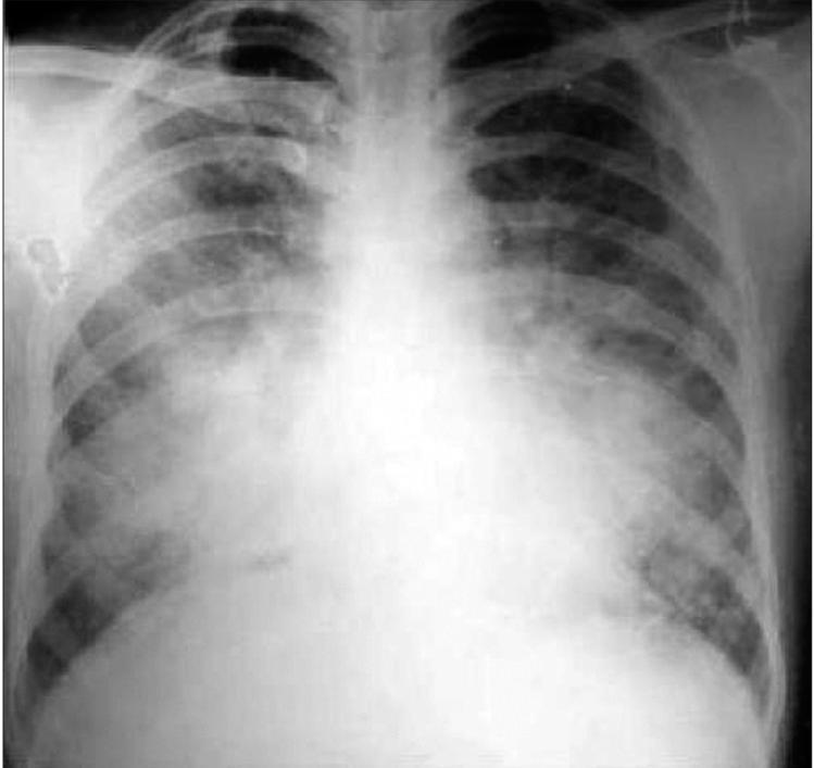
This diagnostic image is a posteroanterior (PA) view chest X-ray demonstrating a bilateral, symmetric alveolar filling pattern. The abnormal opacities are primarily concentrated in the perihilar and mid-lung zones, exhibiting a classic 'bat-wing' or 'butterfly' distribution. These areas show increased density compared to normal aerated lung tissue, while the pulmonary apices and peripheral costophrenic angles appear relatively spared. The cardiac silhouette is of normal size with no signs of cardiomegaly, suggesting a non-cardiogenic etiology for the pulmonary edema or infiltrates. Clinical significance includes the assessment of Acute Respiratory Distress Syndrome (ARDS) or diffuse alveolar hemorrhage, particularly in the context of severe systemic infections like Plasmodium vivax malaria. This image serves as an educational example of bilateral alveolar opacities and the radiographic distinction between cardiogenic and non-cardiogenic pulmonary processes for intermediate to advanced medical learners.
Treatment of Acute Pulmonary Edema
1. Immediate stabilization (ABCs)
- Position: Sit the patient upright/high Fowler's position with legs dependent to reduce venous return and improve ventilation.
- Oxygen: Supplemental O2 to keep SpO2 ≥ 90-94%.
- Non-invasive ventilation (NIV) - CPAP or bilevel (BiPAP) - is first-line respiratory support in most patients with respiratory distress. It reduces preload and afterload, decreases work of breathing, and lowers intubation rates. Evidence shows no significant difference in outcomes between CPAP and bilevel ventilation for cardiogenic pulmonary edema (Fishman's Pulmonary Diseases and Disorders, p. 3533-3535).
- Intubation and mechanical ventilation if the patient is in respiratory failure, has altered mental status, or fails to improve on NIV.
- Continuous cardiac monitoring, IV access, and identification/treatment of the precipitant (e.g., acute MI, arrhythmia, hypertensive emergency) is essential and runs in parallel with symptomatic treatment.
2. Pharmacologic therapy (guided by blood pressure)
- Mainstay of therapy. IV furosemide causes direct venodilation (reducing preload) within minutes, before diuresis even begins, and this venodilatory effect is a key reason for its rapid symptomatic benefit in acute pulmonary edema (Goodman & Gilman's Pharmacological Basis of Therapeutics, p. 1811; Washington Manual of Medical Therapeutics, p. 6388).
- Reduces left ventricular filling pressures and pulmonary congestion (Katzung's Basic and Clinical Pharmacology, p. 2407).
- Caution: in patients with adequate blood pressure, vasodilator therapy should be initiated together with diuretics, since the venodilatory/natriuretic effect alone can transiently raise afterload if used without a vasodilator (Braunwald's Heart Disease, p. 1294).
- IV nitroglycerin is used commonly for afterload and preload reduction, typically started at 5-10 mcg/min IV and titrated up to effect (Rosen's Emergency Medicine, p. 1808-1816).
- Especially useful when pulmonary edema is driven by hypertensive crisis or ischemia, since nitrates also improve coronary blood flow and reduce myocardial oxygen demand.
- Avoid or use cautiously in hypotension, severe aortic stenosis, or recent PDE5-inhibitor use.
- Historically used for anxiolysis and mild venodilation, but is now used sparingly/avoided as first-line therapy because observational data link it to increased mortality and intubation rates; reserve for selected patients with significant anxiety/pain and use cautiously.
- If the patient is hypotensive or in cardiogenic shock, diuretics and nitrates are relatively contraindicated; instead use inotropes (dobutamine, milrinone) or vasopressors (norepinephrine) to support perfusion, with consideration of mechanical circulatory support (IABP, ECMO) in refractory shock.
3. Treat the underlying precipitant
- Acute coronary syndrome → reperfusion therapy
- Hypertensive emergency → aggressive BP control with IV agents
- Tachy/bradyarrhythmia → rate/rhythm control or pacing
- Valvular emergency (e.g., acute mitral regurgitation, aortic regurgitation) → may need urgent surgical evaluation
- Volume overload from renal failure → consider ultrafiltration/dialysis if diuretic-refractory (Harrison's Principles of Internal Medicine 22E, p. 2320)
4. Non-cardiogenic pulmonary edema
Recent evidence note
Create a summary document or quick reference guide for treating acute pulmonary edema
Tachycardiaomyopathy
"tachycardia-induced cardiomyopathy" AND management
mkdir -p /tmp/workspace/pulmonary-edema-tachycardiomyopathy/output && ls -la /tmp/workspace/
~/pulmonary-edema-tachycardiomyopathy/build_doc.py
from docx import Document
from docx.shared import Pt, Inches, RGBColor
from docx.enum.text import WD_ALIGN_PARAGRAPH
from docx.enum.table import WD_TABLE_ALIGNMENT
from docx.oxml.ns import qn
from docx.oxml import OxmlElement

doc = Document()

style = doc.styles["Normal"]
style.font.name = "Arial"
style.font.size = Pt(11)

NAVY = RGBColor(0x1F, 0x3B, 0x57)
RED = RGBColor(0xB0, 0x20, 0x20)
GREY = RGBColor(0x55, 0x55, 0x55)

def shade_cell(cell, hex_color):
    tcPr = cell._tc.get_or_add_tcPr()
    shd = OxmlElement('w:shd')
    shd.set(qn('w:val'), 'clear')
    shd.set(qn('w:color'), 'auto')
    shd.set(qn('w:fill'), hex_color)
    tcPr.append(shd)

def set_cell_text(cell, text, bold=False, color=None, size=10, align=None):
    cell.text = ""
    p = cell.paragraphs[0]
    if align:
        p.alignment = align
    run = p.add_run(text)
    run.bold = bold
    run.font.size = Pt(size)
    if color:
        run.font.color.rgb = color

# Title
title = doc.add_heading("Quick Reference Guide", level=0)
title.alignment = WD_ALIGN_PARAGRAPH.CENTER
sub = doc.add_paragraph()
sub.alignment = WD_ALIGN_PARAGRAPH.CENTER
r = sub.add_run("Acute Pulmonary Edema and Tachycardia-Induced Cardiomyopathy")
r.bold = True
r.font.size = Pt(15)
r.font.color.rgb = NAVY

note = doc.add_paragraph()
note.alignment = WD_ALIGN_PARAGRAPH.CENTER
rn = note.add_run("For clinician educational reference. Always individualize therapy to hemodynamic status, etiology, and local protocols.")
rn.italic = True
rn.font.size = Pt(9)
rn.font.color.rgb = GREY

doc.add_paragraph()

# ============ SECTION 1: ACUTE PULMONARY EDEMA ============
h1 = doc.add_heading("1. Acute Pulmonary Edema", level=1)
for run in h1.runs:
    run.font.color.rgb = NAVY

doc.add_paragraph(
    "Most commonly cardiogenic (acute decompensated heart failure, precipitated by ischemia, "
    "arrhythmia, hypertensive crisis, or valvular disease). Can also be non-cardiogenic "
    "(ARDS, high-altitude, neurogenic, renal volume overload)."
)

h2 = doc.add_heading("1.1 Immediate Stabilization (ABCs)", level=2)
for run in h2.runs:
    run.font.color.rgb = NAVY
bullets = [
    "Position: sit upright, legs dependent, to reduce venous return and improve ventilation.",
    "Oxygen: supplemental O2 to maintain SpO2 90-94%.",
    "Non-invasive ventilation (NIV) - CPAP or bilevel (BiPAP): first-line respiratory support in most patients with respiratory distress; reduces preload/afterload and work of breathing, lowers intubation rates. No significant outcome difference between CPAP and bilevel (Fishman's Pulmonary Diseases and Disorders, p. 3533-3535).",
    "Intubation/mechanical ventilation if respiratory failure, altered mental status, or NIV failure.",
    "Continuous cardiac monitoring, IV access; identify and treat the precipitant in parallel."
]
for b in bullets:
    doc.add_paragraph(b, style="List Bullet")

h2 = doc.add_heading("1.2 Pharmacologic Therapy (Guided by Blood Pressure)", level=2)
for run in h2.runs:
    run.font.color.rgb = NAVY

table = doc.add_table(rows=1, cols=3)
table.style = "Light Grid Accent 1"
table.alignment = WD_TABLE_ALIGNMENT.CENTER
hdr = table.rows[0].cells
set_cell_text(hdr[0], "Agent Class", bold=True, color=RGBColor(0xFF,0xFF,0xFF))
set_cell_text(hdr[1], "Role / Dosing Notes", bold=True, color=RGBColor(0xFF,0xFF,0xFF))
set_cell_text(hdr[2], "Key Cautions", bold=True, color=RGBColor(0xFF,0xFF,0xFF))
for c in hdr:
    shade_cell(c, "1F3B57")

rows_data = [
    ("Loop diuretics (IV furosemide)",
     "Mainstay of therapy. IV furosemide causes direct venodilation (reduces preload) within minutes, before diuresis begins; reduces LV filling pressures and pulmonary congestion.",
     "Use together with a vasodilator in normotensive/hypertensive patients - diuretic-induced natriuresis alone can transiently raise afterload."),
    ("Nitrates (IV nitroglycerin)",
     "Preload and afterload reduction; improves coronary blood flow, reduces myocardial O2 demand. Typical start 5-10 mcg/min IV, titrate to effect.",
     "Avoid/caution in hypotension, severe aortic stenosis, recent PDE5-inhibitor use."),
    ("Morphine",
     "Historically for anxiolysis and mild venodilation.",
     "No longer first-line - associated with increased mortality/intubation in observational data; reserve for select cases of severe anxiety/pain."),
    ("Inotropes / vasopressors (dobutamine, milrinone, norepinephrine)",
     "For hypotensive patients or cardiogenic shock; supports perfusion when diuretics/nitrates are contraindicated.",
     "Consider mechanical circulatory support (IABP, ECMO) if refractory shock."),
]
for a, b, c in rows_data:
    row = table.add_row().cells
    set_cell_text(row[0], a, bold=True, size=10)
    set_cell_text(row[1], b, size=10)
    set_cell_text(row[2], c, size=10, color=RED)

doc.add_paragraph()

h2 = doc.add_heading("1.3 Treat the Underlying Precipitant", level=2)
for run in h2.runs:
    run.font.color.rgb = NAVY
bullets2 = [
    "Acute coronary syndrome -> urgent reperfusion.",
    "Hypertensive emergency -> aggressive IV BP control.",
    "Tachy/bradyarrhythmia -> rate/rhythm control or pacing (see Section 2 for tachycardia-driven cases).",
    "Acute valvular emergency (e.g., acute MR/AR) -> urgent surgical evaluation.",
    "Volume overload from renal failure -> consider ultrafiltration/dialysis if diuretic-refractory."
]
for b in bullets2:
    doc.add_paragraph(b, style="List Bullet")

h2 = doc.add_heading("1.4 Non-Cardiogenic Pulmonary Edema", level=2)
for run in h2.runs:
    run.font.color.rgb = NAVY
doc.add_paragraph(
    "Diuretics/nitrates are far less effective (problem is capillary leak, not hydrostatic pressure). "
    "Management shifts to lung-protective ventilation, treating the underlying cause (sepsis, transfusion "
    "reaction), and conservative fluid management."
)

doc.add_page_break()

# ============ SECTION 2: TACHYCARDIA-INDUCED CARDIOMYOPATHY ============
h1 = doc.add_heading("2. Tachycardia-Induced Cardiomyopathy (Tachycardiomyopathy)", level=1)
for run in h1.runs:
    run.font.color.rgb = NAVY

h2 = doc.add_heading("2.1 Definition and Key Features", level=2)
for run in h2.runs:
    run.font.color.rgb = NAVY
bullets3 = [
    "Prolonged tachycardia causes diastolic and systolic LV dysfunction, even without other structural heart disease.",
    "Diagnosis is retrospective: confirmed when correction of the arrhythmia is followed by improved ventricular function.",
    "Consider in any patient with tachycardia and LV systolic dysfunction who is not in sinus rhythm.",
    "May occur as an isolated condition or superimposed on pre-existing cardiomyopathy (e.g., worsens a patient with mild DCM who develops AF).",
    "\"Purest\" form: incessant/very frequent atrial tachycardia or permanent junctional reciprocating tachycardia (often young patients).",
    "Almost any sustained arrhythmia can cause it - includes very frequent PVCs (typically >20,000/24h) or recurrent nonsustained VT.",
    "Duration of arrhythmia (not just heart rate) is the critical driver - mean symptom duration in one series was 6 years with a mean ventricular response of only 117 bpm.",
    "Always rule out hyperthyroidism, which can independently cause tachycardia and (rarely) its own dilated cardiomyopathy.",
]
for b in bullets3:
    doc.add_paragraph(b, style="List Bullet")
doc.add_paragraph("Source: Braunwald's Heart Disease, p. 1398-1406.", style="Intense Quote")

h2 = doc.add_heading("2.2 Treatment Principles", level=2)
for run in h2.runs:
    run.font.color.rgb = NAVY

table2 = doc.add_table(rows=1, cols=2)
table2.style = "Light Grid Accent 1"
hdr2 = table2.rows[0].cells
set_cell_text(hdr2[0], "Strategy", bold=True, color=RGBColor(0xFF,0xFF,0xFF))
set_cell_text(hdr2[1], "Details", bold=True, color=RGBColor(0xFF,0xFF,0xFF))
for c in hdr2:
    shade_cell(c, "1F3B57")

rows2 = [
    ("Definitive treatment = eliminate the tachycardia",
     "Correcting the causal arrhythmia is the only treatment that reverses the cardiomyopathy. Rate or rhythm control is the goal, not just symptom control."),
    ("Rate control",
     "Beta-blockers, non-dihydropyridine calcium channel blockers (verapamil/diltiazem), or digoxin to slow AV nodal conduction (e.g., in atrial fibrillation)."),
    ("Rhythm control",
     "Antiarrhythmic drugs or cardioversion to restore sinus rhythm when feasible, especially if the patient remains symptomatic on rate control."),
    ("Catheter ablation",
     "Preferred, often curative, for incessant atrial tachycardia, AVNRT/AVRT, atrial flutter, and frequent PVC-induced cardiomyopathy. Ablation of PVCs is generally associated with improvement in ventricular function."),
    ("Anticoagulation",
     "If atrial fibrillation/flutter is the cause, assess thromboembolic risk (e.g., CHA2DS2-VASc) and anticoagulate accordingly (DOAC preferred over warfarin in most nonvalvular AF)."),
    ("Guideline-directed HF therapy",
     "Continue standard heart failure medical therapy (ACE-I/ARB/ARNI, beta-blocker, MRA, SGLT2i as tolerated) while the arrhythmia is being treated, since systolic dysfunction is present."),
    ("Acute presentation with pulmonary edema / cardiogenic shock",
     "Urgent rate or rhythm control (including electrical cardioversion) is the priority intervention - reversal of severe tachycardia-induced cardiomyopathy with cardiogenic shock has been achieved specifically through urgent rhythm/rate control. Combine with supportive pulmonary edema care from Section 1 (oxygen/NIV, cautious diuresis, hemodynamic support) while arrhythmia treatment is arranged."),
]
for a, b in rows2:
    row = table2.add_row().cells
    set_cell_text(row[0], a, bold=True, size=10)
    set_cell_text(row[1], b, size=10)

doc.add_paragraph()

h2 = doc.add_heading("2.3 Monitoring and Prognosis", level=2)
for run in h2.runs:
    run.font.color.rgb = NAVY
bullets4 = [
    "Most cases improve within 3-6 months after arrhythmia correction; occasional late improvement up to 1 year.",
    "Reassess ejection fraction early after restoration of sinus rhythm, then again at 3-6 months (beat-to-beat EF variability in AF makes a single early reading unreliable).",
    "Mild LV dilation or subtle dysfunction may persist even after EF normalizes; recurrence of the arrhythmia can cause deterioration again, so long-term rhythm/rate surveillance is needed.",
    "Use 24-hour ambulatory monitoring to confirm adequate rate control during both activity and rest, not just at a single clinic visit."
]
for b in bullets4:
    doc.add_paragraph(b, style="List Bullet")
doc.add_paragraph("Source: Braunwald's Heart Disease, p. 1400-1406; The Washington Manual of Medical Therapeutics, p. 2093-2119.", style="Intense Quote")

doc.add_page_break()

# ============ SECTION 3: INTEGRATED APPROACH ============
h1 = doc.add_heading("3. Integrated Approach When Both Overlap", level=1)
for run in h1.runs:
    run.font.color.rgb = NAVY
doc.add_paragraph(
    "A patient can present in acute pulmonary edema caused by an incessant tachyarrhythmia that has "
    "produced tachycardia-induced cardiomyopathy. In this scenario:"
)
bullets5 = [
    "Stabilize first: oxygen/NIV, positioning, IV access, monitoring (Section 1.1).",
    "Identify the rhythm: 12-lead ECG; compare to prior ECGs since incessant atrial tachycardia can mimic sinus tachycardia.",
    "Treat the pulmonary edema supportively (cautious diuretic/nitrate use per hemodynamics), but recognize that symptomatic relief will be limited until the tachycardia itself is controlled.",
    "Pursue urgent rate or rhythm control (pharmacologic or electrical cardioversion) as the definitive intervention - this is what reverses both the acute decompensation and the underlying cardiomyopathy.",
    "Arrange definitive rhythm therapy (e.g., catheter ablation) once stabilized, since eliminating the arrhythmia is required for full myocardial recovery.",
    "Rule out hyperthyroidism and other reversible contributors during the same workup."
]
for b in bullets5:
    doc.add_paragraph(b, style="List Bullet")

doc.add_paragraph()
foot = doc.add_paragraph()
fr = foot.add_run(
    "Sources: The Washington Manual of Medical Therapeutics; Lippincott Illustrated Reviews - Pharmacology; "
    "Katzung's Basic and Clinical Pharmacology; Barash, Cullen, and Stoelting's Clinical Anesthesia; "
    "Fishman's Pulmonary Diseases and Disorders; Rosen's Emergency Medicine; Goodman & Gilman's The "
    "Pharmacological Basis of Therapeutics; Braunwald's Heart Disease; Harrison's Principles of Internal Medicine 22E."
)
fr.italic = True
fr.font.size = Pt(8)
fr.font.color.rgb = GREY

out_path = "/tmp/workspace/pulmonary-edema-tachycardiomyopathy/output/Acute_Pulmonary_Edema_and_Tachycardiomyopathy_QuickRef.docx"
doc.save(out_path)
print("Saved:", out_path)
cd /tmp/workspace/pulmonary-edema-tachycardiomyopathy && python3 build_doc.py
Acute Pulmonary Edema and Tachycardiomyopathy QuickRef
Word Document · DOCX
- Acute Pulmonary Edema - ABCs/positioning/oxygen/NIV, a pharmacology table (loop diuretics, nitrates, morphine caution, inotropes/vasopressors), treating the underlying precipitant, and non-cardiogenic edema notes.
- Tachycardia-Induced Cardiomyopathy - definition, diagnostic pearls (retrospective diagnosis, ruling out hyperthyroidism, PVC burden thresholds), and a treatment table (rate control, rhythm control, catheter ablation, anticoagulation, guideline-directed HF therapy, and urgent management when it presents with cardiogenic shock/pulmonary edema).
- Integrated Approach - a step-by-step pathway for when a tachyarrhythmia-driven cardiomyopathy presents as acute pulmonary edema, since stabilization must happen in parallel with urgent rate/rhythm control (the only definitive fix).
Tachycardiaomyopathy Explain
Tachycardia-Induced Cardiomyopathy (Tachycardiomyopathy)
What it is
Who gets it and what causes it
- Can occur as an isolated condition in an otherwise normal heart, or superimposed on pre-existing heart disease (e.g., a patient with mild dilated cardiomyopathy who develops atrial fibrillation gets a further drop in systolic function - both from loss of atrial contribution and from the sustained rapid, irregular rate).
- The "purest" form is caused by incessant or very frequent atrial tachycardia or permanent junctional reciprocating tachycardia, classically seen in children or young adults.
- Almost any sustained arrhythmia can do it, including:
- Atrial fibrillation with poorly controlled rate
- Frequent premature ventricular contractions (PVCs) - typically >20,000 PVCs per 24 hours, though it's been described with fewer
- Recurrent nonsustained ventricular tachycardia
- Incessant atrial tachycardia can be mistaken for ordinary sinus tachycardia on a routine ECG - comparing to a prior ECG and looking closely at P-wave morphology can reveal subtle differences.
- Hyperthyroidism must always be ruled out - it can cause tachycardia directly and, rarely, its own independent dilated cardiomyopathy, confounding the picture.
Why duration matters more than rate
Clinical presentation
Treatment (the core principle)
- Rate control: beta-blockers, non-dihydropyridine calcium channel blockers (verapamil/diltiazem), or digoxin to slow AV nodal conduction.
- Rhythm control: antiarrhythmic drugs or cardioversion to restore sinus rhythm.
- Catheter ablation: often curative and preferred for incessant atrial tachycardia, AVNRT/AVRT, atrial flutter, and PVC-driven cardiomyopathy - ablation of frequent PVCs is generally followed by improvement in ventricular function.
- If AF/flutter is the cause, assess and treat thromboembolic risk with anticoagulation.
- Continue guideline-directed heart failure therapy in parallel while the rhythm problem is being addressed.
- If the patient presents acutely decompensated (pulmonary edema/cardiogenic shock), urgent rate or rhythm control - including electrical cardioversion - takes priority, alongside supportive care (oxygen/NIV, cautious diuresis, hemodynamic support).
Prognosis and follow-up
- Most cases recover within 3-6 months after the arrhythmia is corrected; occasional late recovery up to a year.
- Because ejection fraction fluctuates beat-to-beat during AF, the right way to assess recovery is to check EF early after restoration of sinus rhythm, then recheck at 3-6 months.
- Even after EF normalizes, mild residual LV dilation can persist, and recurrence of the arrhythmia can cause the cardiomyopathy to relapse - so ongoing rhythm/rate surveillance (including 24-hour ambulatory monitoring to confirm control during both activity and rest) is important long-term.
Cardiorenal syndrome
"cardiorenal syndrome" AND management
Cardiorenal Syndrome (CRS)
Definition
Classification (5 subtypes)
| Type | Popular Term | Description |
|---|---|---|
| 1 | Acute cardiorenal syndrome | Acute worsening of cardiac function (e.g., acute decompensated HF, cardiogenic shock) causes acute kidney injury (AKI) |
| 2 | Chronic cardiorenal syndrome | Chronic cardiac dysfunction (chronic HF) causes progressive, permanent chronic kidney disease |
| 3 | Acute renocardiac syndrome | Acute worsening of kidney function (AKI) causes acute cardiac dysfunction (e.g., fluid overload, electrolyte disturbance, uremic cardiac depression) |
| 4 | Chronic renocardiac syndrome | Chronic kidney disease contributes to cardiac dysfunction (LV hypertrophy, diastolic dysfunction, accelerated cardiovascular disease) |
| 5 | Secondary cardiorenal syndrome | A systemic condition (sepsis, amyloidosis, diabetes, lupus) causes simultaneous cardiac and renal dysfunction, not one driving the other |
Pathophysiology (Type 1, the most common in practice)
- Venous congestion - elevated central/renal venous pressure raises interstitial pressure around the renal tubules, directly impairing GFR (arguably more important than reduced cardiac output).
- Reduced effective circulating volume - fluid shifting into the interstitium (edema) further contracts effective circulating volume despite total-body volume overload, worsening renal perfusion.
- Neurohormonal activation - RAAS and sympathetic activation, which initially compensate but drive sodium/water retention and further congestion.
- Elevated intra-abdominal pressure from ascites/bowel edema can add a mechanical component in severe right-sided failure.
Diagnosis
Treatment
- Loop diuretics are first-line. IV administration with dose uptitration on admission (a reasonable starting point is doubling the home oral dose). Dose equivalency: furosemide 80 mg PO = furosemide 40 mg IV = torsemide 20 mg PO = bumetanide 1 mg PO/IV. The DOSE trial found no efficacy difference between bolus and continuous infusion dosing.
- In patients with GFR <30 mL/min/1.73m², bumetanide may be preferred since its excretion is less dependent on kidney function, and it may carry less ototoxicity risk than furosemide.
- Monitor response with daily weights and net fluid balance, though these are imperfect surrogates; emerging practice uses urine sodium output/concentration in the first hours after a diuretic dose to decide faster whether to escalate.
- Diuretic resistance is common in CRS. Management options:
- Add a thiazide (e.g., metolazone) for "sequential nephron blockade" - watch closely for hypokalemia/hypomagnesemia.
- Add a mineralocorticoid receptor antagonist to boost natriuresis, balanced against hyperkalemia risk (especially with concurrent RAAS blockers).
- SGLT2 inhibitors have emerged as an important protective, disease-modifying therapy in heart failure and are increasingly used in CRS.
- Ultrafiltration is reserved for diuretic-refractory congestion; results across trials have been mixed (see recent evidence below) and it's not a routine first-line strategy.
Recent evidence worth flagging
- PMID 39575645 (2025, ESC Heart Failure) - systematic review on the usefulness of ultrafiltration in cardiorenal syndrome; worth checking before assuming ultrafiltration is a reliable second-line option, since results in this space have historically been mixed (e.g., CARRESS-HF showed diuretic-based stepped care outperformed ultrafiltration for renal outcomes).
- PMID 39731023 (2024, BMC Cardiovascular Disorders) - meta-analysis on dapagliflozin (SGLT2i) in acute heart failure, relevant to the SGLT2i point above.
- PMID 41071650 (2025) - systematic review of preclinical models of cardiorenal syndrome, useful for mechanism but not directly clinical.
I'm giving you a scenario help me for Diagnosis You'll aski me question of history and ill give you answer
20 year old female presented with shortness of breath orthopnea
- How long has the shortness of breath been going on (hours, days, weeks, months)?
- Is it getting worse gradually, or did it come on suddenly?
- How many pillows does she need to sleep on now vs. before (orthopnea severity)?
- Does she wake up at night gasping for breath (paroxysmal nocturnal dyspnea)?
- Any palpitations, chest pain, or fainting/dizziness?
- Any cough, fever, or hemoptysis (coughing blood)?
- Any leg/ankle swelling or abdominal swelling?
- Any joint pains, especially in childhood (history of rheumatic fever)?
- Is she pregnant, or has she recently delivered (within the last few months)?
- Any known heart murmur or heart disease diagnosed before?
- Any past history of asthma or other lung disease?
Sob from past few hours It get started suddenly She has breathlessness on lying down No history of paroxysmal nocturnal dyspnoea No any associated symptoms No pregnancy history No recent delivery No any heart murmur or any cardiac diseases No any history of respiratory system
- Respiratory rate, heart rate, blood pressure, temperature, SpO2?
- Is she pale (pallor), cyanosed, or sweaty/diaphoretic?
- Raised JVP (neck vein distension)?
- Any pedal/sacral edema on exam (even mild)?
- Heart sounds - any muffled heart sounds, gallop, or new murmur on exam?
- Chest - any reduced air entry on one side, crepitations, or dullness to percussion?
- Any recent viral illness / flu-like symptoms in the past 2-3 weeks (fever, myalgia, sore throat) before this started?
- Menstrual history - are her periods heavy or prolonged (any chance of significant blood loss/anemia)?
- Any recent trauma to the chest?
- Any history of tuberculosis contact or treatment?
- Any drug use (recreational, diet pills, herbal supplements)?
Rr- 40-50/min HR- 120-130/min Temperature normal Spo2 -70%on RA No pallor no cyanosis diaphoresis No nexk vein distension No pedal/sacral edema Her previous day 2D was normal with EF -60% Today's 2D echo- severe LV dysfunction, EF -15% No murmur RS- Course crepitation onb/l chest whole lung No any previous medical illness No any history of significant blood loss no trauma No tb contact No any drug use But her relatives are giving history of her hectic day schedule from which she's getting less.sleep
- What does the ECG show - any ST elevation/depression, diffuse T wave inversion, low voltage QRS, heart block, or arrhythmia?
- Troponin level - normal, mildly raised, or markedly elevated?
- Any CBC/CRP/ESR done - raised WBC or inflammatory markers?
- What pattern of LV dysfunction - is it global/diffuse hypokinesia, or a specific pattern like apical ballooning with hyperkinetic base (Takotsubo pattern), or a regional wall motion abnormality corresponding to one coronary territory?
- Any pericardial effusion seen?
- Any LV dilation, or is the LV size normal/small despite the low EF?
- Was there one specific acute emotional or physical stressful event right before this (an argument, bad news, extreme exertion), or just the general pattern of a busy few days with poor sleep?
- Any fever, sore throat, muscle aches, or diarrhea in the last 2-4 weeks, even mild and forgotten?
- Any recent vaccination (in the last few weeks)?
- Any family history of sudden cardiac death or cardiomyopathy?
I'll give you photos of 2d echo and ecg
Topics this case touches on
- Differential diagnosis of a previously normal heart (EF 60%) crashing to severe LV dysfunction (EF 15%) within 24 hours in a young female
- Echo pattern interpretation - global hypokinesia vs. regional/apical-ballooning patterns and what that tells us diagnostically
- ECG interpretation (once you share that image)
- Further workup needed to confirm the diagnosis (troponin, inflammatory markers, viral serology, cardiac MRI, possible biopsy)
- Acute management - cardiogenic shock + pulmonary edema stabilization (oxygen/ventilation, inotropes, mechanical support if needed)
- Prognosis and follow-up
Brief overview
- Differential diagnosis reasoning (fulminant myocarditis vs. Takotsubo vs. others) given the global hypokinesia pattern
- ECG interpretation (pending your upload)
- Further workup (troponin, inflammatory markers, viral serology, cardiac MRI/biopsy)
- Acute management (cardiogenic shock + pulmonary edema stabilization)
- Prognosis and follow-up
ECG Findings (Preksha Mandaliya, 22/08/2026)
- HR 118 bpm - sinus tachycardia
- PR interval 84 ms - short PR interval
- QRS 94 ms - normal duration
- QTc 462 ms - mildly/borderline prolonged
- Machine read: Sinus tachycardia, short PR interval, nonspecific T wave abnormality
- No ST-segment elevation, no pathological Q waves, no AV block reported
Putting the full picture together
| Feature | This patient | Fulminant Myocarditis | Takotsubo Cardiomyopathy |
|---|---|---|---|
| Onset | Sudden, over hours, previously normal heart | Classic - rapid, catastrophic | Classic - rapid, stress-triggered |
| Echo pattern | Global LV hypokinesia, mildly dilated LV | Typically global/diffuse hypokinesia, little dilation | Typically apical ballooning with hyperkinetic base, LV size often normal |
| ECG | Sinus tachycardia, short PR, nonspecific T changes, no ST elevation | Often normal or nonspecific ST-T changes; can be dramatic (mimicking MI) in some cases | Usually more striking - ST elevation/depression, deep T inversion, marked QTc prolongation |
| Pericardial effusion | None | Can be present | Usually absent |
| Trigger | Hectic schedule/sleep deprivation (no single acute emotional/physical shock identified) | Often preceded by viral prodrome (may be absent/mild) | Usually a distinct acute emotional or physical stressor |
| Valves | Structurally normal, functional MR/TR from LV failure | Typically normal | Typically normal |
What would still help confirm it
- Troponin (I/T) - typically markedly elevated in fulminant myocarditis
- Inflammatory markers - CBC (WBC), CRP, ESR
- Viral serology / recent illness screen and BNP/NT-proBNP
- Cardiac MRI (once stabilized) - gold-standard non-invasive imaging for myocardial inflammation/edema
- Endomyocardial biopsy - considered if there's rapid deterioration, suspected giant cell/eosinophilic myocarditis, or no improvement with supportive care, since it can change management (immunosuppression)
Immediate priority
Yes
"fulminant myocarditis" AND treatment
Acute Management of Fulminant Myocarditis (Cardiogenic Shock + Pulmonary Edema)
1. Immediate stabilization
- Oxygen/ventilatory support: Given SpO2 70% and RR 40-50, she likely needs more than nasal cannula - NIV (CPAP/BiPAP) if she can protect her airway and cooperate, or early intubation and mechanical ventilation if she's tiring, has altered mentation, or fails NIV.
- Continuous ECG, arterial line, and central access - she's unstable enough to warrant invasive monitoring in an ICU/CCU setting.
- Check blood pressure and perfusion status first - this determines the whole pharmacologic pathway:
- If she's normotensive/hypertensive with adequate perfusion: cautious IV diuretics (loop diuretic) +/- vasodilators for preload/afterload reduction, as in standard acute pulmonary edema.
- If she's hypotensive or in shock (likely, given EF 15-20% and this severity): diuretics/nitrates are relatively contraindicated - move to inotropic support instead.
2. Inotropic and hemodynamic support
- Dobutamine or milrinone are typical first-line inotropes to improve cardiac output; norepinephrine if there's significant vasodilation/hypotension.
- If she remains in refractory cardiogenic shock despite dual inotropes and significant vasopressor requirement, mechanical circulatory support (MCS) should be escalated early:
- IABP (intra-aortic balloon pump) for milder support
- VA-ECMO - the key point for fulminant myocarditis specifically: it's used as a bridge to recovery, not bridge to transplant, because myocarditis has a real chance of full myocardial recovery. Recent meta-analysis data (PMID 36205853, PMID 40211564) confirm ECMO is the standard rescue therapy in this scenario, with reasonable survival particularly when LV unloading strategies are used alongside ECMO.
- Indication for VA-ECMO per Miller's Anesthesia: cardiogenic shock with end-organ hypoperfusion despite dual inotropes and significant vasopressor requirement - this is exactly the trajectory to watch for in her.
3. Guideline-directed heart failure therapy
- Once hemodynamically stabilized, transition toward standard HFrEF therapy (beta-blocker, ACE-I/ARB/ARNI, MRA, SGLT2i) as tolerated - most fulminant myocarditis survivors recover substantial LV function, so early GDMT supports that recovery.
4. The biopsy and immunosuppression question
- Endomyocardial biopsy (EMB) is specifically recommended when:
- There's rapid deterioration or refractory cardiogenic shock (her scenario)
- Ventricular tachyarrhythmias are present
- There's a suspicion of a specific, treatable pathology - most importantly giant cell myocarditis or eosinophilic/necrotizing myocarditis, especially in a young patient
- Why it matters: routine viral lymphocytic myocarditis has no proven benefit from immunosuppression. But if biopsy reveals giant cell myocarditis (more indolent onset than typical fulminant myocarditis, but can overlap), combined immunosuppression (e.g., corticosteroids + cyclosporine/other agents) meaningfully changes prognosis - untreated giant cell myocarditis has a much worse outcome, while treated cases can achieve remission.
- Practical takeaway for her case: if she doesn't stabilize with supportive care/MCS in the expected timeframe, or if there are red flags (persistent ventricular arrhythmias, lack of recovery signal), biopsy becomes more urgent to rule in/out giant cell or eosinophilic myocarditis before deciding on immunosuppression.
5. Prognosis
Recent evidence flag
- PMID 39179417 (2024) - systematic review of clinical practice guidelines for acute myocarditis management, useful if you want a guideline-level summary rather than textbook narrative.
- PMID 40211564 (2025) and PMID 36205853 (2023) - both meta-analyses specifically on ECMO outcomes in fulminant myocarditis (pediatric and VA-ECMO cohorts respectively), supporting the ECMO-as-bridge-to-recovery approach above.
What if i say this patients trop i was normal
Why this changes things
Revised leading differential
- Stress (Takotsubo) cardiomyopathy - global/diffuse variant. Classic Takotsubo is apical ballooning, but a minority of cases present as global or diffuse hypokinesia rather than the typical apical pattern. The normal/low troponin relative to the severity of dysfunction fits the "stunning without necrosis" mechanism well.
- Catecholamine/stress-mediated acute LV dysfunction more broadly (severe sympathetic surge from exhaustion/sleep deprivation) - overlapping mechanism with Takotsubo, sometimes described separately when a discrete emotional trigger isn't identified.
- Fulminant myocarditis is not excluded, but is now less favored - troponin timing matters (see below).
Important caveat before fully ruling out myocarditis
What would help nail this down further
- Was troponin checked once or serially? And exactly how many hours after symptom onset?
- BNP/NT-proBNP level - markedly elevated in Takotsubo, often out of proportion to troponin (reinforces the mismatch pattern if high).
- CRP/ESR/WBC - if elevated, tilts back toward an inflammatory process (myocarditis); if normal, supports a non-inflammatory stunning process (Takotsubo/catecholamine).
- Blood pressure trend during the episode - any paroxysmal hypertensive spikes would raise suspicion for a catecholamine-secreting process (pheochromocytoma) as the trigger, which can also be checked with plasma/urine catecholamines or metanephrines if suspected.
- Coronary angiogram, if done - Takotsubo work-up conventionally requires excluding significant obstructive coronary disease (though it doesn't exclude the diagnosis outright, per Braunwald's).