
Can u give me a simple flow chart of pda pathophysiology it should be a diagram from from any authorized we website
PDA patent ductus arteriosus pathophysiology flowchart diagram authorized medical website
patent ductus arteriosus pathophysiology flowchart diagram site:heart.org OR site:ncbi.nlm.nih.gov OR site:uptodate.com
PDA patent ductus arteriosus pathophysiology diagram flowchart
A clean, simple medical flowchart diagram showing the pathophysiology of Patent Ductus Arteriosus (PDA). The flowchart should flow top to bottom with labeled boxes and arrows. Start with "Patent Ductus Arteriosus (PDA)" at the top, then show: "Ductus arteriosus fails to close after birth" → "Left-to-Right Shunt (Aorta → Pulmonary Artery)" → split into two parallel pathways: Left path: "Increased Pulmonary Blood Flow" → "Pulmonary Vascular Congestion" → "Pulmonary Edema / Respiratory Distress". Right path: "Increased Left Heart Volume Load" → "Left Ventricular Dilation & Hypertrophy" → "Heart Failure". Then both paths converge at the bottom box: "If untreated → Eisenmenger Syndrome (Shunt Reversal → Right-to-Left → Cyanosis)". Use blue for cardiac boxes, light red for lung-related boxes, and yellow for the bottom Eisenmenger box. White background, clean medical diagram style, clear arrows, large readable text.
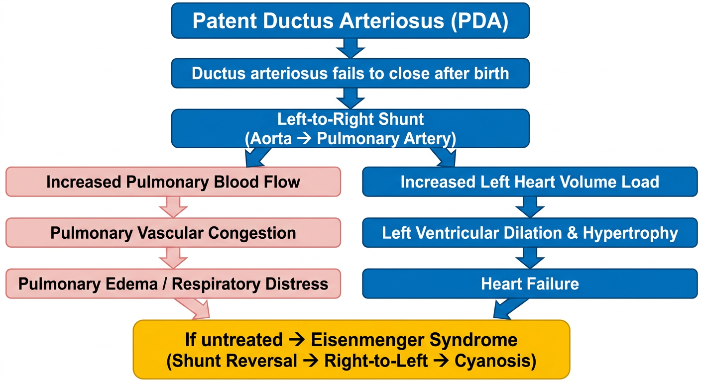
- PDA - the ductus arteriosus (a fetal vessel connecting the aorta and pulmonary artery) fails to close after birth.
- Left-to-right shunt - because aortic pressure is higher than pulmonary artery pressure, blood flows from the aorta into the pulmonary circulation.
- Pulmonary pathway - excess blood in the lungs causes pulmonary overcirculation, congestion, and respiratory distress (tachypnea, diaphoresis with feeds, poor weight gain).
- Cardiac pathway - the left heart is volume-overloaded, leading to left ventricular dilation, hypertrophy, and eventually heart failure. Wide pulse pressure and bounding pulses are classic signs.
- Eisenmenger syndrome (end stage, if untreated) - rising pulmonary vascular resistance eventually reverses the shunt to right-to-left, causing cyanosis.
Note: Web search was temporarily unavailable, so this diagram was generated from textbook content rather than a pre-existing authorized web image. The underlying medical content is directly sourced from Tintinalli's Emergency Medicine, A Comprehensive Study Guide.