
Case study in emergency medicine
Topic pneumonia
What is the most likely diagnosis
pneumonia chest X-ray consolidation
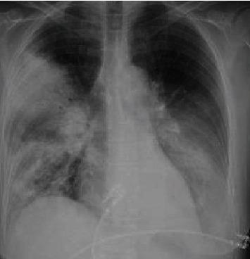
This diagnostic image is a posterior-anterior (PA) view chest X-ray demonstrating multifocal pneumonia with prominent consolidations. In the right lung, there is dense consolidation involving the upper lobe, characterized by increased radiopacity that obscures the regional vascular markings. Patchy, ill-defined airspace opacities are also visible extending into the right lower lobe. The left lung field reveals significant consolidation primarily localized within the lower lobe, partially obscuring the left hemidiaphragm silhouette. The cardiac silhouette appears normal in size, and the trachea is midline. The visual findings are consistent with severe pulmonary infection, such as atypical pneumonia (e.g., Legionella species). This clinical imaging serves as an educational example of multifocal bacterial consolidation and its radiographic distribution in the context of acute respiratory distress and sepsis.
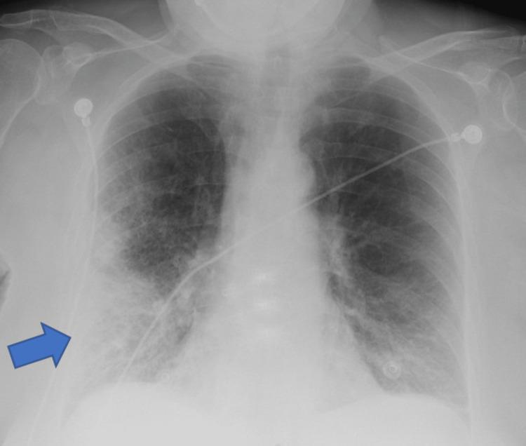
A frontal (anteroposterior) chest X-ray demonstrating clinical signs of pneumonia. A prominent blue arrow indicates a large area of increased opacity in the right lower lung field, suggestive of lobar consolidation or a dense pulmonary infiltrate. The opacity is heterogeneous with ill-defined borders, blending into the surrounding parenchyma. Beyond the focal area, there are diffuse bilateral reticulonodular interstitial markings, more pronounced in the mid and lower lung zones. The right costophrenic angle is relatively sharp, while the left shows mild blunting. Several medical devices are present: multiple radiopaque ECG leads/electrodes are positioned on the upper chest and shoulders, and a radiopaque enteric tube (nasogastric or feeding tube) is visible traversing the mediastinum toward the stomach. The image illustrates typical radiological findings of an infectious process in an acute clinical setting, highlighting focal consolidation against a background of generalized pulmonary congestion or inflammation.
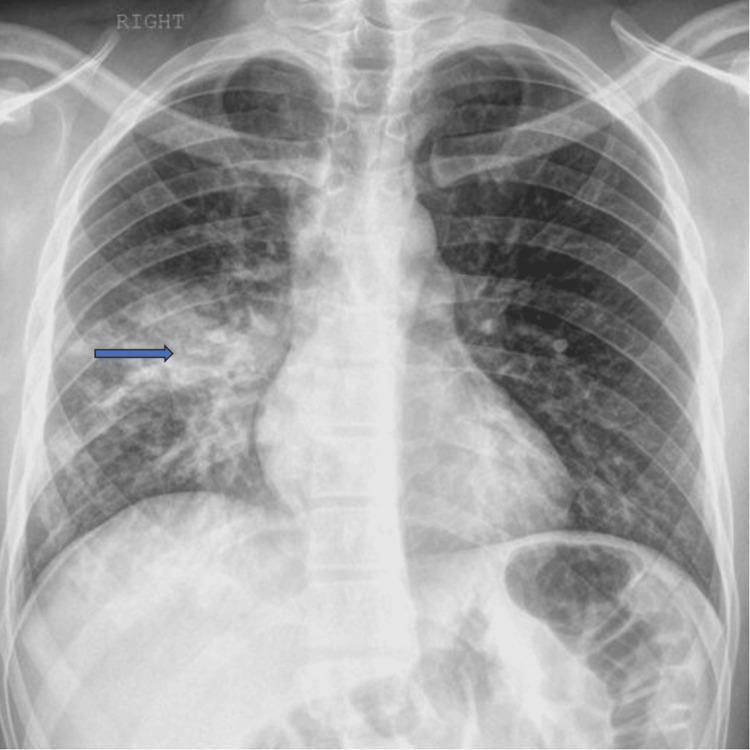
This diagnostic image is a posterior-anterior (PA) chest X-ray demonstrating common radiographic features of focal pneumonia. The primary finding is located in the right lung field, where patchy airspace opacities and an area of pulmonary consolidation are visible, highlighted by a blue arrow. The consolidation is characterized by increased density that obscures the underlying pulmonary vasculature, consistent with fluid or inflammatory exudate replacing air in the alveoli. The left lung field appears clear without similar focal opacities. The mediastinal contours, including the cardiac silhouette and trachea, are within normal limits. The costophrenic angles are sharp, suggesting the absence of significant pleural effusion. Bony structures, including the ribs, clavicles, and vertebrae, as well as the surrounding soft tissues, show no acute abnormalities. This image serves as a clinical example of localized infectious process, such as community-acquired pneumonia, in an adult patient.
Emergency Medicine Case Study: Pneumonia
THE CASE
History of Present Illness
- 3 days of worsening productive cough with rust-colored sputum
- High fever with shaking chills (T 39.6°C at home)
- Acute onset right-sided pleuritic chest pain (sharp, worse with breathing)
- Progressive dyspnea - now short of breath at rest
- No nausea, vomiting, or diarrhea
- No recent hospitalization or travel
Physical Examination
| Parameter | Finding |
|---|---|
| Temp | 39.8°C (103.6°F) |
| HR | 112 bpm |
| BP | 102/68 mmHg |
| RR | 26 breaths/min |
| SpO2 | 88% on room air |
| GCS | 14 (mildly confused) |
- Decreased breath sounds right lower lobe
- Dullness to percussion at right base
- Bronchial breath sounds + coarse crackles right lower lobe
- Egophony (E-to-A change) present at right base
Investigations
- WBC: 18,400/µL with 88% neutrophils, 10% bands (left shift)
- CRP: 210 mg/L (elevated)
- Procalcitonin: 3.8 ng/mL (elevated - suggests bacterial infection)
- BUN/Cr: 32/1.6 (mildly elevated - dehydration)
- Blood glucose: 290 mg/dL
- Blood cultures: sent (×2 sets)
QUESTION: What is the Most Likely Diagnosis?
ANSWER: Community-Acquired Pneumonia (CAP) due to Streptococcus pneumoniae
Diagnostic Reasoning
| Clinical Clue | Points To |
|---|---|
| Sudden onset with shaking rigors | Classic pneumococcal feature |
| Rust-colored (blood-tinged) sputum | Pathognomonic for S. pneumoniae |
| Lobar consolidation on CXR (right lower lobe) | Typical for pneumococcal CAP |
| Pleuritic chest pain | Pleural involvement from pneumococcus |
| High fever (39.8°C) with marked leukocytosis | Typical/bacterial rather than atypical |
| Age 68, COPD, DM, unvaccinated | Classic host risk factors |
Why NOT the Alternatives?
| Organism | Against it in this case |
|---|---|
| Mycoplasma pneumoniae | "Walking pneumonia" - gradual onset, dry cough, younger patients, interstitial pattern on CXR - not lobar |
| Legionella | No diarrhea/GI symptoms, no water-exposure history, no hyponatremia; Legionella also shows multiple patchy non-segmented infiltrates, not lobar consolidation |
| Klebsiella | More common in alcoholics; "currant jelly" sputum; upper lobe with bulging fissure sign |
| S. aureus | Gradual onset post-viral illness; multilobar patchy infiltrates, abscesses |
| H. influenzae | More insidious; patient already has COPD but basilar patchy (not lobar) pattern |
| PE/CHF | No edema, no DVT risk factors, no bilateral involvement |
Severity Assessment - PSI/PORT Score (CURB-65)
| CURB-65 Criterion | This Patient | Points |
|---|---|---|
| Confusion (new) | Yes (GCS 14) | +1 |
| Urea > 7 mmol/L (BUN > 20) | Yes (BUN 32) | +1 |
| Respiratory rate ≥ 30/min | Yes (RR 26 - borderline) | +1 |
| Blood pressure (SBP <90 or DBP <60) | Yes (BP 102/68) | +1 |
| 65 years or older | Yes (68 years) | +1 |
Emergency Management
- Airway/Breathing: Supplemental O2 to maintain SpO2 ≥94%; prepare for possible NIV or intubation given SpO2 88%
- IV Access + Fluids: Aggressive fluid resuscitation for hypotension (30 mL/kg NS bolus)
- Antibiotics within 4 hours (ideally <1 hour for severe CAP):
- IV Beta-lactam (ceftriaxone 1-2g IV) + macrolide (azithromycin 500mg IV) - for typical + atypical coverage
- OR fluoroquinolone monotherapy (levofloxacin)
- Blood cultures × 2 before antibiotics (already sent)
- Urine Legionella/Pneumococcal antigens - especially for severe CAP
- Glucose control - address hyperglycemia (290 mg/dL)
- ICU consultation given CURB-65 ≥4 and hemodynamic instability
- Pneumococcal + influenza vaccination on discharge
Key Teaching Points
- Rust-colored sputum + sudden onset + lobar consolidation = S. pneumoniae until proven otherwise
- Always classify acquiring environment: CAP vs HAP vs VAP vs HCAP - this drives organism prediction and antibiotic choice
- CURB-65 is the EM go-to severity score; PSI/PORT is more detailed
- Atypical presentations (confusion only, no fever) are common in elderly and diabetic patients
- Time-to-antibiotic matters - target <4 hours for CAP, <1 hour for septic patients