
Facial nerve landmarks
facial nerve surgical landmarks parotidectomy anatomy
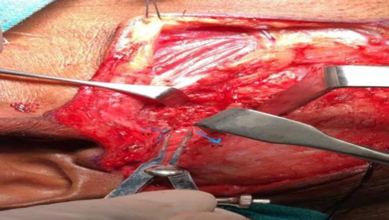
This clinical photograph illustrates an intraoperative view of an open parotidectomy surgical field. The image highlights the identification and measurement of surgical landmarks within the parotid bed. Castroviejo calipers are utilized to measure the shortest distance between the tragal pointer (TP), indicated by blue dotted lines, and the main trunk of the facial nerve (FN), denoted by a blue arrow. The surgical field displays exposed soft tissues, including the posterior belly of the digastric muscle and the sternocleidomastoid muscle, following glandular resection. Self-retaining or manual retractors are visible, providing exposure of the deep neck anatomy and the facial nerve's exit from the stylomastoid foramen. This visual demonstrates critical surgical steps in head and neck surgery, emphasizing the importance of anatomical landmarks for the preservation of the facial nerve during parotid gland procedures. The clinical focus is on surgical anatomy, nerve monitoring, and precise measurement for surgical outcomes.
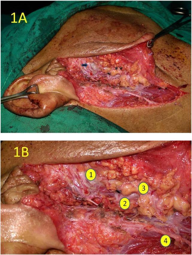
Clinical intraoperative photograph of a right quadrant parotidectomy, presented in two panels (1A and 1B). Panel 1A provides a wide surgical field view showing the post-excision anatomy of the upper quadrant. A blue arrow identifies the skeletonized zygomatic branch of the facial nerve, while a double asterisk marks the rough, nodular transected surface of the lower parotid gland quadrant. Panel 1B is a high-magnification close-up view identifying key neurovascular and glandular landmarks: (1) the zygomatic branch of the upper trunk of the facial nerve, appearing as a thin, white fiber; (2) the origin of the lower trunk of the facial nerve; (3) the yellowish, lobulated parenchyma of the transected lower quadrant; and (4) the great auricular nerve located inferior to the primary surgical site. The images demonstrate the surgical preservation of the facial nerve trunk and its branches during selective salivary gland resection. This material is suited for teaching surgical anatomy, parotid surgery techniques, and nerve-sparing protocols in otolaryngology or surgical oncology.
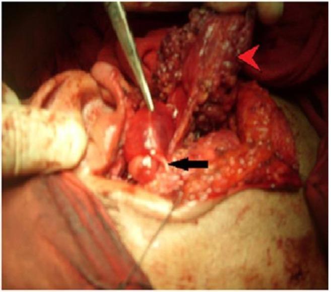
Clinical intraoperative photograph of the left parotid region during a parotidectomy. The surgical field demonstrates the relationship between a focal mass and the facial nerve. A red arrowhead indicates the retracted superficial lobe of the parotid gland, revealing granular glandular tissue. Centrally, a well-circumscribed, smooth-walled mass is visible. A black arrow points to the superior division of the facial nerve, which appears as a thin, white, fibrous strand directly coursing over and becoming encased within the mass. A metal surgical dissector is shown at the superior aspect of the lesion to facilitate exposure. This visual demonstrates the surgical anatomy of a facial nerve neurofibroma and the technical challenge of nerve preservation when a neoplasm incorporates nerve fascicles. Key landmarks include the auricle (left) and the dissected surgical bed with associated soft tissue and vascularity.
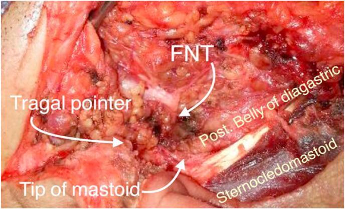
This clinical photograph shows an intraoperative surgical field during a parotidectomy, focusing on the identification of the facial nerve trunk (FNT). The image illustrates key anatomical landmarks used to localize the nerve as it exits the stylomastoid foramen. The FNT is centrally located and annotated in the dissection field. Superior to the nerve is the 'Tragal pointer', a cartilaginous landmark. Inferior and slightly posterior to the FNT is the 'Tip of mastoid' process. Additional visible muscular landmarks include the posterior belly of the digastric muscle, which lies deep and inferior to the nerve trunk, and the sternocleidomastoid muscle, visible at the inferior-most aspect of the surgical site. The photograph demonstrates the spatial relationship between these soft tissue and bony structures, which serve as essential surgical guides for preventing iatrogenic facial nerve injury during head and neck surgery. The educational focus is on the surgical anatomy of the parotid region and the safe identification of the extra-temporal facial nerve.
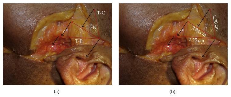
This clinical photograph displays a surgical dissection of the preauricular and parotid regions, illustrating the surgical anatomy and morphometric relationships of the facial nerve. The image is divided into two panels (a and b) showing the same field with anatomical labels and measurements. Key landmarks identified include the tragus (T), the lateral pole of the mandibular condyle (C), the pes anserinus (P) of the facial nerve, and the temporozygomatic division of the facial nerve (FN). Panel (b) specifically quantifies the distances from the posterior apex of the tragus to these landmarks: 2.20 cm to the lateral pole of the condyle (T-C), 2.31 cm to the point where the facial nerve crosses the posterior border of the condylar neck (T-FN), and 2.25 cm to the pes anserinus (T-P). This anatomical diagram serves as a guide for identifying and preserving the facial nerve during preauricular surgical approaches, such as those used in parotidectomy or temporomandibular joint surgery, by providing standardized reference measurements from the tragus.
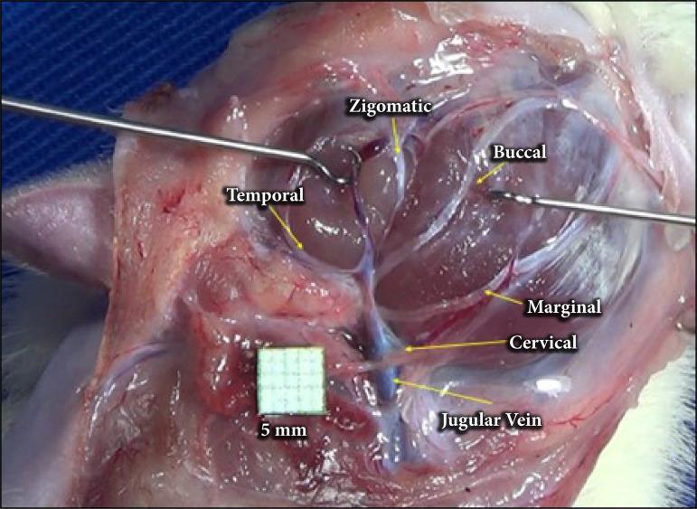
A clinical intraoperative photograph showing the anatomical arrangement of the facial nerve branches in a surgical dissection, following parotidectomy. The image provides a lateral view of the cervicofacial region where the parotid gland has been removed to expose the extratemporal facial nerve plexus. Labeled branches include the temporal branch extending superiorly, the zygomatic and buccal branches traveling toward the mid-face, and the marginal mandibular and cervical branches descending inferiorly. The cervical branch is depicted in close proximity to the jugular vein, which is visible in the lower surgical field. Surgical instruments (retractors) are positioned to maintain exposure of the deep tissues. A 5mm grid scale is included for anatomical measurement. This visual serves as an educational guide for surgical anatomy, identifying key landmarks for nerve preservation during parotid gland surgery or facial nerve decompression.
Facial Nerve Landmarks
Intratemporal Landmarks (Mastoid/Middle Ear Surgery)
- Cochleariform process (processus cochleariformis): The facial nerve passes just superior to this structure and the oval window as it travels from the geniculate ganglion toward the second genu - Shambaugh Surgery of the Ear, p. 533.
- Second genu: Posterior to the oval window, the nerve turns sharply inferior to begin its vertical (mastoid) segment. This genu lies a few millimeters anteromedial to the lateral semicircular canal, making the lateral semicircular canal one of the most reliable landmarks for localizing the nerve during mastoidectomy - Shambaugh Surgery of the Ear, p. 533.
- Fossa incudis / short process of the incus: In the posterior epitympanum, this is an important surgical landmark for the facial nerve as it approaches the second genu - Shambaugh Surgery of the Ear, p. 4019 (block 7).
- Digastric ridge: A bony ridge on the mastoid that points toward the stylomastoid foramen and vertical (mastoid) segment of the nerve - used as a landmark during canal-wall-up mastoidectomy - Shambaugh Surgery of the Ear.
- Pyramidal eminence: The origin of the stapedius tendon; a reliable landmark for the second genu, distal to which the nerve continues as the mastoid segment - Cummings Otolaryngology Head and Neck Surgery, p. 3543-3544.
Extratemporal Landmarks (Parotid/Facial Surgery)
- Tragal pointer: The inferior tip of the cartilaginous external auditory canal. The main trunk of the facial nerve lies approximately 1 cm deep and inferior to this point. It is a mobile landmark, so care is needed when retracting - Scott-Brown's Otorhinolaryngology, p. 167.
- Tip of the mastoid process: Located posteroinferior to the nerve trunk; used together with the tragal pointer to triangulate the stylomastoid foramen exit point.
- Posterior belly of the digastric muscle: Lies deep and inferior to the nerve trunk; its anterior border is a classic landmark, particularly useful during retrograde identification of individual branches (e.g., marginal mandibular).
- Tympanomastoid suture line: The nerve exits roughly 6-8 mm deep to the inferior end of this suture in many descriptions, a landmark used when the tragal pointer is distorted by tumor or scarring.
- Retromandibular vein: Generally lies just deep to the facial nerve and its branches (especially the marginal mandibular branch); a constant landmark used in the retrograde method of nerve identification - Bailey & Love's Short Practice of Surgery, 28th ed., p. 2879-2885.
- Pes anserinus: The point where the nerve trunk divides into its temporofacial and cervicofacial divisions, just within the substance of the gland - used as a reference point in morphometric studies (average ~2.25 cm from the posterior tragus in one series).
- Great auricular nerve: Runs superficial to the sternocleidomastoid and is encountered before the facial nerve trunk; its posterior branch should be preserved when possible during dissection.
Intraoperative photo - key landmarks for finding the facial nerve trunk
Practical Summary
| Landmark | Location relative to nerve | Used in |
|---|---|---|
| Tragal pointer | Nerve trunk ~1 cm deep/inferior | Parotidectomy |
| Tympanomastoid suture | Nerve ~6-8 mm deep to inferior end | Parotidectomy |
| Posterior belly of digastric | Nerve lies superior to it | Parotidectomy |
| Retromandibular vein | Nerve (esp. marginal mandibular branch) lies superficial | Retrograde dissection |
| Second genu / lateral semicircular canal | Nerve turns vertical just anteromedial to canal | Mastoid surgery |
| Pyramidal eminence | Marks second genu, distal mastoid segment begins | Middle ear surgery |
| Cochleariform process | Nerve passes just superior to it | Middle ear surgery |
| Digastric ridge | Points toward stylomastoid foramen | Mastoidectomy |