
Tongue histology 5m answer
Reading File
Now let me search the medical library for tongue histology content in parallel:
Finding Sources
Finding Sources
Finding Sources
Finding Sources
Excellent! Found dedicated tongue histology sections in Junqueira's Basic Histology and in Histology: A Text and Atlas. Let me read both in parallel:
Reading File
Reading File
Good content found. Let me read more from both textbooks to get details on taste buds, papillae types, and glands:
Reading File
Reading File
I now have excellent content from both Junqueira's and Histology A Text and Atlas. Let me fetch the key histology images to include in the answer:
Loading Image
Loading Image
Loading Image
I now have all the content and images needed for a thorough 5-mark answer. Here is the complete answer:
Histology of the Tongue
Sources: Junqueira's Basic Histology, 17e; Histology: A Text and Atlas, 8e
1. General Structure
The tongue is a muscular organ covered by mucous membrane (mucosa). Its bulk consists of striated (voluntary) muscle arranged in three planes - longitudinal, transverse, and vertical - running at right angles to one another. This unique triplanar organization is found only in the tongue and permits its extraordinary mobility for speech, mastication, and swallowing. Variable amounts of adipose tissue are interspersed between the muscle bundles. The lamina propria penetrates between the muscle fascicles, making the mucosa strongly adherent to the muscular core.
- The ventral (inferior) surface has typical smooth lining mucosa - non-keratinized stratified squamous epithelium.
- The dorsal surface is specialized mucosa bearing hundreds of lingual papillae on the anterior two-thirds, and lingual tonsils on the posterior third (root).
- A V-shaped groove, the sulcus terminalis, separates these two regions. At its apex lies the foramen cecum, the embryological remnant of the thyroid diverticulum.
2. Lingual Papillae (Four Types)
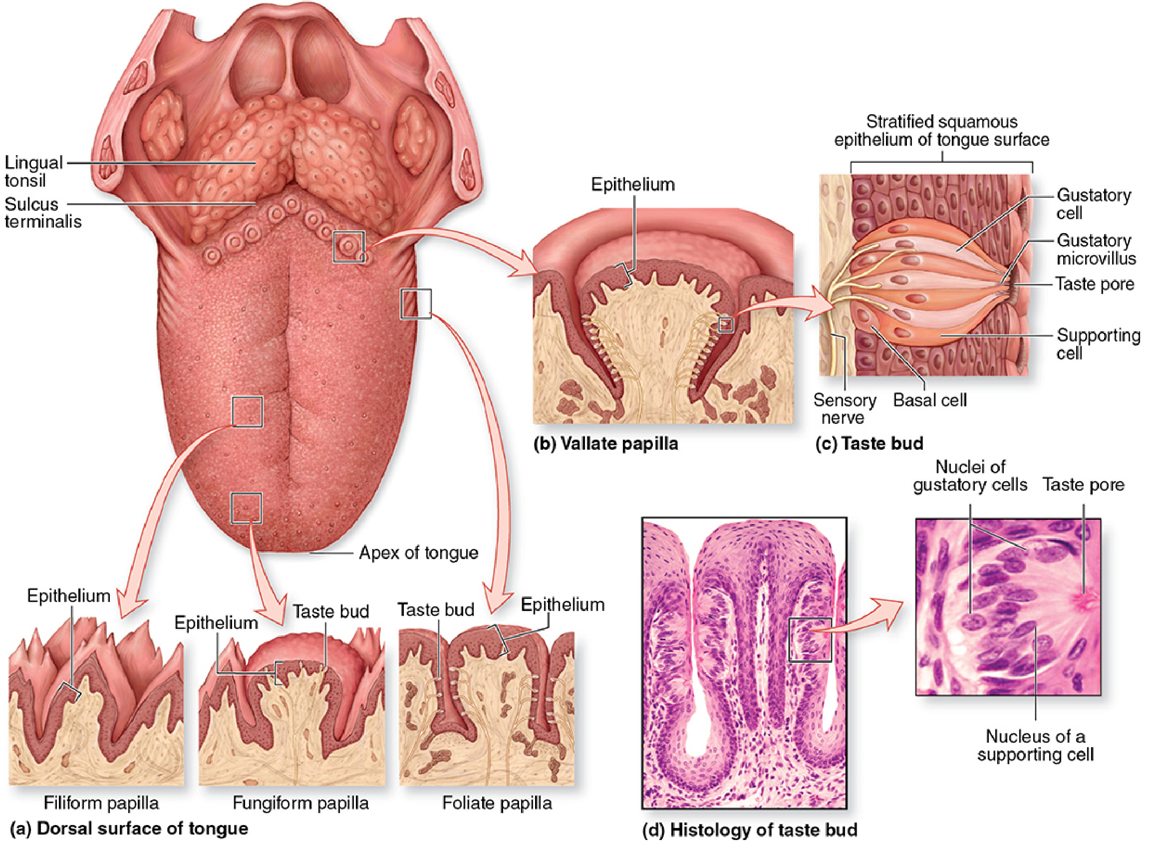
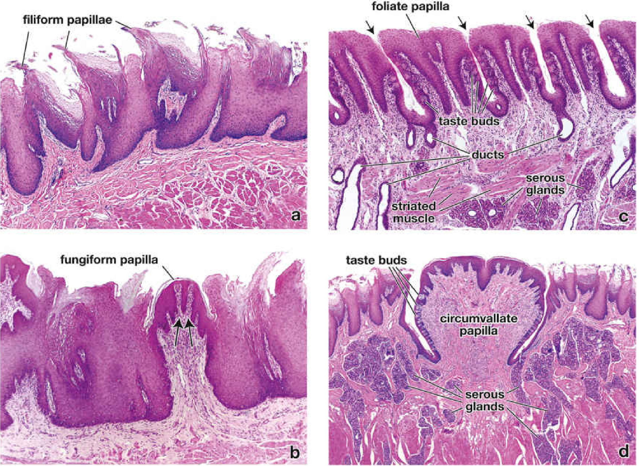
| Papilla | Shape | Epithelium | Taste Buds | Notes |
|---|---|---|---|---|
| Filiform | Elongated, conical, pointed | Heavily keratinized SSE | Absent | Most numerous; mechanical function only; tips point posteriorly |
| Fungiform | Mushroom/bulbous | Lightly keratinized SSE | Present (scattered) | Less numerous; well-vascularized CT core; appear as red dots grossly |
| Circumvallate (Vallate) | Large, flat-topped, surrounded by a moat/trench | Slightly keratinized SSE | Very abundant on lateral walls (~250 per papilla) | Only 8-12 in number; arranged in a V near sulcus terminalis; von Ebner glands drain into moat |
| Foliate | Parallel leaf-like ridges on lateral tongue | Non-keratinized SSE | Present on lateral surfaces | Rudimentary in adult humans; best developed in children |
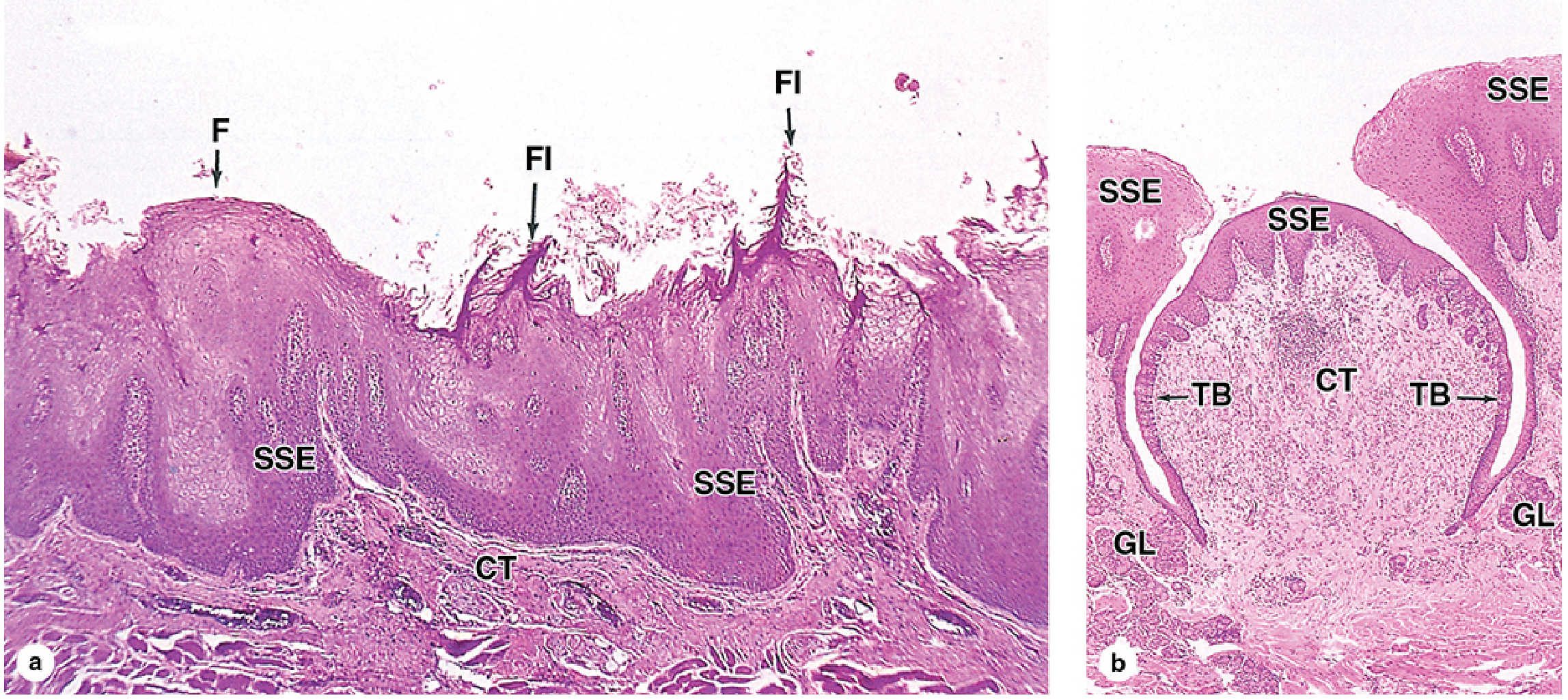
3. Taste Buds
Taste buds are ovoid neuroepithelial structures embedded within the stratified squamous epithelium. They are most abundant on circumvallate papillae, present on fungiform and foliate papillae, but absent from keratinized filiform papillae.
Cell types (50-100 cells per bud):
- Gustatory (taste) cells - elongated cells; bear microvilli that project through the taste pore (a 2-µm apical opening) to sample dissolved molecules (tastants) in saliva. Turn over every 7-10 days.
- Supporting cells - slender; function not fully defined.
- Basal stem cells - slowly dividing; give rise to all other cell types.
- Afferent sensory axons enter at the base through the basal lamina and synapse with gustatory cells.
Five taste modalities detected: salty (Na⁺ ions), sour (H⁺ from acids), sweet (sugars), bitter (alkaloids/toxins), and umami (amino acids e.g. glutamate). Salt and sour use ion channel receptors; sweet, bitter, and umami use G-protein-coupled receptors.
4. Von Ebner (Lingual Serous) Glands
These are pure serous minor salivary glands located in the connective tissue deep to the circumvallate and foliate papillae. Their ducts open into the moat (trench) surrounding each circumvallate papilla. They continuously flush the moat with a watery secretion, washing away food particles so taste buds receive fresh stimuli. Their secretion also contains a lingual lipase that prevents formation of a hydrophobic film over taste buds which would otherwise impair gustation.
5. Posterior Tongue - Lingual Tonsils
The posterior third (root) of the tongue lacks papillae. Instead, the dorsal surface shows an irregular, nodular contour due to underlying lingual tonsils - aggregates of lymphoid nodules within the lamina propria covered by non-keratinized stratified squamous epithelium with deep crypts.
Quick Summary Table
| Feature | Anterior 2/3 | Posterior 1/3 |
|---|---|---|
| Surface | Lingual papillae | Lingual tonsils |
| Epithelium | Keratinized SSE (specialized mucosa) | Non-keratinized SSE |
| Nerve supply | Ant. 2/3: lingual n. (general); chorda tympani (taste) | Post. 1/3: glossopharyngeal n. (general + taste) |
- Junqueira's Basic Histology, 17e, pp. 738-741
- Histology: A Text and Atlas, 8e, pp. 1419-1423
This is a shared conversation. Sign in to Orris to start your own chat.