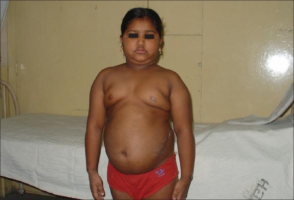
Cushing vs addison
Addison disease hyperpigmentation adrenal insufficiency
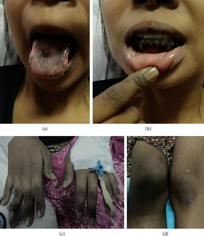
This set of clinical photographs illustrates the hallmark signs of primary adrenal insufficiency (Addison's disease), characterized by generalized mucocutaneous hyperpigmentation. Panel (a) shows significant hyperpigmentation of the tongue, presenting as dark, irregular macules and diffuse speckling primarily on the dorsal surface, accompanied by a faint whitish plaque. Panel (b) depicts mucosal hyperpigmentation of the gingiva and inner lip. Panel (c) demonstrates diffuse hyperpigmentation of the hands, with notable darkening over the extensor surfaces of the interphalangeal joints (knuckle hyperpigmentation), while the nail beds appear spared. Panel (d) shows similar localized hyperpigmentation over the knees. These visual findings result from increased levels of adrenocorticotropic hormone (ACTH) and its precursor, pro-opiomelanocortin, which stimulate melanocytes via alpha-melanocyte-stimulating hormone receptors. This presentation is essential for the clinical diagnosis of chronic adrenal cortical failure and differentiates primary from secondary adrenal insufficiency.
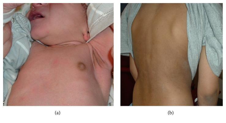
Two-panel clinical photograph demonstrating cutaneous hyperpigmentation associated with pediatric adrenal insufficiency. Panel (a) shows the anterior chest and axilla of a 10-day-old male infant. Notable findings include marked hyperpigmentation of the nipple and areola, as well as the axillary vault, contrasting with the lighter surrounding skin. This presentation is characteristic of salt-wasting congenital adrenal hyperplasia (CAH) due to 21-hydroxylase deficiency. Panel (b) displays the posterior torso of a 9-year-old male with Addison's disease, showing diffuse, generalized bronzing of the skin with subtle mottling. Pathophysiologically, these skin changes result from elevated levels of adrenocorticotropic hormone (ACTH) and melanocyte-stimulating hormone (MSH) due to lack of cortisol negative feedback. The images highlight key diagnostic dermatologic signs of primary adrenal insufficiency in different pediatric age groups, emphasizing the importance of examining skin creases and pigmented areas for early clinical recognition of endocrinological disorders.
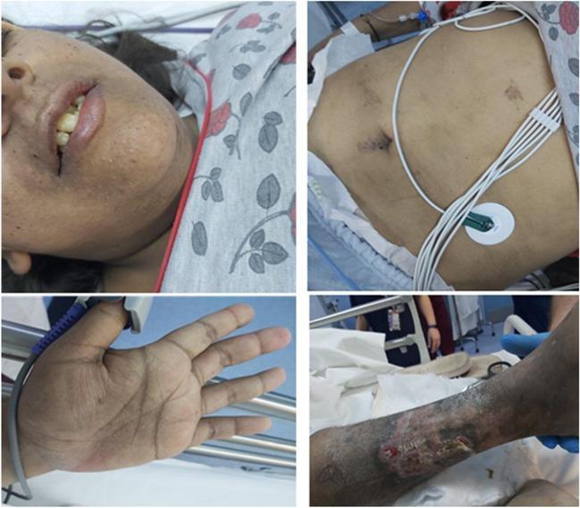
This composite of four clinical photographs illustrates classic dermatological manifestations of Primary Adrenal Insufficiency (PAI). The top-left image displays perioral and labial hyperpigmentation, with increased melanin deposition along the vermilion border. The bottom-left image highlights characteristic hyperpigmentation of the palmar creases, where skin darkening is preferentially concentrated in the flexural lines of the hand. The top-right image shows the patient's abdomen with hyperpigmented surgical scars from a recent laparoscopic cholecystectomy, demonstrating the tendency for pigmentation to accentuate at sites of trauma or friction; medical monitoring via ECG leads is also visible. The bottom-right image shows the left lower extremity featuring a chronic skin ulcer with surrounding hyperpigmentation and stasis-like changes. Collectively, these findings—generalized hyperpigmentation with accentuation in creases, scars, and mucous membranes—are hallmark signs of elevated adrenocorticotropic hormone (ACTH) levels, aiding in the clinical diagnosis of Addison's disease or adrenal crisis in a hospital setting.
Cushing Syndrome vs. Addison Disease
The Adrenal Axis (Quick Orientation)
Cushing Syndrome - Hypercortisolism
Definition & Causes
| Category | Details |
|---|---|
| Exogenous (most common overall) | Iatrogenic - oral, topical, inhaled steroids. Always take a thorough drug history. |
| ACTH-dependent (80-85% of endogenous) | Pituitary adenoma (Cushing disease, ~70%) or ectopic ACTH from malignancy (~10%) |
| ACTH-independent | Adrenal adenoma, adrenal carcinoma, or bilateral hyperplasia |
Cushing disease specifically refers to a pituitary ACTH-secreting corticotroph adenoma (described by Harvey Cushing in 1932). Cushing syndrome is the broader term for hypercortisolism from any source.
Clinical Features
| System | Features |
|---|---|
| Adipose/Body habitus | Central (truncal) obesity, moon facies, buffalo hump (dorsocervical fat pad), supraclavicular fat pads |
| Skin | Wide purple striae (>1 cm), easy bruising, thin skin, acne |
| Metabolic | Hyperglycemia (steroid diabetes), hypertension, hyperlipidemia |
| Musculoskeletal | Proximal muscle wasting/weakness, osteoporosis, pathological fractures |
| Reproductive | Virilization in women, menstrual irregularity, decreased libido |
| Neuropsychiatric | Depression, emotional lability, psychosis |
| Immune | Immunosuppression, increased infection susceptibility |
Lab Findings
- ACTH: LOW (in primary adrenal/exogenous) or HIGH (in pituitary/ectopic)
- Elevated serum cortisol (loss of diurnal rhythm - elevated midnight cortisol is pathologic)
- Hyperglycemia, hypokalemia (especially ectopic ACTH)
- Elevated 24-hour urine free cortisol
Diagnosis
- 24-hour urine cortisol (>90 mcg/24h)
- Midnight salivary cortisol
- 1 mg overnight dexamethasone suppression test (failure to suppress = Cushing's)
- ACTH levels to classify ACTH-dependent vs. independent
Addison Disease - Primary Adrenocortical Insufficiency
Definition & Causes
- Autoimmune destruction of all adrenal cortex zones (most common in developed world)
- Infections (TB, fungal, HIV)
- Bilateral adrenal hemorrhage (Waterhouse-Friderichsen syndrome)
- Infiltration (metastases, amyloid)
- Part of polyglandular autoimmune syndrome types I, II, IV (with hypoparathyroidism, candidiasis, vitiligo, thyroiditis, diabetes)
Clinical Features
| System | Features |
|---|---|
| Metabolic | Hypoglycemia, hyponatremia, hyperkalemia, metabolic acidosis |
| Cardiovascular | Hypotension, orthostatic hypotension, salt craving |
| GI | Anorexia, weight loss, nausea, vomiting, diarrhea |
| Musculoskeletal | Profound weakness, fatigue |
| Skin (hallmark!) | Hyperpigmentation - diffuse, most prominent in sun-exposed areas, pressure sites, mucous membranes, palmar creases, new scars, nipples, areolae, nail beds |
| Reproductive (women) | Decreased pubic/axillary hair, decreased libido (loss of adrenal androgens) |
Hyperpigmentation occurs because low cortisol removes negative feedback on the pituitary → ACTH rises dramatically. ACTH is cleaved from POMC, the same precursor as alpha-MSH, which stimulates melanocytes. This sign only appears in PRIMARY adrenal failure (not in secondary/pituitary causes). - Costanzo Physiology; Andrews' Dermatology
Lab Findings
- ACTH: HIGH (primary insufficiency)
- Low cortisol, low aldosterone
- Hyponatremia, hyperkalemia, metabolic acidosis
- Hypoglycemia
Diagnosis
- Paired serum cortisol + plasma ACTH
- Cosyntropin (ACTH) stimulation test: failure to raise cortisol above 550 nmol/L at 1 hour is diagnostic
- CT adrenals to rule out infiltration/infection
- Normal to low ACTH in secondary (pituitary/hypothalamic) insufficiency
Treatment
Side-by-Side Comparison
| Feature | Cushing Syndrome | Addison Disease |
|---|---|---|
| Cortisol | Excess | Deficient |
| Aldosterone | Often excess | Deficient (primary) |
| ACTH | Low (primary/exogenous) / High (pituitary/ectopic) | High (primary) / Low (secondary) |
| Body weight | Increased (central obesity) | Decreased (weight loss) |
| Blood pressure | Hypertension | Hypotension |
| Blood glucose | Hyperglycemia | Hypoglycemia |
| Sodium | Normal/high | Low (hyponatremia) |
| Potassium | Low/normal | High (hyperkalemia) |
| Skin | Thin, striae, bruising | Hyperpigmentation |
| Muscle | Wasting/weakness | Weakness |
| Immune | Suppressed | Not primarily affected |
| Key skin clue | Purple striae, moon face, buffalo hump | Diffuse bronze hyperpigmentation |
Clinical Pearl: Why Hyperpigmentation in Addison?
Clinical Images