
Give detailed notes on anatomy of female genital tract,including reproductive system and external genitalia,development and embryology ,placenta and fetal membrane for mbbs
Reading File
This request covers about four chapters of an MBBS anatomy and embryology syllabus, so I'll split it up and go deep on one part at a time. Pick a topic below.
Topic map
- Female external genitalia (vulva): mons pubis, labia majora and minora, clitoris, vestibule, vestibular bulbs, Bartholin's and Skene's glands, blood supply, nerve supply and lymphatics.
- Internal reproductive organs: vagina, uterus (parts, ligaments, supports, relations, blood supply, lymphatics, nerve supply), uterine tubes and ovaries, with applied anatomy.
- Development and embryology of the female genital tract: gonadal differentiation, paramesonephric (Müllerian) ducts, uterovaginal canal, vaginal plate, external genitalia, descent of the ovary, and anomalies such as septate and bicornuate uterus, Mayer-Rokitansky-Küster-Hauser syndrome, androgen insensitivity syndrome, hymen and vaginal atresia.
- Placenta and fetal membranes: decidua, chorionic villi, placental structure, circulation and barrier, functions, amnion, chorion, umbilical cord, and anomalies such as placenta previa, accreta, and single umbilical artery.
Brief overview
- Structure: The female genital tract has external parts (the vulva) and internal parts (vagina, uterus, uterine tubes, ovaries). The vulva sits in the perineum. The internal organs lie in the pelvis and are supported by the pelvic floor, the cardinal (transverse cervical) ligaments and the uterosacral ligaments.
- Development: The paramesonephric ducts form the uterine tubes, uterus and upper vagina. The mesonephric (Wolffian) ducts regress in females. The lower vagina comes from the sinovaginal bulbs of the urogenital sinus. The external genitalia stay female by default when there is no testosterone or dihydrotestosterone.
- Placenta and membranes: The placenta has a fetal part (chorion frondosum) and a maternal part (decidua basalis). It is discoid, weighs about 500 g and is haemochorial. The fetal membranes are the amnion, chorion and decidua capsularis. The umbilical cord normally contains two arteries and one vein.
Asking for Preferences
Which topic do you want detailed MBBS notes on first?
This request covers about four chapters of an MBBS anatomy and embryology syllabus, so I'll split it up and go deep on one part at a time. Pick a topic below.
Topic map
- Female external genitalia (vulva): mons pubis, labia majora and minora, clitoris, vestibule, vestibular bulbs, Bartholin's and Skene's glands, blood supply, nerve supply and lymphatics.
- Internal reproductive organs: vagina, uterus (parts, ligaments, supports, relations, blood supply, lymphatics, nerve supply), uterine tubes and ovaries, with applied anatomy.
- Development and embryology of the female genital tract: gonadal differentiation, paramesonephric (Müllerian) ducts, uterovaginal canal, vaginal plate, external genitalia, descent of the ovary, and anomalies such as septate and bicornuate uterus, Mayer-Rokitansky-Küster-Hauser syndrome, androgen insensitivity syndrome, hymen and vaginal atresia.
- Placenta and fetal membranes: decidua, chorionic villi, placental structure, circulation and barrier, functions, amnion, chorion, umbilical cord, and anomalies such as placenta previa, accreta, and single umbilical artery.
Brief overview
- Structure: The female genital tract has external parts (the vulva) and internal parts (vagina, uterus, uterine tubes, ovaries). The vulva sits in the perineum. The internal organs lie in the pelvis and are supported by the pelvic floor, the cardinal (transverse cervical) ligaments and the uterosacral ligaments.
- Development: The paramesonephric ducts form the uterine tubes, uterus and upper vagina. The mesonephric (Wolffian) ducts regress in females. The lower vagina comes from the sinovaginal bulbs of the urogenital sinus. The external genitalia stay female by default when there is no testosterone or dihydrotestosterone.
- Placenta and membranes: The placenta has a fetal part (chorion frondosum) and a maternal part (decidua basalis). It is discoid, weighs about 500 g and is haemochorial. The fetal membranes are the amnion, chorion and decidua capsularis. The umbilical cord normally contains two arteries and one vein.
Finding Sources
Finding Sources
Searching Images
female internal genital organs uterus tubes ovary coronal section
Searching Images
development of female genital tract paramesonephric ducts uterovaginal canal
Searching Images
structure of placenta chorionic villi maternal fetal circulation
Reading File
Reading File
Finding Sources
Reading File
Reading File
Reading File
Searching PubMed
"placenta accreta"[MeSH Terms] OR "placenta previa"[MeSH Terms]
Female genital tract, development, placenta and fetal membranes (MBBS notes)
These notes draw on Gray's Anatomy for Students, The Developing Human, and Langman's Medical Embryology. Where I added standard MBBS detail from my own knowledge, I haven't tied it to a page. Check numerical values against your prescribed textbook.
PART 1. EXTERNAL GENITALIA (VULVA / PUDENDUM)
The vulva is the external genitalia lying in the urogenital triangle of the perineum.
Components
- Mons pubis
- A fatty pad over the pubic symphysis, covered by pubic hair.
- Labia majora
- Two thick, hair-bearing skin folds.
- They are homologous to the scrotum.
- They contain the termination of the round ligament of the uterus (and a possible patent processus vaginalis, the canal of Nuck).
- They are rich in sebaceous and sweat glands.
- Labia minora
- Two thin, hairless folds medial to the labia majora.
- Anteriorly they split to form the prepuce (above) and frenulum (below) of the clitoris.
- Posteriorly they fuse to form the frenulum of the labia (fourchette).
- They are homologous to the ventral penile skin and the penile urethra.
- Clitoris
- Homologous to the penis, but it is not traversed by the urethra.
- It has a glans, a body, and two crura.
- It is formed of two corpora cavernosa. There is no corpus spongiosum.
- The crura attach to the ischiopubic rami and are covered by the ischiocavernosus muscle.
- Vestibule of the vagina
- The space between the labia minora.
- It receives the openings of the urethra, vagina, ducts of the greater vestibular glands, and Skene's (paraurethral) glands.
- Bulbs of the vestibule
- Two erectile masses on either side of the vaginal orifice, covered by bulbospongiosus.
- They are homologous to the bulb of the penis.
- Greater vestibular glands (Bartholin's)
- They lie posterolateral to the vaginal orifice, in the posterior part of the bulb.
- Their ducts open at the 5 and 7 o'clock positions in the groove between the hymen and the labium minus.
- They are homologous to the bulbourethral (Cowper's) glands.
- They secrete mucus for lubrication.
- Duct blockage causes a Bartholin's cyst or abscess. Treatment is marsupialisation.
- Hymen
- A thin mucosal fold at the vaginal orifice.
- After rupture its remnants are the carunculae hymenales.
Blood supply
- Branches of the internal pudendal artery (main supply) and the external pudendal arteries (from the femoral artery).
Nerve supply
- Anterior part: ilioinguinal nerve and genital branch of the genitofemoral nerve (L1).
- Posterior part: pudendal nerve (S2 to S4) and the posterior femoral cutaneous nerve.
Lymphatic drainage
- Superficial inguinal nodes, which are the main group.
- The clitoris and glans may also drain to deep inguinal and internal iliac nodes.
PART 2. INTERNAL GENITAL ORGANS
A. Vagina
- A fibromuscular tube about 8 cm long. The posterior wall (about 9 cm) is longer than the anterior wall (about 7.5 cm).
- It lies between the bladder and urethra in front, and the rectum and anal canal behind.
- The upper end surrounds the cervix, forming the fornices: anterior, posterior and two lateral.
- The posterior fornix is the deepest. It is related to the pouch of Douglas (rectouterine pouch).
- Culdocentesis and drainage of a pelvic abscess are done through the posterior fornix.
- Relations of the lateral fornix: the ureter and uterine artery (the "water under the bridge" relation).
- Epithelium: non-keratinised stratified squamous, with no glands.
- Lubrication is by transudation. The cervix and Bartholin's glands contribute.
- Blood supply: vaginal artery (from the internal iliac), plus the uterine artery (cervicovaginal branch) and the internal pudendal artery.
- Nerve supply: the lower one third is somatic (pudendal nerve, pain sensitive). The upper two thirds is autonomic (uterovaginal plexus) and is pain insensitive.
- Lymphatics: the upper third drains to the internal and external iliac nodes. The middle third drains to the internal iliac nodes. The lower third drains to the superficial inguinal nodes.
B. Uterus
Per Gray's, the uterus is a thick-walled muscular organ in the midline between the bladder and rectum. It consists of a body and a cervix, and inferiorly it joins the vagina. The uterine tubes open into the peritoneal cavity next to the ovaries. The body is flattened anteroposteriorly and has a rounded top, the fundus. The cavity of the body is a narrow slit, an inverted triangle when viewed from the front. Each upper corner is continuous with a uterine tube, and the lower corner is continuous with the cervical canal (Gray's Anatomy for Students, p. 554).
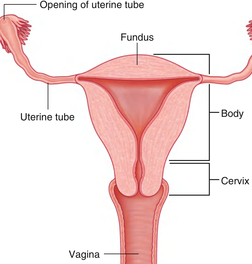
Size and shape (nulliparous)
- Pear shaped.
- Length 7.5 cm, width 5 cm, thickness 2.5 cm.
- Weight 50 to 80 g.
- Cavity capacity about 5 mL.
Parts
- Fundus: the part above the openings of the tubes.
- Body: the part from the fundus to the isthmus.
- Isthmus: about 1 cm. It becomes the lower uterine segment in pregnancy, which is the site of the lower segment caesarean section (LSCS) incision.
- Cervix: about 2.5 cm, with a supravaginal and a vaginal part. The internal os is at the top and the external os at the bottom.
- The external os is round in a nullipara and a transverse slit in a parous woman.
- The cervical canal is fusiform, with palmate folds (arbor vitae).
- The squamocolumnar junction at the external os is the transformation zone, the site of cervical carcinoma.
Position
- Anteverted: the long axis of the uterus is tilted forward relative to the vagina (angle about 90 degrees).
- Anteflexed: the body is bent forward on the cervix (angle about 125 degrees).
Layers of the wall
- Perimetrium (peritoneum).
- Myometrium (three smooth muscle layers).
- Endometrium (functional layer and basal layer).
Peritoneal relations
- The anterior surface is covered by peritoneum and forms the vesicouterine pouch.
- The posterior surface is covered down to the posterior fornix and forms the rectouterine pouch (of Douglas).
Supports of the uterus
| Type | Structures |
|---|---|
| Main (primary) | Pelvic floor (levator ani), perineal body, urogenital diaphragm |
| Ligamentous (secondary) | Transverse cervical (cardinal / Mackenrodt's) ligaments (the most important), uterosacral ligaments, pubocervical ligaments |
| Others | Round ligament (maintains anteversion), broad ligament (peritoneal fold only, minimal support) |
- Round ligament: runs from the uterine cornea, through the deep inguinal ring and inguinal canal, to the labium majus. It is a remnant of the gubernaculum.
- Broad ligament: a double peritoneal fold with the mesosalpinx, mesovarium and mesometrium. The parts are mesosalpinx, mesovarium, and mesometrium.
- Ligament of the ovary: attaches the ovary to the uterus.
Blood supply
- Uterine artery (from the anterior division of the internal iliac). It crosses over the ureter about 1.5 to 2 cm lateral to the cervix.
- It anastomoses with the ovarian artery.
- Veins form the uterine venous plexus, which drains to the internal iliac vein.
Lymphatic drainage
- Fundus: para-aortic nodes, via the ovarian vessels.
- Body: external iliac nodes.
- Cervix: internal iliac, external iliac and sacral nodes.
- A part of the fundus also drains along the round ligament to the superficial inguinal nodes.
Nerve supply
- Sympathetic (T12 to L2) and parasympathetic (S2 to S4), via the inferior hypogastric plexus (uterovaginal plexus).
- Uterine pain travels with sympathetic fibres (T10 to L1). Cervical pain travels with parasympathetic fibres (S2 to S4).
Applied
- Hysterectomy: the main hazard is injury to the ureter while ligating the uterine artery (Gray's Anatomy for Students, p. 554).
- Prolapse: caused by weakness of the cardinal and uterosacral ligaments and the pelvic floor.
- Pap smear: taken from the transformation zone.
C. Uterine (fallopian) tubes
Per Gray's, the tubes extend from the upper body of the uterus to the lateral pelvic wall. They are enclosed in the upper margin of the mesosalpinx of the broad ligament (Gray's Anatomy for Students, p. 555).
- Length about 10 cm.
- Four parts, from medial to lateral:
- Intramural (interstitial): about 1 cm, the narrowest part.
- Isthmus: about 2.5 cm, narrow and thick walled. It is the site for tubal ligation.
- Ampulla: about 5 cm, the widest and longest part. It is the usual site of fertilisation and the commonest site of ectopic pregnancy.
- Infundibulum: the funnel-shaped end with finger-like fimbriae. The ovarian fimbria is the longest, and it attaches to the ovary.
- Histology: ciliated and secretory (peg) cells.
- Blood supply: uterine and ovarian arteries.
- Applied: ectopic pregnancy (commonest in the ampulla), salpingitis, and tubal ligation.
D. Ovary
- Almond shaped, about 3 × 2 × 1 cm.
- It lies in the ovarian fossa on the lateral pelvic wall.
- The fossa is bounded by the external iliac vessels (above), the internal iliac vessels (behind), and the obturator nerve at its floor.
- It is the only intraperitoneal organ that is not covered by peritoneum. It is covered by germinal epithelium.
- Attachments:
- Mesovarium, a peritoneal fold from the broad ligament.
- Ligament of the ovary, running to the uterus.
- Suspensory ligament (infundibulopelvic ligament), carrying the ovarian vessels and nerves.
- Blood supply: the ovarian artery, a branch of the abdominal aorta at L2.
- The right ovarian vein drains to the IVC. The left ovarian vein drains to the left renal vein.
- Lymphatics: para-aortic nodes.
- Nerve supply: T10 sympathetic, via the ovarian plexus. Ovarian pain is referred to the umbilical region.
- Applied: an ovarian pedicle torsion involves the infundibulopelvic ligament. An ovarian cyst or a pelvic mass may press on the obturator nerve and cause referred pain to the medial thigh.
PART 3. DEVELOPMENT AND EMBRYOLOGY
A. Sexual differentiation
- Indifferent stage (up to week 6): gonads, two pairs of ducts (mesonephric/Wolffian and paramesonephric/Müllerian), and common external genital primordia.
- Genetic sex is set at fertilisation.
- Gonadal sex: the SRY gene on the Y chromosome drives testis development. In its absence, ovaries develop.
- In the female (no testosterone, no AMH):
- The mesonephric ducts regress (remnants: epoophoron, paroophoron, Gartner's duct).
- The paramesonephric ducts persist and develop.
B. Development of the ovary
- Gonadal ridges form from the intermediate mesoderm and coelomic epithelium.
- Primordial germ cells migrate from the yolk sac wall (hindgut region) to the gonadal ridge during week 4 to 6.
- Primary sex cords break up into cell clusters, and the cortical cords form primordial follicles.
- Descent of the ovary: the ovary descends to the pelvis, guided by the gubernaculum. The gubernaculum becomes the ligament of the ovary and the round ligament of the uterus.
C. Development of the uterine tubes, uterus and vagina
- The paramesonephric ducts form as invaginations of coelomic epithelium lateral to the mesonephric ducts.
- The cranial (vertical) parts stay unfused and become the uterine tubes. The funnel-shaped opening into the coelom becomes the fimbriated end.
- The caudal (horizontal) parts cross the mesonephric ducts and fuse in the midline.
- The fused ducts form the uterovaginal primordium. This gives the uterus (body and cervix) and the upper part of the vagina.
- The uterine stroma and myometrium come from the surrounding mesenchyme.
- The uterovaginal primordium contacts the posterior wall of the urogenital sinus and induces two outgrowths, the sinovaginal bulbs.
- The bulbs fuse to form the solid vaginal plate, which canalises by the fifth month to make the vaginal lumen.
- The vaginal epithelium therefore comes from at least two origins (the upper part from the paramesonephric ducts and the lower part from the urogenital sinus), but the boundary is unclear (Color Atlas of Human Anatomy, Vol 2).
- The hymen forms from the invaginated posterior wall of the urogenital sinus. Normally it perforates before birth.
Quick mnemonic
- Paramesonephric ducts: tubes, uterus, upper third of the vagina.
- Sinovaginal bulbs (urogenital sinus): lower two thirds of the vagina (this varies between textbooks, as noted above).
D. External genitalia
By default (no androgen), the indifferent external structures develop as female.
| Indifferent structure | Female derivative |
|---|---|
| Genital tubercle | Clitoris |
| Urogenital folds | Labia minora |
| Labioscrotal swellings | Labia majora |
| Urogenital sinus | Vestibule, urethra, vaginal lower part, greater vestibular (Bartholin) glands, paraurethral glands |
| Gubernaculum | Round ligament and ovarian ligament |
E. Anomalies
Per The Developing Human, uterine and vaginal duplications result from arrested development of the uterovaginal primordium in week 8. The causes are incomplete development of a paramesonephric duct, failure of parts of one or both ducts to develop, incomplete fusion of the ducts, and incomplete canalisation of the vaginal plate (The Developing Human, p. 749).
| Anomaly | Cause |
|---|---|
| Uterus didelphys (double uterus) | Failure of fusion of the lower parts of the paramesonephric ducts. There may be a double or single vagina. |
| Bicornuate uterus | Duplication of only the upper part of the uterine body. |
| Bicornuate uterus with a rudimentary horn | One duct is retarded and does not fuse. The horn may not communicate with the cavity. |
| Septate uterus | A thin septum divides the cavity, and the outside appears normal. |
| Unicornuate uterus | One paramesonephric duct fails to develop, leaving one uterine tube. Women may be fertile, but with higher rates of preterm delivery and recurrent pregnancy loss (The Developing Human, p. 750). |
| Absent vagina and uterus | About 1 in 5000 births. The sinovaginal bulbs fail to develop and form the vaginal plate. Because the developing uterus induces the bulbs, absence of the vagina usually means absence of the uterus (The Developing Human, p. 750). This is the Mayer-Rokitansky-Küster-Hauser (MRKH) syndrome. |
| Vaginal atresia | Failure of canalisation of the vaginal plate. |
| Transverse vaginal septum | About 1 in 80,000 women, usually at the junction of the middle and upper thirds. |
| Imperforate hymen | The most common obstructive anomaly of the female tract (The Developing Human, p. 750). It presents at puberty with cryptomenorrhoea / haematocolpos and cyclical lower abdominal pain with primary amenorrhoea. |
Disorders of sex development (DSD) relevant to the female tract
- Androgen insensitivity syndrome (46,XY): testes are present and make AMH, so there is no uterus or tubes. The external genitalia are female because the tissue cannot respond to androgen.
- Congenital adrenal hyperplasia (46,XX): excess androgen virilises the external genitalia.
- Turner syndrome (45,X): streak gonads.
PART 4. PLACENTA AND FETAL MEMBRANES
A. Development of the placenta
Per Langman's, the fetal component of the placenta comes from the trophoblast and extraembryonic mesoderm (chorionic plate). The maternal component comes from the uterine endometrium (Langman's Medical Embryology, p. 129).
Stages of villus development
- Primary villi: a core of cytotrophoblast covered by syncytiotrophoblast (about day 13).
- Secondary villi: with an extraembryonic mesoderm core.
- Tertiary villi: with fetal blood vessels (by about day 21).
- By the second month the trophoblast has many secondary and tertiary villi, giving a radial appearance. Stem (anchoring) villi run from the chorionic plate mesoderm to the cytotrophoblast shell.
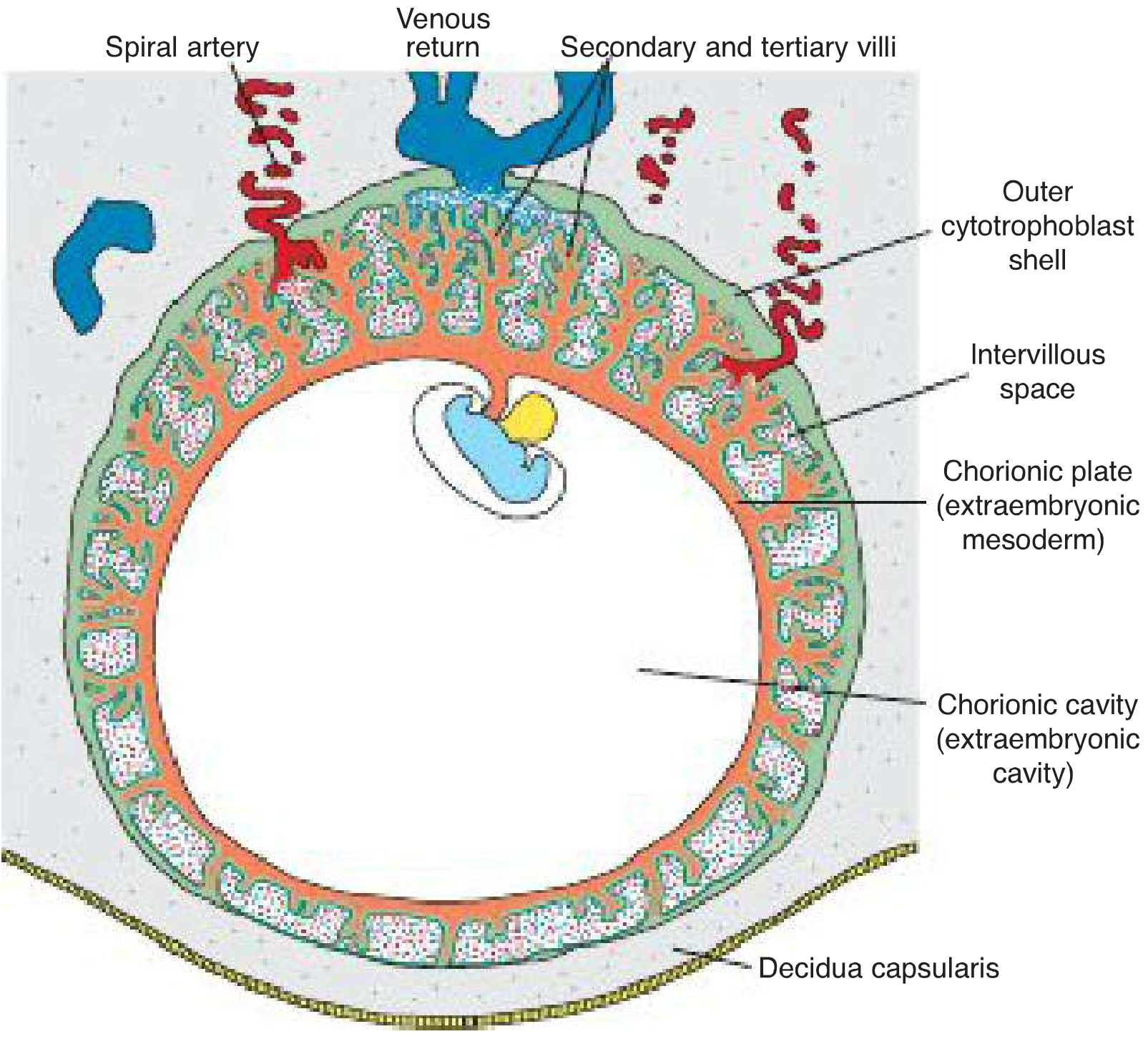
Spiral artery remodelling
- Cytotrophoblast cells invade the end of the spiral arteries and replace the maternal endothelium.
- This converts them from small, high-resistance vessels to large, low-resistance vessels, which increases maternal blood flow to the intervillous space (Langman's Medical Embryology, p. 130).
- Failure of this invasion is implicated in preeclampsia (about 5% of pregnancies), which presents with hypertension and proteinuria from about 20 weeks (Langman's Medical Embryology, p. 131).
Villi at 4 months
- Cytotrophoblast and some connective tissue cells disappear, so only syncytium and the fetal capillary endothelium separate the maternal and fetal blood (Langman's Medical Embryology, p. 130).
- Syncytial knots can break off into the maternal circulation, usually without symptoms.
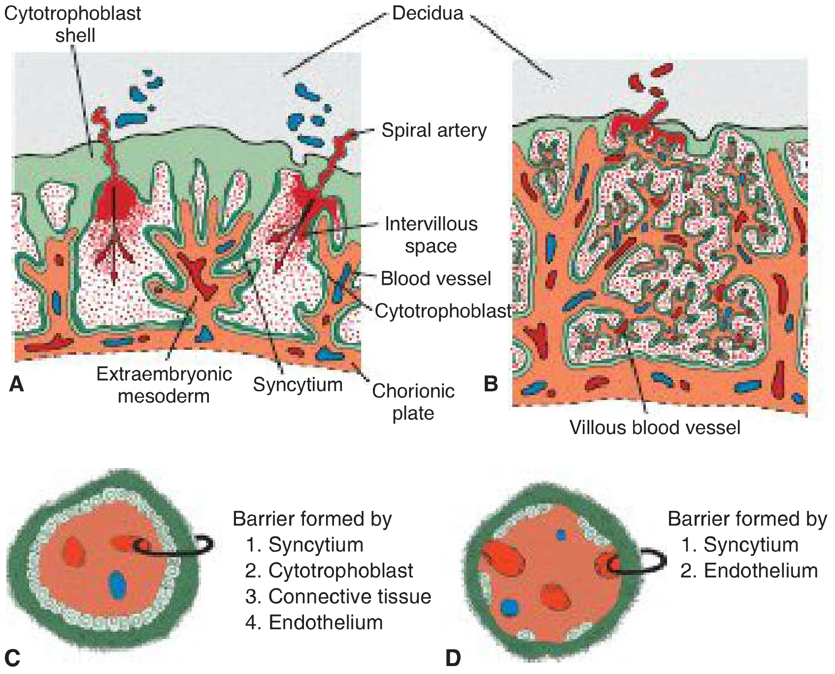
Chorion frondosum and chorion laeve
- Villi at the embryonic pole (towards the decidua basalis) proliferate to form the chorion frondosum, which becomes the fetal part of the placenta.
- Villi at the abembryonic pole degenerate to form the chorion laeve, which is smooth.
Decidua (the endometrium of pregnancy)
- Decidua basalis: beneath the conceptus, forms the maternal placenta.
- Decidua capsularis: overlies the conceptus.
- Decidua parietalis: lines the rest of the uterus.
- The capsularis fuses with the parietalis at about 22 to 24 weeks, obliterating the uterine cavity.
B. The mature placenta (at term)
Per The Developing Human:
- Discoid, 15 to 20 cm in diameter, 2 to 3 cm thick.
- Weighs 500 to 600 g, about one sixth of the fetal weight (The Developing Human, p. 372).
- Has a fetal surface (smooth, covered by amnion, with the umbilical cord inserted) and a maternal surface (rough, divided into 15 to 20 cotyledons by placental septa).
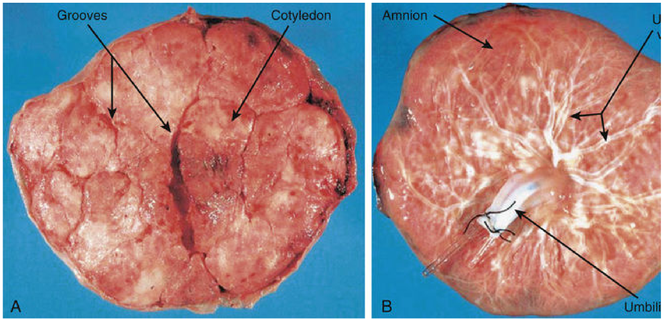
Layers from the fetal to the maternal side
- Amnion, chorionic plate, intervillous space with villi, cytotrophoblastic shell, decidua basalis (basal plate).
Placental circulation
- Maternal side: blood enters the intervillous space through the spiral arteries, bathes the villi, and leaves through the endometrial veins. There is no mixing of the two blood supplies.
- Fetal side: deoxygenated blood goes to the placenta in two umbilical arteries. Oxygenated blood returns in the single umbilical vein.
Placental barrier (membrane)
- In early pregnancy it has four layers: syncytiotrophoblast, cytotrophoblast, villous connective tissue, and fetal capillary endothelium.
- In late pregnancy it is thin, with only syncytiotrophoblast and capillary endothelium.
- Placenta type: haemochorial (maternal blood directly contacts the chorion). The shape is discoid.
Functions
- Respiration and gas exchange (by diffusion).
- Nutrition and excretion.
- Transfer of IgG antibodies (passive immunity).
- Endocrine: hCG (maintains the corpus luteum), hPL, progesterone, oestrogens (oestriol), relaxin.
- Barrier function, which is incomplete. Viruses (rubella, CMV, HIV, Zika), many drugs, alcohol and teratogens cross.
- Metabolism, storage, and transport.
Placental shape variants
- Membranous, accessory (succenturiate), bidiscoid, and horseshoe placenta. Most are of little clinical significance (The Developing Human, p. 373).
Third stage of labour
- It lasts about 15 minutes in 90% of pregnancies. A retained placenta is one not expelled within 60 minutes.
- The placenta separates through the spongy layer of the decidua basalis. Myometrial contraction constricts the spiral arteries, which prevents excessive bleeding (The Developing Human, p. 372).
C. Abnormal placentation
| Condition | Key points |
|---|---|
| Placenta previa | The placenta implants over or near the internal os. It presents with painless, recurrent antepartum haemorrhage in the third trimester. Digital vaginal examination is contraindicated. |
| Placenta accreta spectrum | Villi attach abnormally deep, from accreta (to the myometrium) to increta and percreta. It is linked to previous caesarean section and previa. A recent meta-analysis found conservative management was associated with better surgical outcomes than cesarean hysterectomy (Hessami 2025, PMID 39884567). |
| Abruptio placentae | Premature separation of a normally sited placenta. It is painful, with a rigid uterus. |
| Gestational trophoblastic disease | Hydatidiform mole and choriocarcinoma. |
| Preeclampsia | Failure of cytotrophoblast invasion. |
| IUGR | Causes include placental insufficiency, infection, smoking and hypertension. |
D. Fetal membranes
- Amnion
- A thin, tough, avascular membrane lining the amniotic cavity. It is derived from the epiblast (ectoderm) and extraembryonic mesoderm.
- It covers the umbilical cord and fuses with the chorion to obliterate the chorionic cavity.
- Amniotic fluid: about 800 to 1000 mL at term.
- Functions: protects the fetus from trauma, allows movement, keeps temperature constant, and helps lung development.
- Sources: initially a maternal filtrate, then fetal urine from week 10 or so, plus lung fluid.
- The fetus swallows about 400 mL a day.
- Polyhydramnios (more than 2000 mL) is associated with oesophageal atresia, anencephaly, and maternal diabetes.
- Oligohydramnios is associated with renal agenesis (Potter sequence), obstructive uropathy and leaking membranes. It causes pulmonary hypoplasia.
- Chorion
- Formed of trophoblast and extraembryonic somatic mesoderm.
- The chorion frondosum becomes the placenta. The chorion laeve is the smooth membrane.
- Yolk sac
- Early haematopoiesis, source of primordial germ cells, and gut contribution.
- Its remnant is a Meckel's diverticulum if the vitelline duct persists.
- Allantois
- Its remnant is the urachus (median umbilical ligament).
- Its vessels become the umbilical vessels.
- Decidua
- See above.
E. Umbilical cord
- About 50 cm long and 1 to 2 cm in diameter.
- Contains two umbilical arteries and one umbilical vein, embedded in Wharton's jelly (mucoid connective tissue), covered by amnion.
- Single umbilical artery occurs in about 1% of births and is associated with congenital anomalies (cardiac, renal).
- Abnormal insertions: velamentous insertion (vessels run in the membranes, risk of vasa previa) and marginal insertion.
- Remnants after birth: umbilical vein becomes the ligamentum teres hepatis, the arteries become the medial umbilical ligaments, and the ductus venosus becomes the ligamentum venosum.
- Cord prolapse is an obstetric emergency.
F. Twin placentation
- Dichorionic diamniotic: seen in dizygotic twins and monozygotic twins if splitting is before day 3.
- Monochorionic diamniotic: splitting at day 4 to 8 (the commonest type of monozygotic twinning).
- Monochorionic monoamniotic: splitting at day 8 to 13.
- Conjoined twins: splitting after day 13.
High-yield viva points
- The ureter lies below the uterine artery ("water under the bridge").
- The posterior fornix is the deepest and is related to the pouch of Douglas.
- The ampulla is the commonest site of fertilisation and ectopic pregnancy.
- The ovarian artery comes from the aorta at L2.
- Paramesonephric (Müllerian) ducts form the tubes, uterus and upper vagina. The imperforate hymen is the commonest obstructive anomaly.
- The placenta is haemochorial and discoid, weighs 500 to 600 g, with 15 to 20 cotyledons.
- The cord has 2 arteries and 1 vein.
I can expand any part (for example, a labelled-diagram guide, MCQs, or a long-answer exam format) if you tell me which.
Note on sources: the library image search returned no extra figures, so the images above come from the textbook pages I read. The placenta accreta result is the only recent systematic review I found for this topic. It does not contradict the textbook anatomy.
Now create for physiology of ovulation and menstruation
Reading File
Your credits are exhausted. You can purchase a top-up or subscribe to a plan to continue using Orris. If you're already subscribed, your daily credits will reset tomorrow.
This is a shared conversation. Sign in to Orris to start your own chat.