
Explain the complete histology of lips, cheeks, palate, whole tongue, whole teeth, gums, oropharynx, tubular digestive tract and enteric nervous system
Reading File
This is a broad, visual histology question covering many oral/GI structures. I'll run parallel searches across all major topics simultaneously.
Finding Sources
Finding Sources
Finding Sources
Finding Sources
Finding Sources
Let me try simpler searches:
Finding Sources
Finding Sources
Excellent! Found the key histology textbook. Let me read the digestive system histology sections in parallel:
Reading File
Reading File
Excellent content found. Now let me read the detailed histology sections in parallel:
Reading File
Reading File
Let me read the detailed oral cavity content and block 6 for tubular GI/ENS:
Reading File
Reading File
Excellent content. Let me now read more of block 5 (detailed oral histology) and block 6 (tubular GI/ENS):
Reading File
Reading File
Now let me read the detailed oral cavity histology (lips, cheeks, palate) and the GI wall layers section in block 6:
Reading File
Reading File
I have extensive content. Let me read the detailed tubular GI layers and ENS content:
Reading File
I now have rich, detailed content from the textbook. Let me read the specific ENS and large intestine section:
Reading File
I now have comprehensive content from the textbook. Let me now compile the full, detailed answer.Here is the complete histology of each structure, drawn from Histology: A Text and Atlas with Correlated Cell and Molecular Biology (Pawlina/Ross, 9th ed.).
Complete Histology of the Oral Cavity, Digestive Tract, and Enteric Nervous System
1. Lips
The lip is a transition zone between external skin and internal oral mucosa, and this is reflected in its layered structure:
Outer skin surface:
- Thin skin with epidermis (keratinized stratified squamous), hair follicles, sebaceous glands, and sweat glands.
Vermilion border (red zone):
- Stratified squamous keratinized epithelium, but the keratin layer is very thin and transparent, so the underlying rich capillary plexus shows through, giving the red color.
- No hair follicles, sweat glands, or sebaceous glands here.
- Numerous tall connective tissue papillae project into the epithelium, carrying capillary loops close to the surface.
Inner oral mucosal surface:
- Stratified squamous non-keratinized epithelium (lining mucosa).
- Lamina propria of loose connective tissue with blood vessels and lymphatics.
- Submucosa contains labial minor salivary glands (mixed mucoserous), which open directly into the vestibule.
- Beneath the submucosa lies the orbicularis oris skeletal muscle, the core of the lip.
2. Cheeks (Buccae)
The cheeks form the lateral walls of the oral vestibule and share structural features with lips:
- Inner surface: stratified squamous non-keratinized epithelium (lining mucosa). This is thick and highly resistant to mechanical trauma.
- The epithelium rests on a lamina propria of loose connective tissue.
- The submucosa contains buccal minor salivary glands (mixed type) and adipose tissue.
- The parotid (Stensen) duct opens onto the buccal mucosa opposite the second upper molar as a small papilla.
- The core of the cheek is the buccinator skeletal muscle, enclosed in deep fascia.
- Stratified squamous cells at this site show no keratinization because they are not subject to masticatory friction.
3. Palate
The palate is divided into hard and soft regions, each with different histology.
Hard Palate
- Epithelium: stratified squamous keratinized or parakeratinized (masticatory mucosa) - reflects the high mechanical stress of chewing.
- The lamina propria (mucoperiosteum) is densely adherent to the underlying bone of the maxilla with no separate submucosa in most areas.
- The submucosa, where present (lateral regions), contains palatine minor salivary glands (predominantly mucous) and fat.
- A midline raphe runs antero-posteriorly where the mucosa binds directly to bone.
Soft Palate
- Epithelium transitions to stratified squamous non-keratinized on the oral surface.
- Nasopharyngeal surface is lined by pseudostratified ciliated columnar epithelium (respiratory type).
- Core: interlacing bundles of skeletal muscle (palatoglossus, palatopharyngeus, tensor/levator veli palatini, uvular muscle).
- Submucosa contains numerous mucous minor salivary glands and elastic fibers.
- Taste buds may be found on the oral surface epithelium of the soft palate.
4. Tongue
The tongue is a muscular organ whose mucosa and internal structure differ between dorsal, ventral, and posterior regions.
Internal Musculature
- Both intrinsic (longitudinal, transverse, vertical fibers) and extrinsic (genioglossus, etc.) skeletal muscles are present.
- The intrinsic fibers are arranged in three interwoven planes at right angles to each other - unique to the tongue. This gives it extraordinary mobility for speech, mastication, and swallowing.
- Between the muscle bundles lie connective tissue septa, vessels, nerves, and glandular acini.
Ventral (under) surface
- Thin stratified squamous non-keratinized epithelium.
- Smooth surface, no papillae.
Dorsal Surface - Body (Anterior 2/3)
Contains four types of lingual papillae, all projections of the lamina propria covered by epithelium:
1. Filiform papillae
- Most numerous; thread-like, conical projections pointing posteriorly.
- Stratified squamous keratinized epithelium, giving rough texture.
- No taste buds.
- Function: mechanical - grip food.
2. Fungiform papillae
- Scattered among filiform papillae; mushroom-shaped, slightly raised red dots.
- Stratified squamous non-keratinized (thin keratin layer).
- Large primary connective tissue core + secondary smaller papillae project up into the epithelium, bringing capillaries close to the surface (causes the red color on gross inspection).
- Bear taste buds on their superior surface.
3. Circumvallate (vallate) papillae
- 7-12 large papillae arranged in a V-shape (sulcus terminalis) dividing anterior 2/3 from posterior 1/3.
- Each papilla is surrounded by a deep circular trench (moat/cleft).
- Side walls of the papilla (not the top surface) bear numerous taste buds.
- Stratified squamous non-keratinized epithelium lines the cleft walls.
- Serous glands of von Ebner (Ebner glands) are pure serous glands whose ducts open into the base of the trench. Their watery secretion washes the cleft, refreshing taste buds for new stimuli.
4. Foliate papillae
- Parallel ridges on the lateral edges of the posterior tongue; prominent in infants, rudimentary in adults.
- Stratified squamous non-keratinized epithelium with taste buds on the lateral walls.
- Adjacent connective tissue clefts separate the folds.
- Serous minor salivary glands also open here.
Posterior 1/3 (Root/Base of Tongue)
- No papillae.
- Stratified squamous non-keratinized epithelium.
- Lamina propria contains large lymphoid aggregates - the lingual tonsil (part of Waldeyer's ring).
- Mucous glands (lingual mucous glands) present in the submucosa.
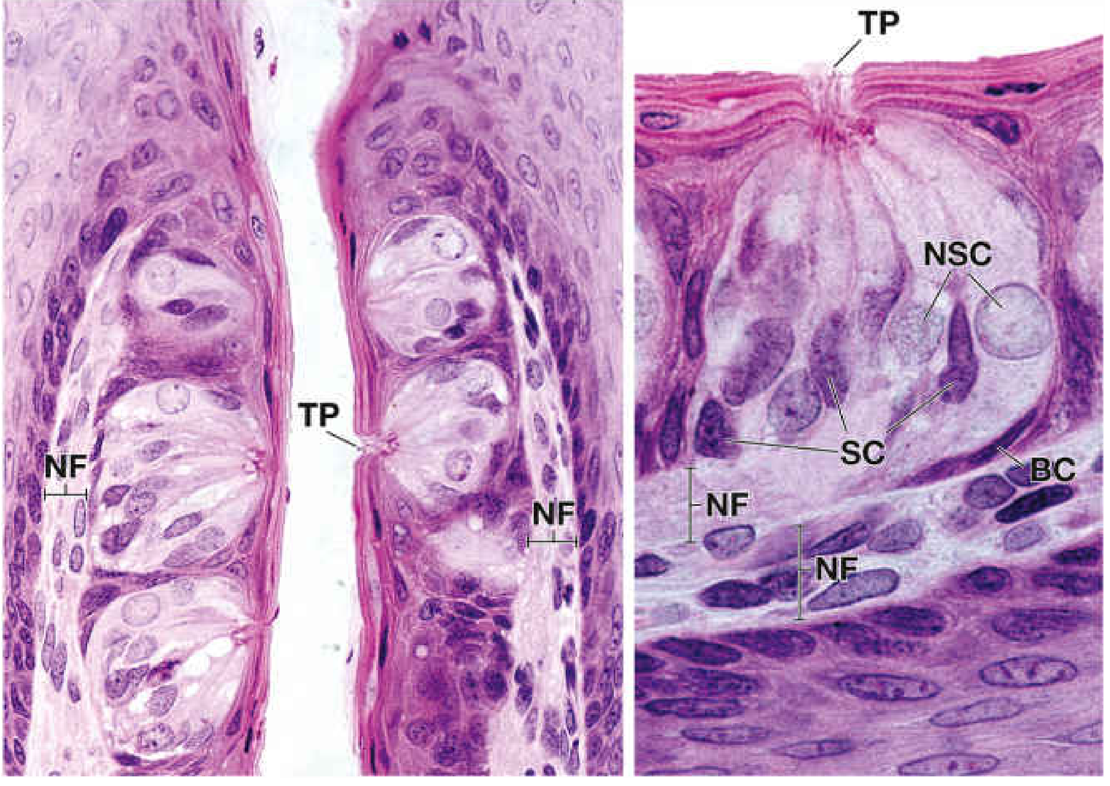
Taste Buds
- Oval epithelial organelles embedded in the surface epithelium of fungiform, circumvallate, foliate papillae, and soft palate.
- Open at the surface via a small taste pore.
- Three cell types:
- Neuroepithelial (type II) sensory cells: elongated, with apical microvilli extending into the taste pore; form synapses with afferent nerve fibers at their base.
- Supporting (type I) cells: interspersed with sensory cells; have microvilli.
- Basal cells: at the base; act as stem cells replacing sensory and supporting cells every ~10 days.
5. Teeth
Overall Tooth Structure
Each tooth consists of a crown (projects into oral cavity) and root (embedded in alveolar bone). The central pulp cavity (pulp chamber in crown, root canal in root) contains the dental pulp. Three specialized mineralized tissues form the tooth proper.
Enamel
- Covers the anatomic crown.
- The hardest biological substance in the body: 96-98% inorganic (hydroxyapatite crystals), 1-2% organic matrix.
- Composed of parallel enamel rods (prisms) ~4-6 μm in diameter, each rod formed by a single ameloblast during development.
- Rods run from the dentinoenamel junction (DEJ) to the outer surface, often in a slightly twisted ("Hunter-Schreger bands" pattern in cross section).
- Enamel-regulating proteins: amelogenins, ameloblastins, enamelins.
- Once formed and the tooth erupts, ameloblasts are lost - enamel cannot regenerate.
Dentin
- Forms the bulk of the tooth; lies deep to enamel (in crown) and deep to cementum (in root).
- Less hard than enamel: ~70% inorganic (hydroxyapatite), ~30% organic (mainly type I collagen).
- Contains dentinal tubules (3-5 μm diameter) running from pulp to DEJ; each tubule houses a thin odontoblast process (Tomes' fiber).
- Initially deposited as predentin (unmineralized) by odontoblasts; under the influence of dentin phosphoprotein (DPP) and dentin sialoprotein (DSP), predentin mineralizes to dentin.
- Odontoblast cell bodies remain in the pulp, lining the pulp cavity wall - they are the only cells that persist within a mature tooth.
- Peritubular dentin (more mineralized) surrounds the tubules; intertubular dentin lies between tubules.
- The dentinoenamel junction (DEJ) is scalloped, interdigitating between enamel and dentin.
Cementum
- Covers the root surface; resembles bone in composition (~50% inorganic).
- Two types:
- Acellular (primary) cementum: formed first, no cementocytes, covers cervical and mid-root.
- Cellular (secondary) cementum: formed later, contains cementocytes in lacunae; covers apical root.
- Collagen fibers (Sharpey fibers) project from the cementum surface to form the periodontal ligament.
Dental Pulp
- Fills the pulp chamber and root canal.
- Loose connective tissue with fibroblasts, undifferentiated mesenchymal cells, macrophages, lymphocytes, plasma cells, mast cells.
- Odontoblasts line the periphery, adjacent to predentin.
- Rich vascularity and innervation (sensory unmyelinated C fibers + myelinated A-delta fibers).
- Contains ground substance rich in proteoglycans; collagen fibers (mainly type I and III).
- The pulp-dentin complex acts as a sensory and reparative unit.
6. Gums (Gingiva)
- Gingiva is the masticatory mucosa surrounding the necks of teeth and covering the alveolar bone.
- Epithelium: stratified squamous keratinized or parakeratinized - can withstand the forces of mastication.
- Divided into:
- Free gingiva: forms the gingival margin and the inner wall of the gingival sulcus. The junctional epithelium at the base of the sulcus attaches to the tooth enamel via hemidesmosomes and is non-keratinized.
- Attached gingiva: firmly bound to the periosteum; no submucosa - the mucoperiosteum is directly adherent to bone (like the hard palate).
- Interdental papillae: fill the spaces between adjacent teeth.
- The lamina propria is dense connective tissue with abundant collagen bundles (gingival fibers) organized into groups (dentogingival, alveologingival, circular, transseptal).
- No submucosa and no submucosal glands.
- A crevicular/junctional epithelium lines the gingival sulcus and is permeable to fluid (gingival crevicular fluid).
7. Oropharynx
The oropharynx is the portion of the pharynx behind the oral cavity, between the soft palate and the epiglottis.
- Epithelium: stratified squamous non-keratinized on all oral surfaces (posterior pharyngeal wall, lateral walls, anterior surface of epiglottis, valleculae). This contrasts with the nasopharynx, which has pseudostratified ciliated columnar (respiratory) epithelium.
- Lamina propria: loose connective tissue, rich in lymphoid aggregates.
- Palatine tonsils (the main tonsils):
- Located in tonsillar fossae between the palatoglossal and palatopharyngeal arches.
- Covered by stratified squamous non-keratinized epithelium.
- The epithelium invaginates deeply into the lymphoid tissue forming 10-20 blind-ended crypts. The crypts accumulate shed epithelial cells, lymphocytes, and bacteria.
- Beneath the epithelium: dense aggregates of secondary lymphoid follicles with germinal centers; diffuse lymphoid tissue fills the interfollicular regions.
- A fibrous capsule (partial) of dense connective tissue forms the lateral wall of each tonsil.
- Mucous glands are present in the connective tissue deep to the capsule; their ducts bypass (not open into) the crypts.
- Waldeyer's tonsillar ring: the complete ring of lymphoid tissue guarding the oropharyngeal inlet, composed of palatine tonsils (lateral), pharyngeal tonsil/adenoid (posterior superior nasopharynx), lingual tonsil (base of tongue), and tubal tonsils (near eustachian tube orifices).
- Muscularis: pharyngeal constrictor muscles (skeletal), with overlying pharyngobasilar fascia.
- Adventitia: fibrous connective tissue blending with surrounding fascial planes.
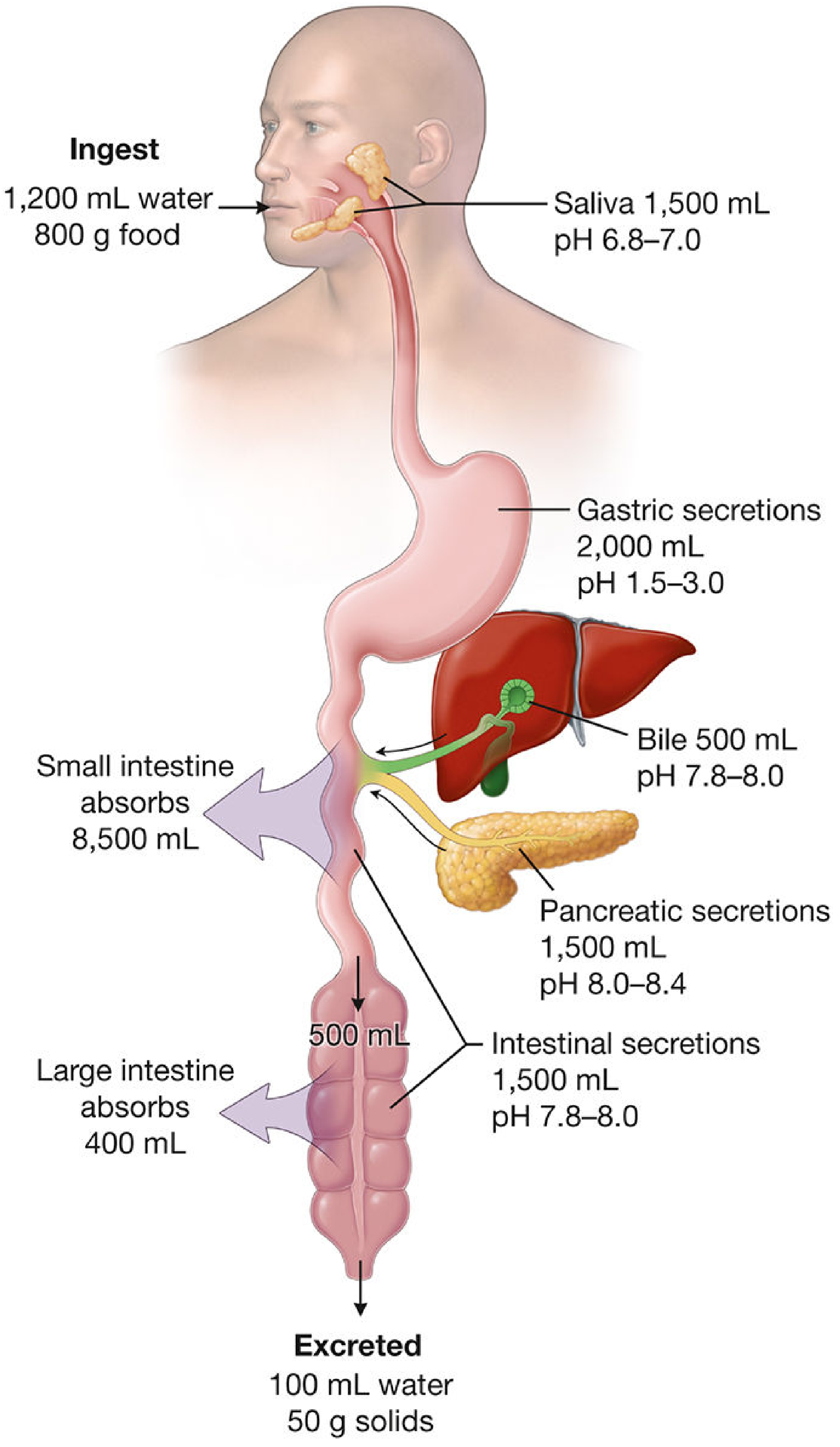
8. Tubular Digestive Tract - General Wall Architecture
The esophagus, stomach, small and large intestine share a common four-layer plan (from lumen outward):
Layer 1: Mucosa
Three components:
- Epithelium: varies by segment (see below).
- Lamina propria: loose connective tissue with fibroblasts, smooth muscle cells, lymphocytes, plasma cells, eosinophils, mast cells, lymphatic capillaries (lacteals in small intestine), capillaries. Contains glands in some segments.
- Muscularis mucosae: a thin layer of smooth muscle (usually inner circular + outer longitudinal). Its contractions create local folds and movements of the mucosa, aiding secretion.
Layer 2: Submucosa
- Dense irregular connective tissue with larger blood vessels, lymphatics, adipose tissue, and nerve elements.
- Contains the submucosal (Meissner) plexus (part of ENS).
- May contain glands (esophageal submucosal glands proper; Brunner glands of duodenum).
Layer 3: Muscularis Externa
- Usually inner circular + outer longitudinal smooth muscle layers.
- The myenteric (Auerbach) plexus lies between these two muscle layers.
- Coordinates peristalsis.
- In the stomach: three layers (oblique, circular, longitudinal).
- In upper 1/3 esophagus: skeletal muscle; middle 1/3: mixed; lower 1/3 and rest of GI: smooth muscle.
Layer 4: Adventitia or Serosa
- Adventitia: fibrous connective tissue blending with surrounding structures (esophagus, retroperitoneal parts of colon).
- Serosa: mesothelium (simple squamous epithelium) + thin connective tissue = the visceral peritoneum (covers intraperitoneal organs).
8a. Esophagus
- Mucosa: stratified squamous non-keratinized epithelium (like the oral cavity it connects to). At the gastroesophageal junction, this abruptly transitions to simple columnar - the Z-line (ora serrata), visible endoscopically.
- Lamina propria: loose connective tissue; esophageal cardiac glands (mucous) present near both ends of esophagus.
- Muscularis mucosae: present but thin; single longitudinal smooth muscle layer.
- Submucosa: dense connective tissue with esophageal glands proper - small compound tubuloalveolar mucous glands distributed along the esophagus (more concentrated in upper half); their excretory ducts (lined by stratified squamous epithelium) open onto the luminal surface.
- Muscularis externa: inner circular + outer longitudinal. Upper 1/3 = skeletal muscle; middle 1/3 = mixed skeletal + smooth; lower 1/3 = smooth muscle only.
- Outer coat: adventitia along most of its course; serosa only in the short intra-abdominal segment.
8b. Stomach
Gastric Mucosa:
- Simple columnar epithelium of surface mucous cells covers the entire gastric surface, dipping into gastric pits (foveolae). Multiple gastric glands open into each pit.
- The mucous cells secrete neutral mucus forming a thick protective gel layer (the gastric mucous barrier).
- Regional glands differ:
- Cardiac glands (cardia): simple/branched tubular, mucous cells; transition from esophageal epithelium.
- Fundic (oxyntic) glands (body + fundus): most important and most numerous. Contain:
- Mucous neck cells: in the isthmus/neck; secrete acidic mucus; are also the stem cells.
- Parietal (oxyntic) cells: large, pyramidal, intensely eosinophilic; secrete HCl (via H+/K+-ATPase) and intrinsic factor; have extensive intracellular canaliculi lined by microvilli (increase surface area for acid secretion).
- Chief (peptic/zymogenic) cells: basophilic, basal granules; secrete pepsinogen (pro-enzyme) and gastric lipase.
- Enteroendocrine cells: scattered; secrete gastrin (G cells, antrum), ghrelin, histamine, somatostatin, etc.
- Pyloric glands (antrum/pylorus): branched coiled tubular glands; mostly mucous cells + G cells (gastrin-secreting enteroendocrine cells); deeper gastric pits.
- Lamina propria: sparse between glands; smooth muscle fibers from muscularis mucosae extend up between glands.
- Muscularis mucosae: prominent; inner circular + outer longitudinal smooth muscle.
- Submucosa: dense connective tissue; no glands.
- Muscularis externa: uniquely three layers - inner oblique, middle circular (forms pyloric sphincter), outer longitudinal. Myenteric plexus between layers.
- Serosa: visceral peritoneum.
8c. Small Intestine
The small intestine is specialized for digestion and absorption via three major surface-amplifying mechanisms: plicae circulares (permanent circular folds of mucosa + submucosa), villi, and microvilli (brush border). Together they increase absorptive area ~600-fold.
Mucosa:
- Epithelium: simple columnar with a brush border of microvilli.
- Enterocytes (absorptive cells): tall columnar cells with dense apical microvilli (brush border; each cell has ~3,000 microvilli). Absorb sugars, amino acids, lipids, and electrolytes.
- Goblet cells: unicellular mucous glands; increase in number from duodenum to ileum. Secrete mucus to lubricate and protect.
- Enteroendocrine cells: scattered; secrete CCK, secretin, GIP, motilin, somatostatin, etc.
- Paneth cells: located at the base of intestinal crypts (of Lieberkühn); pyramidal, with apical eosinophilic granules containing lysozyme, defensins, and other antimicrobial proteins. Regulate gut microbiome.
- M cells (Microfold cells): overlying Peyer's patches; no brush border; sample antigens from the lumen and deliver them to underlying lymphoid tissue.
- Stem cells: located in the crypts of Lieberkühn; continuously renew the epithelium (turnover ~3-5 days).
- Villi: finger-like projections (0.5-1.5 mm) of the lamina propria covered by epithelium. Core contains loose connective tissue, fenestrated capillaries, a central lacteal (blind-ended lymphatic capillary for fat absorption), and smooth muscle strands from muscularis mucosae.
- Crypts of Lieberkühn (intestinal glands): straight tubular glands extending from the villus base to the muscularis mucosae; all epithelial cell types arise here.
- Regional differences:
- Duodenum: villi broad/leaf-shaped; Brunner glands in submucosa (branched tubuloalveolar, mucous; secrete alkaline mucus to neutralize gastric acid). Short crypts.
- Jejunum: tallest villi (finger-like); most goblet cells and enterocytes. Maximal absorption zone.
- Ileum: shorter villi; numerous Peyer's patches (aggregated lymphoid follicles) in the lamina propria and submucosa; more goblet cells; M cells overlying follicles.
- Lamina propria: loose connective tissue; plasma cells (especially IgA-producing), lymphocytes, eosinophils, mast cells.
- Muscularis mucosae: inner circular + outer longitudinal smooth muscle.
- Submucosa: dense connective tissue; Brunner glands in duodenum only.
- Muscularis externa: inner circular + outer longitudinal smooth muscle.
- Serosa: visceral peritoneum.
8d. Large Intestine (Colon, Cecum, Appendix, Rectum, Anal Canal)
Colon:
- No villi (unlike small intestine).
- Mucosa: simple columnar epithelium with numerous, tightly packed, straight crypts of Lieberkühn (tubular glands extending full thickness of mucosa).
- Crypts are longer and more straight than in small intestine.
- Predominantly goblet cells (abundant mucus production - the main secretory function) and absorptive (colonocyte) cells (absorb water and electrolytes).
- Paneth cells rare (normally absent in colon except cecum and right colon).
- Enteroendocrine cells present.
- Stem cells at crypt base; colonocyte turnover ~3-4 days.
- Lamina propria: lymphocytes, plasma cells, eosinophils; lymphoid nodules may be present.
- Muscularis mucosae: prominent, two layers.
- Submucosa: dense connective tissue; lymphatic vessels. Lymph node pattern relevant to colorectal cancer spread.
- Muscularis externa: inner circular (complete) + outer longitudinal layer organized into three flat bands: taeniae coli (instead of a complete longitudinal layer). The taeniae are shorter than the colon, causing the wall to pucker into haustra (sacculations).
- Serosa or adventitia: intraperitoneal portions have serosa with appendices epiploicae (fat-filled peritoneal outpouchings).
Appendix:
- Short, tubular, blind-ended.
- Mucosa: simple columnar with crypts; relatively fewer crypts, more irregular.
- Lamina propria massively infiltrated by lymphoid tissue (secondary follicles extending across muscularis mucosae into submucosa) - the appendix functions as a lymphoid organ.
- Muscularis externa: two complete smooth muscle layers (no taeniae).
Rectum:
- Transitional zone; mucosa similar to colon (simple columnar, long crypts, goblet cells).
- No taeniae; complete outer longitudinal layer.
- Prominent internal hemorrhoidal plexus in submucosa of lower rectum.
Anal Canal:
- Pectinate line (dentate line) marks the junction between endodermal and ectodermal tissues:
- Above pectinate line: simple columnar epithelium (rectal-type), then transitional zone with stratified columnar/cuboidal.
- At pectinate line: sudden transition.
- Below pectinate line: stratified squamous non-keratinized, then keratinized at the anal verge (true skin with hair follicles and sebaceous glands).
- Internal anal sphincter: smooth muscle (involuntary); external anal sphincter: skeletal muscle (voluntary).
9. Enteric Nervous System (ENS)
The ENS is an intrinsic, semi-autonomous nervous system embedded in the wall of the digestive tube from the esophagus to the anal canal. It contains more neurons (~100-500 million) than the spinal cord.
Submucosal Plexus (Meissner Plexus)
- Located in the submucosa.
- Composed of scattered ganglia (clusters of neurons) connected by nerve fiber strands.
- Neurons are multipolar; surrounded by satellite cells (glial cells).
- Small ganglia (2-10 neurons typically).
- Functions: regulates secretion, absorption, and local blood flow; controls the muscularis mucosae.
- Receives input from intrinsic sensory neurons (IPANs) and from the sympathetic and parasympathetic extrinsic systems.
Myenteric Plexus (Auerbach Plexus)
- Located between the inner circular and outer longitudinal layers of the muscularis externa.
- Larger ganglia than Meissner plexus; contains more neurons.
- Neurons: multipolar, with dendrites and axons forming a lattice network surrounding the circumference of the gut.
- Functions: primary controller of peristalsis and segmentation (the "pace-maker" of gut motility). Coordinates the ascending excitatory and descending inhibitory reflexes of the peristaltic reflex.
- In the esophagus, myenteric plexus is present in both the striated and smooth muscle segments.
- Neurotransmitters used: acetylcholine (excitatory), VIP, substance P, nitric oxide (inhibitory), neuropeptide Y, somatostatin, serotonin (5-HT).
Neuronal Cell Types
- IPANs (intrinsic primary afferent neurons): sensory neurons detecting chemical and mechanical stimuli in the mucosa.
- Interneurons: connect IPANs to motor neurons.
- Excitatory motor neurons: release ACh + substance P → smooth muscle contraction.
- Inhibitory motor neurons: release NO + VIP → smooth muscle relaxation.
- Secretomotor neurons: regulate glandular secretion and mucosal ion transport.
Enteric Glia
- Morphologically similar to CNS astrocytes; surround neurons and nerve fibers in ganglia.
- Express S-100 protein and GFAP (glial fibrillary acidic protein).
- Functions: structural support, modulate neurotransmission, maintain gut mucosal barrier integrity.
ENS-Extrinsic Connections
- Parasympathetic (vagus and pelvic nerves): generally excitatory; preganglionic fibers synapse onto ENS neurons.
- Sympathetic (prevertebral ganglia): generally inhibitory; adrenergic fibers largely bypass ENS ganglia to act directly on muscle and blood vessels.
- Enteroendocrine cells: when stimulated, can activate afferent sensory neurons, sending signals to both the ENS and CNS (gut-brain axis). Paracrine hormones (e.g., somatostatin) also modulate ENS activity.
Summary Table
| Structure | Epithelium | Key Features |
|---|---|---|
| Lip (outer skin) | Keratinized stratified squamous | Hair follicles, sweat glands |
| Lip (vermilion) | Thin keratinized SS | No glands; rich capillary papillae → red color |
| Lip (inner) | Non-keratinized SS | Labial salivary glands in submucosa |
| Cheek | Non-keratinized SS | Buccal salivary glands; buccinator muscle core |
| Hard palate | Keratinized/parakeratinized SS | Mucoperiosteum; palatine mucous glands |
| Soft palate | Non-keratinized SS (oral) / Ciliated columnar (nasal) | Skeletal muscle core; taste buds possible |
| Tongue (dorsal body) | Keratinized SS (filiform); Non-keratinized SS (fungiform, circumvallate) | 4 papilla types; taste buds; von Ebner glands |
| Tongue (ventral) | Non-keratinized SS | Smooth; no papillae |
| Tongue (root) | Non-keratinized SS | Lingual tonsil; lingual mucous glands |
| Enamel | N/A (no epithelium in mature tooth) | Hardest body substance; enamel rods; amelogenins |
| Dentin | N/A | Dentinal tubules with odontoblast processes; predentin precursor |
| Cementum | N/A | Bone-like; acellular + cellular types; Sharpey fibers |
| Dental pulp | N/A | Loose CT; odontoblasts lining; rich NV supply |
| Gingiva | Keratinized/parakeratinized SS | Mucoperiosteum; gingival fibers; junctional epithelium |
| Oropharynx | Non-keratinized SS | Palatine tonsils; crypts; Waldeyer's ring |
| Esophagus | Non-keratinized SS | Mixed skeletal/smooth muscularis; esophageal glands; adventitia |
| Stomach | Simple columnar | Gastric pits + glands; parietal cells (HCl); chief cells (pepsinogen) |
| Small intestine | Simple columnar | Villi + crypts; brush border; Paneth cells; Brunner glands (duodenum); Peyer's patches (ileum) |
| Large intestine | Simple columnar | No villi; long straight crypts; many goblet cells; taeniae coli |
| Rectum/anal canal | Columnar → stratified squamous | Pectinate line transition |
| ENS - Meissner plexus | N/A | Submucosa; regulates secretion/blood flow |
| ENS - Auerbach plexus | N/A | Between muscularis externa layers; controls peristalsis |
Source: Histology: A Text and Atlas with Correlated Cell and Molecular Biology (Pawlina & Ross, 9th ed.), Chapters 16 & 17This is a complete, systematic histology reference covering every structure you asked about. Here's a quick orientation:
What this covers:
-
Lips - Three zones (outer skin, vermilion border, inner mucosa) with the vermilion's rich capillary papillae explaining the red color; labial salivary glands in submucosa.
-
Cheeks - Non-keratinized stratified squamous lining with buccal glands; buccinator as core muscle; parotid duct opening opposite upper 2nd molar.
-
Palate - Hard palate = keratinized masticatory mucosa fused to bone; soft palate = non-keratinized oral surface / respiratory epithelium on nasal surface; skeletal muscle core; taste buds possible.
-
Tongue - Unique triplanar interwoven skeletal muscle; 4 papilla types (filiform = keratinized, no taste buds; fungiform/foliate/circumvallate = non-keratinized, carry taste buds); von Ebner serous glands flush circumvallate trenches; lingual tonsil at base.
-
Teeth - Enamel (96% mineral, ameloblast-derived, irreversible); dentin (odontoblast processes in tubules, predentin precursor); cementum (acellular + cellular types); pulp (loose CT, odontoblasts lining, nerve supply).
-
Gums - Masticatory mucosa; mucoperiosteum adherent to bone; junctional epithelium attaches to enamel via hemidesmosomes.
-
Oropharynx - Non-keratinized SS; palatine tonsils with deep crypts + secondary follicles; Waldeyer's ring; skeletal muscle wall.
-
Tubular GI tract - Shared 4-layer plan (mucosa/submucosa/muscularis externa/serosa-adventitia) with regional specializations: esophagus = non-keratinized SS with submucosal mucous glands; stomach = simple columnar with oxyntic glands (parietal + chief cells); small intestine = villi + crypts + Paneth cells + Brunner glands (duodenum) + Peyer's patches (ileum); large intestine = no villi, long straight crypts, predominantly goblet cells, taeniae coli.
-
ENS - Two plexuses: Meissner (submucosa, controls secretion) and Auerbach (between muscle layers, controls peristalsis); IPANs, interneurons, motor neurons, enteric glia; 100-500 million neurons; semi-autonomous gut-brain.
This is a shared conversation. Sign in to Orris to start your own chat.