
Glucocorticoids
Reading File
Searching sources...
Finding Sources
Finding Sources
Good - I have strong leads. Let me read the most comprehensive sections from Goodman & Gilman's and Katzung's (the gold-standard pharmacology texts), plus the Rheumatology text for mechanisms.
Finding Sources
Reading File
Reading File
Excellent - I found a mechanism diagram. Let me get the dedicated glucocorticoid pharmacology section from Goodman & Gilman's and read the Rheumatology/Katzung sections in parallel.
Finding Sources
Loading Image
Reading File
Excellent - I have the mechanism diagram and a potency comparison table. Now let me get the Goodman & Gilman's dedicated glucocorticoid section and the adverse effects section.
Finding Sources
Reading File
Now let me get the adverse effects and the earlier mechanism sections from the Rheumatology text, plus the Goodman & Gilman's glucocorticoid block.
Finding Sources
Reading File
Now let me get the adverse effects and clinical uses sections.
Reading File
Finding Sources
Reading File
Reading File
I now have comprehensive information. Let me also get the adverse effects chart image and the Lippincott adverse effects section.
Loading Image
Reading File
I now have all the material needed for a comprehensive, well-illustrated answer. Let me also fetch the adverse effects diagram.
Loading Image
Reading File
I now have all the information needed for a thorough, well-illustrated answer. Let me compose it.
Glucocorticoids
Overview
Glucocorticoids are steroid hormones produced by the zona fasciculata of the adrenal cortex. Cortisol (hydrocortisone) is the principal endogenous glucocorticoid. Synthetic analogs - prednisone, dexamethasone, methylprednisolone, and others - are among the most widely used drugs in medicine, with applications ranging from replacement therapy to anti-inflammatory and immunosuppressive treatment.
The term "corticosteroid" is often used synonymously with glucocorticoid, but the preferred designation when using exogenous agents therapeutically is "glucocorticoid," since corticosteroid encompasses both glucocorticoid and mineralocorticoid hormones. (Rheumatology, 2-Volume Set)
Endogenous Physiology
Cortisol is released under the control of the hypothalamic-pituitary-adrenal (HPA) axis:
- Hypothalamus releases corticotropin-releasing hormone (CRH)
- Anterior pituitary releases ACTH
- Adrenal cortex releases cortisol, which feeds back to suppress both CRH and ACTH
Cortisol shows a diurnal rhythm: peak levels around 7-8 AM and a nadir around midnight. Endogenous daily production averages approximately 5.7 mg/m²/day of hydrocortisone. (Rheumatology, 2-Volume Set)
Structure-Activity Relationships
The 11β and 17α hydroxyl groups are critical for glucocorticoid activity. Synthetic analogs are built by modifying the cortisol scaffold:
- Adding a double bond in ring A (prednisone/prednisolone) enhances glucocorticoid activity without increasing mineralocorticoid activity
- 6α methylation produces methylprednisolone (further increases glucocorticoid activity)
- 9α fluorination yields triamcinolone
- Both 6α methylation and 9α fluorination together yield dexamethasone
Prednisone is a prodrug - it is rapidly converted to the active form prednisolone in the liver by CYP3A4. Prednisolone is preferred in patients with significant hepatic dysfunction. (Rheumatology, 2-Volume Set)
Potency Comparison
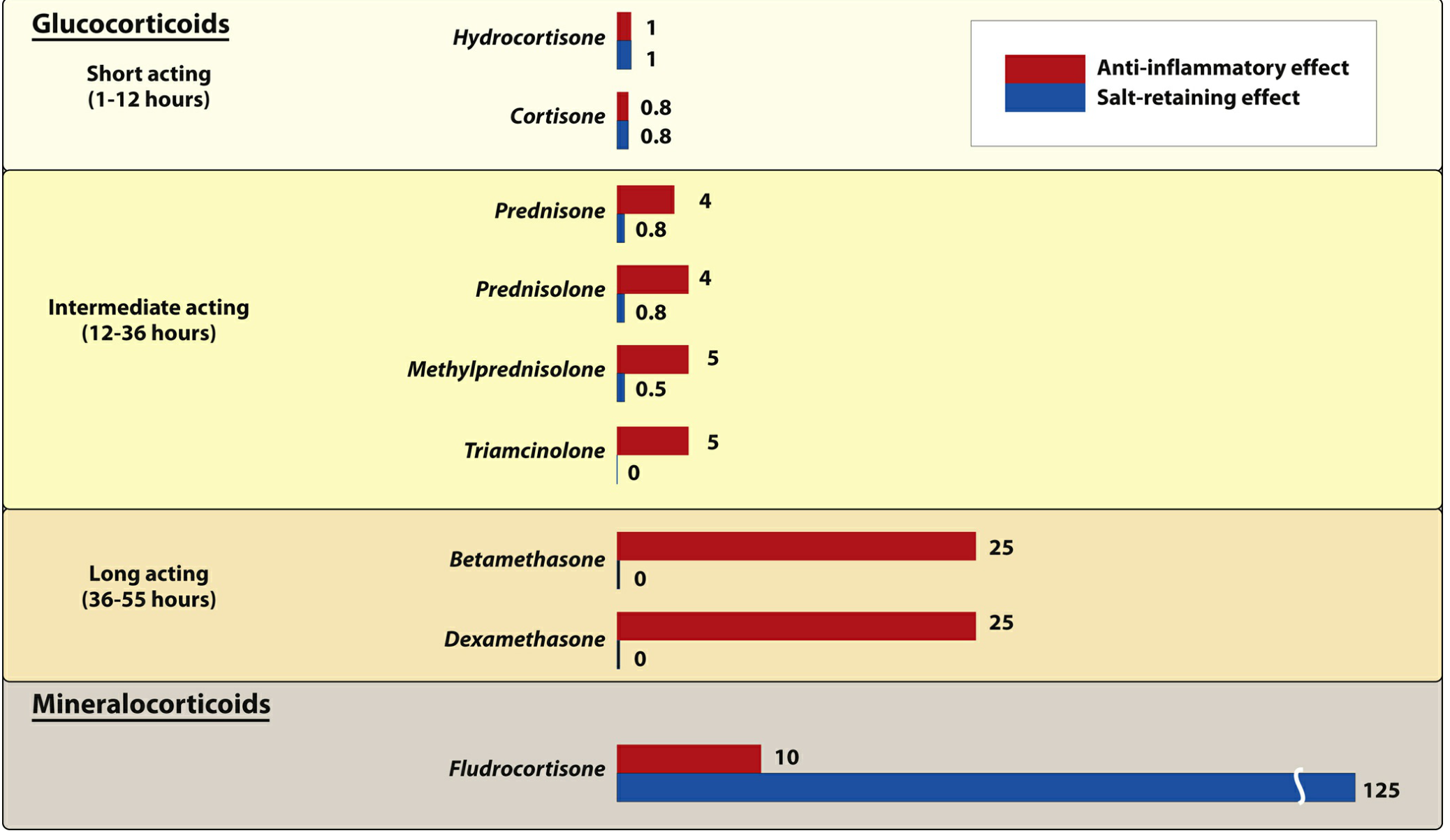
Figure: Pharmacologic effects and duration of action of commonly used natural and synthetic corticosteroids (relative to hydrocortisone = 1). (Lippincott Illustrated Reviews: Pharmacology)
| Glucocorticoid | Equivalent Oral Dose (mg) | Plasma t½ (min) | Anti-inflammatory Potency | Mineralocorticoid Potency |
|---|---|---|---|---|
| Cortisol (hydrocortisone) | 20 | 90 | 1 | 1 |
| Prednisone / Prednisolone | 5 | 200 | 4 | 0.8 |
| Methylprednisolone | 4 | 200 | 5 | 0.5 |
| Triamcinolone | 4 | 200 | 5 | 0 |
| Dexamethasone | 0.75 | 300 | 25 | 0 |
(Rheumatology, 2-Volume Set, Table 62.1)
Mechanism of Action
Glucocorticoids act through genomic and nongenomic pathways:
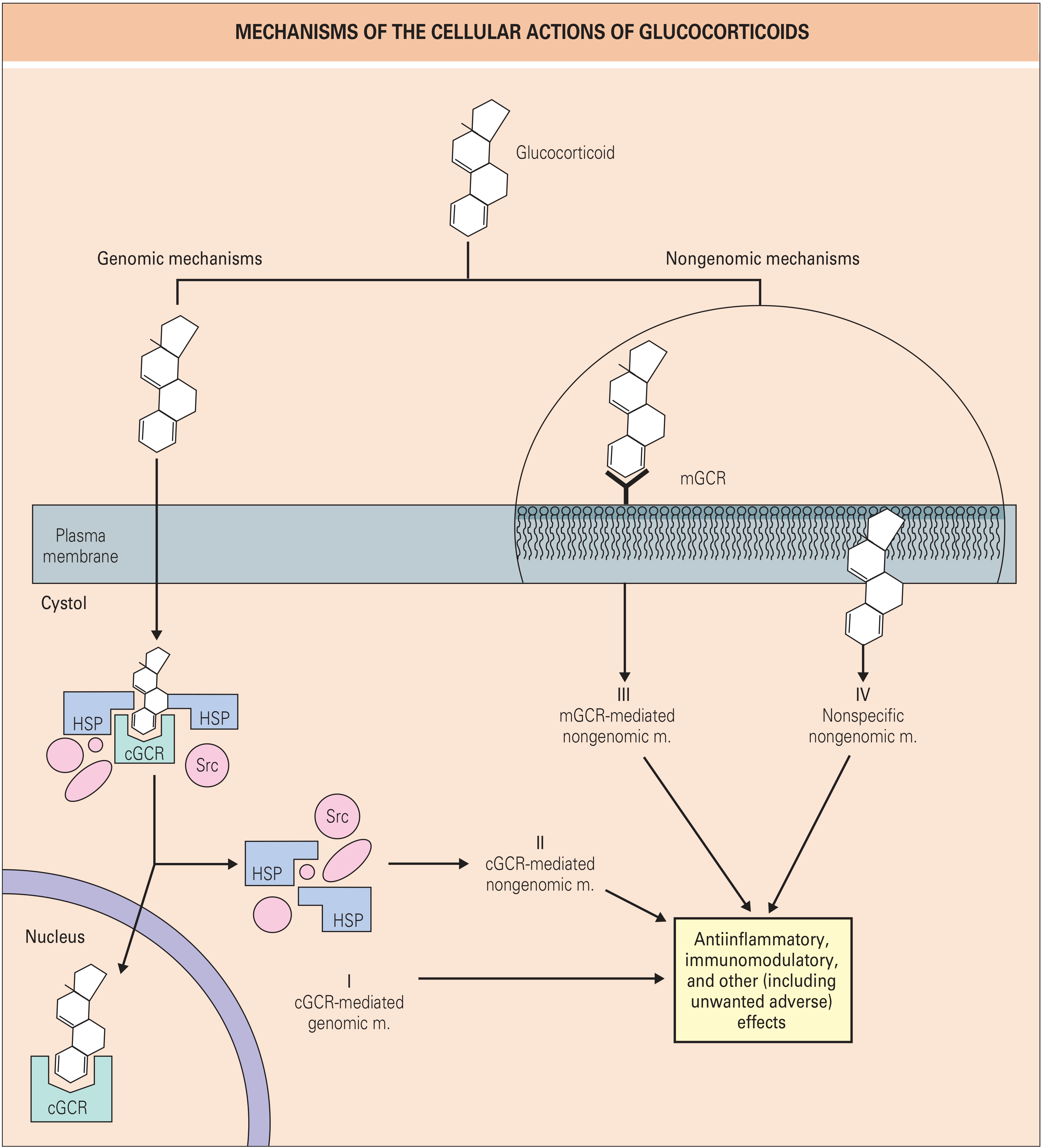
Figure: Mechanisms of the cellular actions of glucocorticoids. Four distinct pathways (I-IV) lead to anti-inflammatory, immunomodulatory, and other effects. (Rheumatology, 2-Volume Set)
Genomic Pathways
-
cGCR-mediated genomic mechanism (Pathway I): Glucocorticoids are lipophilic and freely cross the cell membrane. In the cytosol, they bind to the cytosolic glucocorticoid receptor (cGCR), which is held in an inactive state by heat shock proteins (HSP90, HSP70) and Src kinase. Ligand binding causes dissociation of the chaperone complex. The glucocorticoid-receptor complex is chaperoned to the nucleus, where it binds to glucocorticoid response elements (GREs) in gene promoters or suppressors to activate or repress transcription.
-
Transrepression via NF-κB and AP-1: The glucocorticoid-receptor complex directly inhibits key pro-inflammatory transcription factors - NF-κB and AP-1 - thereby suppressing transcription of genes encoding cytokines (IL-1, IL-6, TNF-α), COX-2, iNOS, and adhesion molecules. This is considered a primary anti-inflammatory mechanism.
-
cGCR-mediated nongenomic mechanism (Pathway II): Released Src kinase from the HSP complex triggers rapid cytoplasmic signaling independently of gene transcription.
Nongenomic Pathways
- Membrane glucocorticoid receptor (mGCR, Pathway III): Glucocorticoids bind membrane-associated receptors, producing rapid effects within seconds to minutes.
- Nonspecific membrane effects (Pathway IV): High-dose glucocorticoids intercalate into cell membranes, altering membrane biophysics. This is the likely basis for the pronounced nongenomic effects of IV pulse methylprednisolone (which has >3-fold more nongenomic effects than prednisolone at equivalent doses).
(Rheumatology, 2-Volume Set)
Anti-Inflammatory and Immunological Effects
Glucocorticoids produce broad suppression of the inflammatory response:
- Inhibit pro-inflammatory cytokines: IL-1β, IL-2, IL-6, IL-8, TNF-α, IFN-γ
- Inhibit pro-inflammatory enzymes: COX-2, phospholipase A2 (via lipocortin-1/annexin A1 induction), collagenase, elastase, plasminogen activator
- Decrease T-cell function and reduce circulating T-cell numbers
- Inhibit Fc receptor expression, reducing clearance of antibody-coated blood cells
- Increase circulating neutrophils (demargination from vessel walls) while impairing their migration to sites of inflammation
- Inhibit leukocyte adhesion to endothelial cells
The transcriptional response to glucocorticoids is highly cell-type specific - the individual genes and pathways affected, as well as the magnitude and direction of regulation, differ between cell types. (Rheumatology, 2-Volume Set)
Dosing Concepts
EULAR definitions for oral prednisone equivalent dosing:
- Low dose: ≤7.5 mg/day
- Medium dose: >7.5 to ≤30 mg/day
- High dose: >30 to ≤100 mg/day
- Very high / pulse therapy: 250-1000 mg/day IV methylprednisolone for 3 days (for life- or organ-threatening disease)
Timing matters: Prednisolone and methylprednisolone clearance is up to 25% lower in the morning than in the evening. Modified-release prednisone (taken at night, releasing 4 hours later) reduces morning stiffness in RA more effectively than immediate-release forms. (Rheumatology, 2-Volume Set)
Therapeutic Uses
| Indication | Notes |
|---|---|
| Replacement therapy (Addison's disease) | Hydrocortisone mimics diurnal cortisol; 2/3 dose in morning, 1/3 afternoon |
| Rheumatoid arthritis | Low-dose reduces disease activity and slows radiographic progression |
| Polymyalgia rheumatica / Giant cell arteritis | Mainstay of therapy |
| SLE, inflammatory myopathy, systemic vasculitis | Usually 1 mg/kg/day; pulse therapy for severe CNS/renal involvement |
| Asthma / allergic rhinitis | Inhaled steroids (fluticasone, budesonide) for long-term control |
| Organ transplant rejection prophylaxis | Immunosuppressive effect |
| Cerebral edema | Dexamethasone (no mineralocorticoid activity) |
| Cushing syndrome diagnosis | Dexamethasone suppression test |
| Septic shock (select cases), ARDS | Hydrocortisone ± fludrocortisone |
| Intra-articular injection | Osteoarthritis flares |
(Lippincott Illustrated Reviews: Pharmacology; Rheumatology, 2-Volume Set)
Adverse Effects
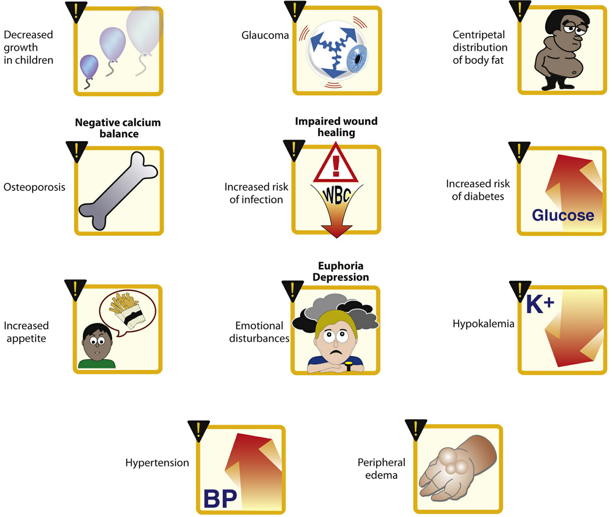
Figure: Commonly observed effects of long-term corticosteroid therapy. (Lippincott Illustrated Reviews: Pharmacology)
Key Adverse Effects (dose-dependent)
Musculoskeletal
- Glucocorticoid-induced osteoporosis (GIOP): The most devastating long-term complication. Up to 40% of users develop bone loss leading to fracture. Predominantly affects trabecular bone initially (vertebrae), later cortical bone (femoral neck). First-year bone loss can be 1.5-20% at ≤10 mg/day. Risk mitigation requires calcium/vitamin D supplementation and bisphosphonate therapy.
- Proximal myopathy: Muscle weakness, particularly in the thighs and shoulder girdle
Metabolic
- Hyperglycemia / steroid-induced diabetes mellitus
- Central (centripetal) obesity, moon facies, buffalo hump (Cushing-like features)
- Hypertriglyceridemia
- Negative calcium balance
Cardiovascular
- Hypertension (sodium and water retention)
- Peripheral edema
Immunological
- Increased susceptibility to infection (bacterial, fungal, viral)
- Impaired wound healing
Endocrine
- HPA axis suppression (adrenal suppression) - risk of adrenal crisis on withdrawal
- Diminished sex hormone production
Neuropsychiatric
- Euphoria, emotional disturbances, depression, psychosis
- Insomnia
Ocular
- Posterior subcapsular cataracts
- Glaucoma (raised intraocular pressure)
Pediatric
- Growth retardation (mechanism unknown, partly via growth hormone suppression; alternate-day dosing reduces this effect)
Skin (topical)
- Skin atrophy, ecchymosis, striae, telangiectasia
(Rheumatology, 2-Volume Set; Lippincott Illustrated Reviews: Pharmacology)
Discontinuation and Adrenal Suppression
Abrupt discontinuation after prolonged therapy can precipitate acute adrenal insufficiency (adrenal crisis), which may be fatal. Manifestations include nausea, vomiting, fever, dehydration, hypotension, shock, hypoglycemia, and hyperkalemia.
Key rules:
- Doses must be tapered slowly according to individual tolerance
- Risk is present any time the HPA axis has been suppressed (typically courses >3 weeks or doses above physiologic replacement)
- Patients on chronic steroids may need stress dosing (increased dose) during surgery or serious illness
Drug Interactions
- Phenobarbital, phenytoin, rifampin: Induce CYP3A4, increasing glucocorticoid metabolism and reducing efficacy
- Concurrent aspirin: Accelerates salicylate metabolism, reducing salicylate levels
- NSAIDs: Additive GI risk (peptic ulceration)
- Antidiabetic drugs: Glucocorticoids antagonize their effects, requiring dose adjustments
Special Considerations
Glucocorticoid resistance: Reported in severe RA, associated with higher glucocorticoid receptor β levels, MAPK activation, and antibodies to lipocortin-1. The 11β-hydroxysteroid dehydrogenase isoform balance also plays a central role.
Selective GR agonists (SEGRAs): Under development to dissociate transrepression (anti-inflammatory) from transactivation (adverse metabolic effects). Fosdagrocorat in phase IIb trials showed similar efficacy to prednisone 10 mg/day with metabolic safety closer to prednisone 5 mg/day, with reduced HbA1c elevations.
Liposomal glucocorticoids: In development to provide prolonged drug concentrations at the site of inflammation with lower systemic exposure. (Rheumatology, 2-Volume Set)
Glucocorticoids Physiology
Searching sources...
Finding Sources
Finding Sources
Finding Sources
Finding Sources
Found the Medical Physiology textbook with dedicated sections. Let me read both sections in parallel.
Reading File
Reading File
Excellent content. Let me now get the metabolic actions, steroidogenesis pathway, and the adrenal anatomy image, plus continue reading the physiological effects.
Loading Image
Reading File
Excellent material. Let me now read the metabolic actions section and get the steroidogenesis/HPA axis diagrams.
Reading File
Reading File
Now let me get the HPA axis regulation diagram and the metabolic actions of cortisol sections.
Reading File
Loading Image
Now let me get the HPA axis diagram and the physiological metabolic effects section.
Finding Sources
Reading File
Excellent - rich physiological content. Let me get the HPA feedback diagram URL and the metabolic actions text.
Reading File
Now let me get the HPA axis image and the detailed physiological effects (glucose, protein, fat, immune, cardiovascular, etc.).
Finding Sources
Reading File
Now let me get the HPA axis diagram and the section on cortisol's physiological effects on metabolism and the immune system.
Finding Sources
Reading File
Found Costanzo Physiology with detailed metabolic actions. Let me read that and get the HPA axis figure.
Reading File
Loading Image
I now have all the core physiological content. Let me read the remaining metabolic and immune actions from Costanzo.
Reading File
I now have comprehensive physiological content from Medical Physiology (Boron & Boulpaep) and Costanzo's Physiology. I have enough to write an outstanding, well-illustrated answer.
Glucocorticoids - Physiology
1. The Adrenal Gland: Source of Glucocorticoids
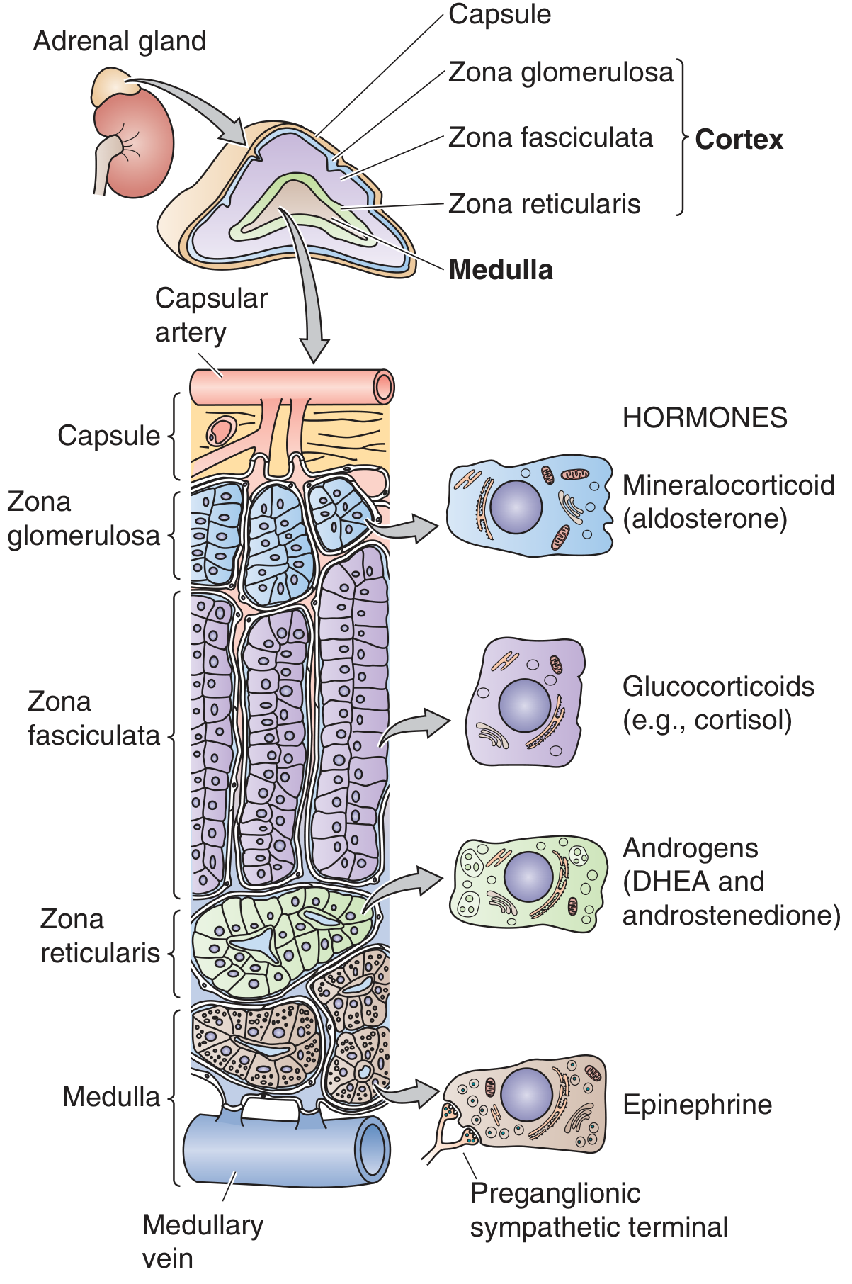
Figure: Anatomy of the adrenal gland. Each gland consists of an outer cortex (3 zones) and an inner medulla. The blood supply flows from the subcapsular region through the cortex into the medulla. (Medical Physiology, Boron & Boulpaep)
The adrenal cortex has three distinct zones, each producing different steroids:
| Zone | Product |
|---|---|
| Zona glomerulosa (outermost) | Mineralocorticoids (aldosterone) |
| Zona fasciculata (middle, largest) | Glucocorticoids (cortisol) |
| Zona reticularis (inner) | Adrenal androgens (DHEA, androstenedione) |
| Medulla | Catecholamines (epinephrine) |
Cortisol is the primary endogenous glucocorticoid in humans (corticosterone is the major glucocorticoid in rodents). Most body tissues are targets for glucocorticoid action - liver, fat, muscle, bone, skin, hematopoietic/lymphoid tissue, and the CNS all express glucocorticoid receptors. (Medical Physiology)
2. Biosynthesis: From Cholesterol to Cortisol
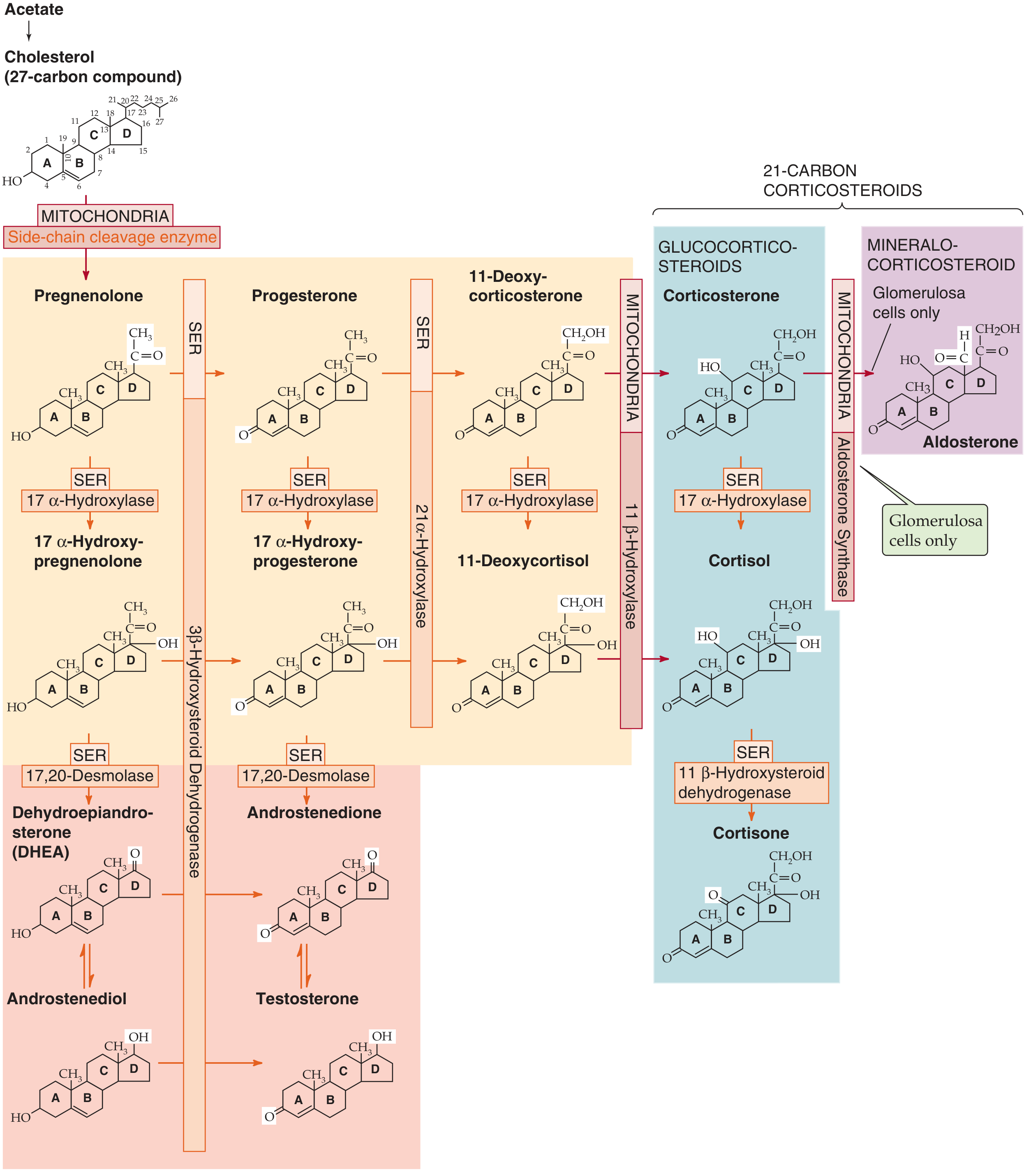
Figure: Biosynthesis of adrenal steroids. All steroids derive from cholesterol via a series of cytochrome P-450 enzyme reactions in the SER and mitochondria. (Medical Physiology)
Cortisol synthesis begins with cholesterol from two sources:
- Circulating LDL taken up by receptor-mediated endocytosis (quantitatively dominant)
- De novo synthesis from acetate
Five key enzymatic steps (zona fasciculata):
-
Cholesterol → Pregnenolone (mitochondria): Side-chain cleavage enzyme (P-450scc / 20,22-desmolase) removes carbons 22-27. This is the rate-limiting step.
-
Pregnenolone → Progesterone (SER): 3β-HSD oxidizes the hydroxyl group at C-3 to a ketone.
-
Progesterone → 17α-Hydroxyprogesterone (SER): 17α-hydroxylase (P-450c17/CYP17) adds -OH at C-17.
-
17α-Hydroxyprogesterone → 11-Deoxycortisol (SER): 21α-hydroxylase (P-450c21/CYP21A2) adds -OH at C-21.
-
11-Deoxycortisol → Cortisol (mitochondria): 11β-hydroxylase (P-450c11/CYP11B1) adds -OH at C-11.
Key enzyme absent from zona glomerulosa: 17α-hydroxylase is not substantially present in the glomerulosa, which is why that zone cannot make cortisol and instead makes aldosterone (using aldosterone synthase/CYP11B2). (Medical Physiology)
Clinical relevance - 21α-hydroxylase deficiency (most common congenital adrenal enzyme defect): Blocks synthesis of both cortisol and aldosterone. The resulting low cortisol removes negative feedback → ACTH rises → drives excessive production of adrenal androgens. Clinically: salt-wasting, hypoglycemia, and virilization (ambiguous genitalia in female infants).
3. Transport and Metabolism
-
~90% of circulating cortisol is protein-bound:
- ~75% bound to corticosteroid-binding globulin (CBG / transcortin) - high affinity, low capacity
- ~15% loosely bound to albumin
- Only ~10% is free (biologically active)
-
Synthetic glucocorticoids (dexamethasone, betamethasone, triamcinolone) have <1% affinity for CBG - they circulate largely free, contributing to greater biological activity per dose.
-
Cortisol is metabolized in the liver. Prednisone (prodrug) requires hepatic conversion to active prednisolone by 11β-HSD. In severe liver disease, prednisolone is preferred over prednisone. (Rheumatology, 2-Volume Set)
-
The enzyme 11β-hydroxysteroid dehydrogenase type 2 (11β-HSD2) inactivates cortisol to cortisone in tissues like the kidney, protecting mineralocorticoid receptors from activation by cortisol (which has equal affinity for the mineralocorticoid receptor). Liquorice inhibits 11β-HSD2, causing apparent mineralocorticoid excess.
4. The HPA Axis: Regulation of Cortisol Secretion
The hypothalamic-pituitary-adrenal (HPA) axis controls cortisol secretion through a classic endocrine hierarchy:
Hypothalamus (paraventricular nucleus)
↓ CRH (41-amino acid neuropeptide)
Anterior Pituitary (corticotroph cells)
↓ ACTH (39-amino acid peptide)
Adrenal Cortex (zona fasciculata)
↓ Cortisol
⟲ Negative feedback → inhibits CRH & ACTH
CRH (Corticotropin-Releasing Hormone)
- Secreted by small-bodied neurons of the paraventricular nucleus (PVN) of the hypothalamus
- Released into the hypophyseal portal venous plexus → reaches anterior pituitary
- Binds CRH-R1 (GPCR) on corticotrophs → Gαs → ↑cAMP → PKA → opens L-type Ca²⁺ channels → exocytosis of pre-formed ACTH
- Over longer time: increases POMC gene transcription
- Arginine vasopressin (AVP) is a co-secretagogue from the PVN that potentiates CRH-induced ACTH release (important during stress involving dehydration or trauma)
ACTH (Adrenocorticotropic Hormone)
- A 39-amino acid peptide secreted by anterior pituitary corticotrophs
- Derived from pro-opiomelanocortin (POMC), a large precursor that also yields:
- β-lipotropin (β-LPH)
- α-MSH, γ-MSH (melanocyte-stimulating hormones - cause skin hyperpigmentation in Addison's disease via excess ACTH/POMC)
- CLIP (corticotropin-like intermediate lobe peptide)
- β-endorphin
- ACTH acts on adrenal fasciculata cells to stimulate cholesterol uptake and activate the rate-limiting step (P-450scc), rapidly driving cortisol synthesis
- Chronic ACTH excess causes adrenal hyperplasia; chronic ACTH deficiency causes adrenal atrophy
Negative Feedback
Cortisol exerts negative feedback at two levels:
- Anterior pituitary: Cortisol binds cytosolic GR → nucleus → binds GREs → inhibits POMC transcription and blocks synthesis of the CRH receptor; also inhibits release of pre-formed ACTH from vesicles.
- Hypothalamus: Cortisol decreases CRH mRNA levels in PVN neurons and inhibits release of pre-formed CRH (less dominant than pituitary feedback).
(Medical Physiology, Boron & Boulpaep)
5. Circadian Rhythm and Pulsatility
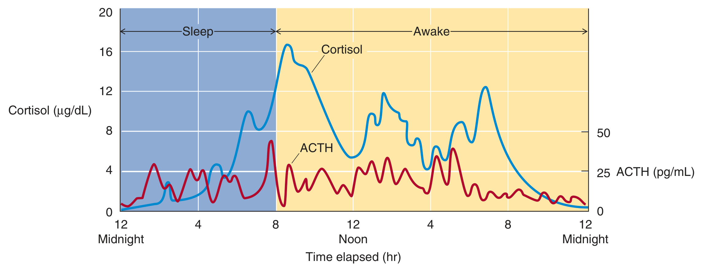
Figure: 24-hour rhythm of ACTH (red, pg/mL) and cortisol (blue, μg/dL). ACTH peaks in early morning around 8 AM with pulsatile bursts. Cortisol follows with its longer half-life, damping the pulsatile oscillations. Both approach nadir around midnight. (Medical Physiology)
Key features:
- ACTH secretory activity is greatest in the early morning and lowest late afternoon/evening
- Cortisol peaks at ~16 μg/dL around 8 AM and falls to near zero by midnight
- The rhythm is driven by the suprachiasmatic nucleus (receives retinal light input) - blind individuals lose circadian rhythms
- Superimposed on the circadian rhythm is pulsatile secretion driven by pulsatile CRH release
- ACTH pulses are sharper and briefer (shorter half-life); cortisol oscillations are broader and damped
6. Stress Response
Higher CNS centers stimulate the HPA axis during physical, psychological, and biochemical stress:
- Hypoglycemia → stimulates CRH and ACTH → ↑cortisol → gluconeogenesis raises blood glucose
- Physical trauma, infection, surgery, major illness → activate ascending noradrenergic pathways → stimulate PVN CRH neurons
- The amplitude of CRH secretory bursts increases during stress (not frequency)
- Chronic stress → chronic ACTH elevation → adrenal hypertrophy
- Glucocorticoids are sometimes called "stress hormones" because they allow the body to cope with acute threats - but chronic activation is harmful
7. Physiological Actions of Cortisol
A. Carbohydrate Metabolism - "Diabetogenic"
Cortisol is overall catabolic and diabetogenic:
- ↑ Gluconeogenesis in liver (key survival function during fasting)
- ↑ Protein catabolism in muscle → provides amino acids to liver as gluconeogenic substrates
- ↑ Lipolysis → provides glycerol to liver for gluconeogenesis; free fatty acids used as alternative fuel
- ↓ Glucose utilization by peripheral tissues (antagonizes insulin action)
- ↓ Insulin sensitivity of adipose tissue
In hypocortisolism (Addison's disease): hypoglycemia In hypercortisolism (Cushing's syndrome): hyperglycemia (Costanzo Physiology)
B. Protein Metabolism
- Catabolic in muscle, bone, connective tissue, skin - mobilizes amino acids
- Anabolic in the liver - stimulates synthesis of enzymes for gluconeogenesis
- Net effect of prolonged excess: muscle wasting, skin thinning, striae, poor wound healing
C. Fat Metabolism - Redistribution
- Excess cortisol causes characteristic fat redistribution:
- ↑ Fat deposition: face (moon facies), trunk, supraclavicular, dorsal interscapular (buffalo hump)
- ↓ Fat in extremities
- Mechanism: differential insulin sensitivity across fat depots; visceral fat is more glucocorticoid-sensitive
D. Anti-Inflammatory Actions
Three major mechanisms:
- Induces lipocortin (annexin A1) - inhibits phospholipase A2, blocking release of arachidonic acid from membrane phospholipids → suppresses prostaglandin and leukotriene synthesis
- Inhibits IL-2 production and T lymphocyte proliferation
- Inhibits histamine and serotonin release from mast cells and platelets
(Costanzo Physiology)
E. Immune Suppression
- Suppresses cellular immunity via T-cell inhibition (IL-2 ↓)
- Reduces Fc receptor expression on macrophages
- Promotes neutrophilia (demargination from vessel walls) while impairing neutrophil migration to inflammatory sites
- Basis for glucocorticoid use in transplant rejection and autoimmune diseases
F. Cardiovascular - Permissive Role
Cortisol is required for normal vascular tone and blood pressure maintenance:
- Up-regulates α₁-adrenergic receptors on arterioles (permissive effect)
- Allows arterioles to respond normally to catecholamines (norepinephrine, epinephrine)
- Without cortisol → hypotension (hallmark of Addison's disease - refractory to vasopressors)
- Excess cortisol → hypertension (Cushing's syndrome)
(Costanzo Physiology)
G. Bone and Calcium Metabolism
Cortisol has anti-anabolic/catabolic effects on bone:
- Decreases synthesis of type I collagen (major bone matrix component)
- Decreases osteoblast function (bone formation ↓)
- Decreases intestinal Ca²⁺ absorption (interferes with vitamin D action)
- Net result: negative calcium balance and osteoporosis with chronic excess
H. Renal Effects
- Increases GFR by causing afferent arteriole vasodilation → ↑ renal blood flow
- Without cortisol, GFR falls; excess cortisol can contribute to hypertension via this mechanism and via sodium retention (weak mineralocorticoid effect of cortisol)
I. CNS Effects
- Glucocorticoid receptors are widely distributed in the CNS (hippocampus, prefrontal cortex, hypothalamus)
- Normal cortisol: needed for normal mood, cognition, and arousal
- Excess cortisol: euphoria, emotional lability, depression, psychosis, insomnia, decreased REM sleep
- Deficiency: fatigue, depression, cognitive impairment
8. Summary Table: Physiological Actions of Glucocorticoids
| System | Effect |
|---|---|
| Carbohydrate | ↑ Gluconeogenesis, ↓ peripheral glucose uptake, ↑ glycogen storage (liver) |
| Protein | ↑ Catabolism (muscle), ↑ hepatic protein synthesis for gluconeogenesis |
| Fat | ↑ Lipolysis; redistribution of fat (central deposition) |
| Immune | ↓ IL-2, ↓ T-cell proliferation, ↓ histamine release, ↓ prostaglandins |
| Cardiovascular | Permissive for catecholamine vasoconstriction (↑ α₁-adrenergic receptors) |
| Bone | ↓ Osteoblast activity, ↓ collagen synthesis, ↓ intestinal Ca²⁺ absorption → osteoporosis |
| Kidney | ↑ GFR (afferent arteriole vasodilation) |
| CNS | Mood, cognition, arousal; excess → emotional disturbance, ↓ REM sleep |
| HPA axis | Negative feedback on CRH and ACTH secretion |
(Costanzo Physiology 7th ed.; Medical Physiology, Boron & Boulpaep)
9. Clinical Correlates
| Condition | Mechanism | Key Features |
|---|---|---|
| Addison's disease (primary adrenal insufficiency) | Adrenal destruction (autoimmune most common today; formerly TB) | Fatigue, hypoglycemia, hypotension, hyponatremia, hyperkalemia, hyperpigmentation (↑ACTH/MSH), salt craving |
| Cushing's syndrome | Glucocorticoid excess (exogenous steroids, pituitary ACTH tumor, adrenal tumor, ectopic ACTH) | Central obesity, moon facies, buffalo hump, hypertension, hyperglycemia, osteoporosis, striae, easy bruising |
| Cushing's disease | Pituitary ACTH-secreting adenoma specifically | Bilateral adrenal hyperplasia, non-suppressible on low-dose dexamethasone test |
| Congenital adrenal hyperplasia | 21α-hydroxylase deficiency (most common) | Salt-wasting, hypoglycemia, virilization (↑adrenal androgens) |
| Exogenous steroid withdrawal | HPA axis suppression → adrenal atrophy | Adrenal crisis on abrupt cessation; must taper slowly |
Lung Surfactant
Searching sources...
Reading File
Finding Sources
Finding Sources
Finding Sources
Reading File
Excellent leads. Let me now read all three major sections in parallel - Murray & Nadel's respiratory medicine (most comprehensive), Ganong's physiology, and the histology textbook.
Reading File
Reading File
Reading File
Excellent content. Now let me get the Murray & Nadel section on surfactant proteins, synthesis/secretion, the inhibition section, and fetch key diagrams.
Reading File
Reading File
Excellent. Now let me get the surfactant proteins section from Murray & Nadel, fetch key images, and get the biochemistry section on DPPC synthesis.
Reading File
Loading Image
Finding Sources
Excellent - the air vs. saline pressure-volume curve is retrieved. Now let me get the SP-B/C section and the type II cell / lamellar body content, plus the RDS clinical section.
Reading File
Finding Sources
Reading File
Finding Sources
Let me get the type II alveolar cell histology image and the RDS/clinical section.
Finding Sources
Reading File
Reading File
Reading File
Excellent. Let me fetch the two key diagrams - the surfactant alveolus diagram and the RDS pathogenesis diagram.
Loading Image
Loading Image
I have all the content needed for a comprehensive, well-illustrated answer. Let me now compose it.
Lung Surfactant
1. Historical Discovery
The story of surfactant begins with a deceptively simple experiment. In 1929, von Neegaard observed that inflating excised lungs with air required more pressure than inflating them with saline - but once inflated, alveoli stayed open even at low volumes. He proposed an "anti-atelectasis factor" lowering alveolar surface tension below what a simple air-water interface would predict. In the 1950s, Clements and Pattle used lung extracts to prove that a surface-active phospholipid was responsible. Shortly after, Avery and Mead causally linked a deficiency of this material to respiratory failure in premature infants - establishing the foundation of our understanding of neonatal RDS. (Murray & Nadel's Textbook of Respiratory Medicine)
2. The Physics: Surface Tension and the Law of Laplace
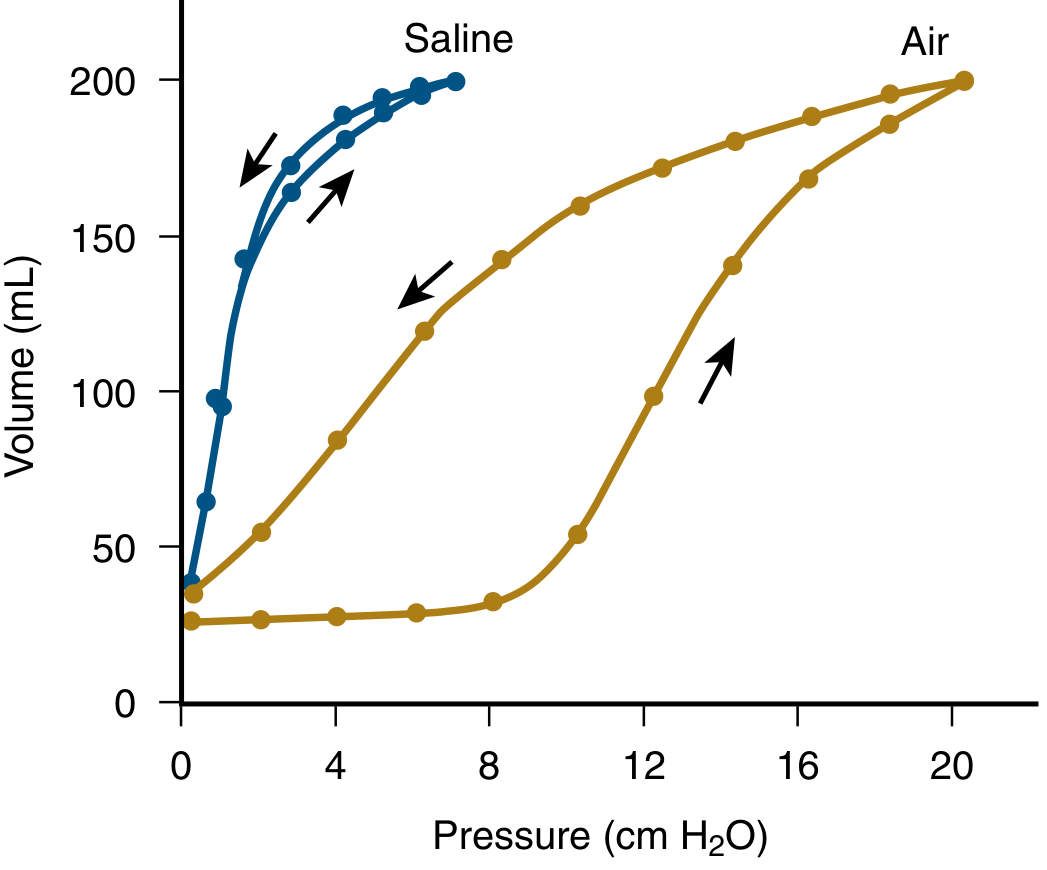
Figure: Air vs. saline pressure-volume curves in an excised cat lung. Much more pressure is needed to inflate with air (gold) than saline (blue), and there is marked hysteresis in the air-filled lung. These features disappear when surface tension is eliminated with saline, demonstrating the dominant role of surfactant at air-liquid interfaces. (Murray & Nadel's Textbook of Respiratory Medicine)
The Law of Laplace
For a spherical alveolus, the distending pressure needed to keep it open is:
$$P = \frac{2T}{r}$$
Where:
- P = distending pressure
- T = surface tension of the lining fluid
- r = radius of the alveolus
Without surfactant, as an alveolus shrinks during expiration (↓ r), if T stays constant, the inward collapse pressure increases. Small alveoli would collapse (atelectasis). Surfactant breaks this problem by reducing T as r decreases - the more DPPC molecules are compressed together as the alveolus shrinks, the lower the surface tension. This keeps the distending pressure manageable at all lung volumes.
Why Surfactant Prevents Pulmonary Edema
Without surfactant, the unopposed surface tension in alveoli would generate an estimated 20 mmHg inward force, strongly favoring transudation of fluid from the blood into alveolar spaces. Surfactant reduces this force, helping keep alveoli dry. (Ganong's Review of Medical Physiology)
3. What Surfactant Does: Functional Summary
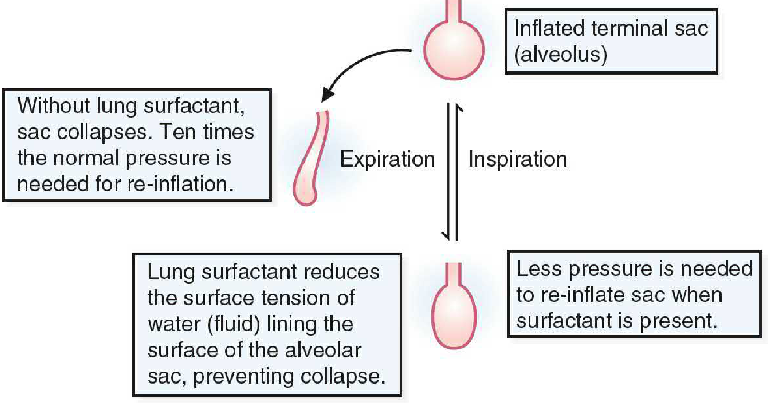
Figure: Without surfactant, alveoli collapse on expiration and require 10× normal pressure to re-inflate. With surfactant present, the sac stays partially inflated and requires much less pressure to re-open. (Basic Medical Biochemistry)
| Function | Mechanism |
|---|---|
| Prevents alveolar collapse (atelectasis) | Reduces surface tension at air-liquid interface, especially at low lung volumes |
| Reduces work of breathing | Less inspiratory effort needed to re-open alveoli |
| Stabilizes alveoli of different sizes | Laplace relationship: surface tension falls as radius falls |
| Prevents pulmonary edema | Opposes fluid transudation from capillaries |
| Innate immune defense | SP-A and SP-D opsonize pathogens and modulate inflammation |
4. Composition of Surfactant
Surfactant is secreted by type II alveolar epithelial cells (AT2 cells / type II pneumocytes) and consists of approximately:
- ~85% phospholipids
- ~5% neutral lipids (cholesterol, free fatty acids)
- ~10% proteins (four surfactant-specific proteins: SP-A, SP-B, SP-C, SP-D)
Phospholipid Composition (Murray & Nadel's Textbook of Respiratory Medicine, Table 3.2)
| Phospholipid | % of Total Phospholipids |
|---|---|
| Dipalmitoylphosphatidylcholine (DPPC) | 47% |
| Unsaturated phosphatidylcholine | 29.3% |
| Phosphatidylglycerol | 11.6% |
| Phosphatidylinositol | 3.9% |
| Phosphatidylethanolamine | 3.3% |
| Sphingomyelin | 1.5% |
| Other | 3.4% |
DPPC - The Critical Lipid
Dipalmitoylphosphatidylcholine (DPPC) is unique among phosphatidylcholines: both fatty acid chains are saturated palmitate (C16:0). This is highly unusual for biological membranes (which typically contain one unsaturated chain) and is essential to surfactant function. Because DPPC is amphiphilic (hydrophilic phosphocholine head + hydrophobic palmitate tails), it spontaneously forms a monomolecular film at the air-liquid interface. At low alveolar volumes, DPPC molecules are compressed together, the surface becomes nearly pure DPPC (~95%) (unsaturated phospholipids and proteins are squeezed out), and surface tension falls to as low as 1-2 mN/m (compared to ~70 mN/m for pure water). (Murray & Nadel's)
5. The Surfactant Proteins
Four unique proteins, designated SP-A through SP-D, are essential components of surfactant:
SP-A (Surfactant Protein A)
- Most abundant surfactant protein
- A large secreted octadecameric collagenous glycoprotein forming a "bouquet-like" structure of 18 monomers (6 trimers)
- Contains a C-type lectin / carbohydrate recognition domain (CRD) - recognizes carbohydrates on pathogens
- Primary role: innate immunity - not biophysical activity
- Opsonizes bacteria, viruses, fungi
- Regulates cytokine production in macrophages (suppresses inflammation in health; enhances it during infection - the "inflammatory paradox")
- Regulates feedback uptake/recycling of surfactant by AT2 cells
- Together with SP-B, needed for formation of tubular myelin (an organized extracellular surfactant intermediate)
- Member of the collectin family (like mannose-binding lectin, C1q)
- Two human genes: SFTPA1 and SFTPA2 on chromosome 10
SP-B (Surfactant Protein B)
- Extremely hydrophobic (18-kDa disulfide-linked homodimer); residues predominantly valine and leucine
- Member of the saposin-like peptide family
- Critical for biophysical surfactant activity:
- Promotes adsorption and surface spreading of phospholipids onto the air-liquid interface
- Enables DPPC to form the stable monomolecular film
- Essential for formation of lamellar bodies (LBs) within AT2 cells - the storage organelles for surfactant
- Required for processing of SP-C (see below)
- SP-B deficiency is uniformly fatal (or requires lung transplantation): homozygous loss-of-function mutations cause absence of both SP-B and SP-C, since SP-B is required for LB formation and thus posttranslational processing of the SP-C proprotein
- Encoded by the SFTPB gene on chromosome 2
SP-C (Surfactant Protein C)
- Extremely hydrophobic 4.2-kDa mature peptide; only ~1% of total surfactant protein by mass
- Works together with SP-B to orient DPPC within the surfactant film and maintain the thin film layer
- Encoded by SFTPC on chromosome 8
- Complex processing: synthesized as a 21-kDa proprotein (proSP-C), processed through the ER, Golgi, and multivesicular bodies into the mature form stored in lamellar bodies
- Mutations in SFTPC (especially in the BRICHOS domain) cause misfolding, ER retention, and are linked to familial interstitial lung disease (ILD) and pulmonary fibrosis in adults and children
SP-D (Surfactant Protein D)
- Hydrophilic collectin glycoprotein, structurally similar to SP-A
- Primary role: host defense (not biophysical)
- Binds to Gram-negative bacteria, fungi, and other pathogens
- Binds lipopolysaccharide (LPS)
- Modulates innate immune and inflammatory responses to inhaled antigens and acute lung injury
- Works with SP-A to organize SP-B and SP-C into the surfactant layer
- Gene located on chromosome 10 near SFTPA1 and SFTPA2
(Murray & Nadel's Textbook of Respiratory Medicine; Ganong's Review of Medical Physiology; Histology - A Text and Atlas)
6. The Type II Pneumocyte and Lamellar Bodies
Surfactant is synthesized and secreted exclusively by type II alveolar epithelial cells (AT2 cells / type II pneumocytes):
- Cuboidal cells found at alveolar corners (accounting for only ~5% of alveolar surface area but ~60% of alveolar cells by number)
- Characterized by distinctive lamellar bodies (LBs) - electron-dense lysosome-related organelles that store packaged surfactant as densely stacked phospholipid bilayers
- Lamellar bodies require SP-B for their formation
- Upon stimulation, lamellar bodies fuse with the plasma membrane and are secreted by exocytosis into the alveolar space
- After secretion, lamellar body contents unroll into an extracellular structured form called tubular myelin (an organized lattice of surfactant bilayers at right angles), which serves as a reservoir for rapid replenishment of the surface monolayer during inspiration
- Surfactant is cleared from the alveolar space by:
- Re-uptake by AT2 cells (major route) - regulated by SP-A
- Macrophage phagocytosis
- Mucociliary clearance
Hormonal Regulation of Synthesis
Surfactant synthesis begins in the fetus around 35 weeks of gestation and is modulated by:
| Hormone | Effect |
|---|---|
| Corticosteroids (cortisol) | ↑ Synthesis of surfactant lipids AND proteins - the major inducer |
| Insulin | Suppresses surfactant synthesis (explains increased RDS risk in infants of diabetic mothers) |
| Prolactin | ↑ Synthesis |
| Thyroxine (T4) | ↑ Synthesis |
| Labor contractions | ↑ Synthesis (explains why elective cesarean before labor ↑ RDS risk) |
(Robbins & Kumar Basic Pathology; Histology - A Text and Atlas)
7. Clinical Conditions
A. Neonatal Respiratory Distress Syndrome (NRDS / Infant RDS / Hyaline Membrane Disease)
Epidemiology:
- Most common cause of respiratory insufficiency in newborns
- ~60% of infants born at <28 weeks gestation
- ~30% born at 28-34 weeks gestation
- <5% born after 34 weeks gestation
- Additional risk factors: male sex, maternal diabetes, cesarean section before labor onset
Pathogenesis:
The immature lung cannot synthesize sufficient surfactant → alveoli collapse on expiration → massive inspiratory effort required → infant tires → generalized atelectasis → hypoxia → epithelial and endothelial damage → plasma proteins leak into alveolar space → hyaline membranes form (eosinophilic membranes lining respiratory bronchioles and alveolar ducts, composed of necrotic type II pneumocytes + fibrin from extravasated fibrinogen).
Morphology (Robbins & Kumar Basic Pathology):
- Lungs are normal size but heavy and airless, with mottled purple color
- Microscopy: diffuse atelectasis with characteristic eosinophilic hyaline membranes lining respiratory bronchioles, alveolar ducts, and alveoli
- Survivors show type II pneumocyte proliferation and interstitial fibrosis as reparative changes
Treatment:
- Antenatal corticosteroids (betamethasone/dexamethasone) given to mother at 24-34 weeks threatened preterm delivery → stimulates fetal surfactant synthesis → reduces RDS incidence and severity
- Exogenous surfactant replacement (intratracheal) at birth: natural (poractant alfa from porcine lungs, beractant from bovine lungs) or synthetic preparations
- Positive pressure ventilation / CPAP (continuous positive airway pressure) to maintain alveolar patency
B. Adult/Acute Respiratory Distress Syndrome (ARDS)
Surfactant dysfunction plays an important role in ARDS:
- Edema fluid containing serum proteins (albumin, globulin, fibrinogen, CRP) leaks into the alveolar space and competitively inhibits surfactant adsorption and film formation
- Phospholipases A2 and C released during lung injury degrade surfactant phospholipids
- Oxidants (reactive oxygen species, peroxynitrite) from activated alveolar macrophages damage both surfactant lipids and proteins
- Inflammatory cytokines (TNF-α, TGF-β1) suppress SP-A, SP-B, SP-C synthesis in AT2 cells
- Exogenous surfactant has been effective in animal models but has not shown consistent benefit in clinical ARDS trials (unlike NRDS); current treatment focuses on lung-protective ventilation and prone positioning
C. Inherited Surfactant Disorders
| Condition | Mutation | Clinical Presentation |
|---|---|---|
| SP-B deficiency | SFTPB (homozygous loss-of-function) | Neonatal respiratory failure; absent LBs; also misprocesses SP-C; requires lung transplant |
| SP-C mutations | SFTPC (BRICHOS domain) | Familial interstitial lung disease in children/adults; pulmonary fibrosis |
| ABCA3 mutations (null) | ABCA3 (transporter for lamellar body lipid loading) | Neonatal respiratory failure; absent LBs; decreased surfactant phospholipids |
| GM-CSF receptor mutations | Common β-chain or α-chain of GM-CSF receptor | Pulmonary alveolar proteinosis (hereditary PAP) - surfactant accumulates due to impaired macrophage clearance |
D. Pulmonary Alveolar Proteinosis (PAP)
- Characterized by accumulation of surfactant-like material (PAS-positive) in alveoli
- Most common form (autoimmune PAP): GM-CSF autoantibodies impair alveolar macrophage function → macrophages cannot clear surfactant → alveolar flooding
- CT: characteristic "crazy paving" pattern
- Treatment: whole-lung lavage; inhaled or systemic GM-CSF
8. Surfactant Inhibition
Surfactant activity can be inhibited by:
| Inhibitor | Source | Mechanism |
|---|---|---|
| Serum albumin, fibrinogen, globulins | Edema fluid (ARDS, IRDS) | Compete with surfactant lipids for air-liquid interface |
| Phospholipase A2 and C products (lysoPC, fatty acids) | Lung injury | Irreversible disruption of surfactant lipid organization |
| Oleic acid | Lung injury | Integrates into monolayer, disrupts DPPC film |
| Peroxynitrite (from macrophage NO + superoxide) | Activated immune cells | Oxidizes and inactivates surfactant proteins and lipids |
| High-concentration oxygen (FiO₂ > 0.85) | Oxygen therapy | Reduces synthesis of surfactant lipids; variable effects on proteins |
| TNF-α, TGF-β1 | Inflammatory cytokines | Suppress SP-A, SP-B, SP-C gene expression |
| E. coli endotoxin | Infection | Inhibits surfactant synthesis in AT2 cells |
SP-B and SP-C together provide the greatest resistance to inhibition by plasma proteins. SP-A can reverse inhibition by soluble proteins but not by lysoPC. (Fishman's Pulmonary Diseases and Disorders)
Quick Reference Summary
| Feature | Detail |
|---|---|
| Produced by | Type II alveolar epithelial cells (AT2 / type II pneumocytes) |
| Storage organelle | Lamellar bodies |
| Key lipid | DPPC (dipalmitoylphosphatidylcholine) - ~47% of phospholipids |
| Biophysical proteins | SP-B, SP-C (hydrophobic; form surface film) |
| Immune proteins | SP-A, SP-D (hydrophilic collectins; innate defense) |
| Fetal onset of synthesis | ~35 weeks gestation |
| Primary inducer | Glucocorticoids (cortisol) |
| Surface tension achieved | 1-2 mN/m at end-expiration (vs ~70 mN/m for water) |
| Deficiency → | Neonatal RDS (hyaline membrane disease) |
| Inactivation → | ARDS, post-infectious lung injury |
| Excess/accumulation → | Pulmonary alveolar proteinosis |
Embryology of Somites
Searching sources...
Finding Sources
Finding Sources
Finding Sources
Finding Sources
Found the primary source - "The Developing Human" (Moore's embryology). Let me read all three key sections in parallel.
Reading File
Reading File
Reading File
Excellent content from three sources. Now let me get more detail from The Developing Human on somite differentiation and derivatives, plus fetch the key diagrams.
Reading File
Finding Sources
Loading Image
Excellent. Now let me get the detailed somite differentiation, sclerotome/dermomyotome/dermatome content and the vertebral column formation details.
Finding Sources
Reading File
Finding Sources
Finding Sources
Reading File
Finding Sources
Excellent - very rich molecular genetics content. Let me now fetch the segmentation clock diagram and get more detail on somite derivatives from the Developing Human.
Loading Image
Loading Image
Reading File
I now have comprehensive, well-sourced content from four textbooks plus three diagrams. Let me compile the definitive answer.
Embryology of Somites
1. Overview
Somites (Greek: soma = body) are paired, segmented blocks of paraxial mesoderm that appear flanking the developing neural tube during the third and fourth weeks of embryonic development. They are among the most visually prominent features of the early embryo and are the precursors of the axial skeleton, skeletal musculature, and dermis of the back. Their orderly, craniocaudal formation is controlled by a molecular "segmentation clock" - one of the most precisely timed events in all of vertebrate development.
2. Timeline: From Gastrulation to Somite Formation
| Stage | Event |
|---|---|
| Week 2 | Bilaminar disc (epiblast + hypoblast) |
| Week 3 (Day 15-16) | Gastrulation: primitive streak appears; epiblast cells ingress to form intraembryonic mesoderm and endoderm |
| Week 3 (Day 17-19) | Paraxial mesoderm condenses as thick longitudinal columns flanking the notochord |
| Week 3 (end) | Paraxial mesoderm begins segmenting into somitomeres, then into distinct somites |
| Day 20 | First pair of somites appears (caudal to the future otic vesicle) |
| Day 26-32 | "Somite period": 38-39 pairs form |
| End of Week 5 | 42-44 pairs of somites present |
(The Developing Human, Moore)
3. Origin: Paraxial Mesoderm
During gastrulation, cells from the primitive node and primitive streak migrate cranially and laterally to form the intraembryonic mesoderm. The portion closest to the midline on either side of the notochord condenses into a thick longitudinal column called paraxial mesoderm (also called the presomitic mesoderm, PSM, before segmentation).
The mesoderm is organized in three lateral tiers:
Midline (notochord)
│
├── Paraxial mesoderm ← SOMITES form here
│ (thick, bilateral columns)
│
├── Intermediate mesoderm ← urogenital system
│ (thinner)
│
└── Lateral mesoderm ← body wall, gut wall, coelom
(thinnest; splits into somatic + splanchnic layers)
(The Developing Human; Fischer's Mastery of Surgery)
4. Somite Numbers by Region
| Region | Number of Somite Pairs |
|---|---|
| Occipital | 4-5 |
| Cervical | 8 |
| Thoracic | 12 |
| Lumbar | 5 |
| Sacral | 5 |
| Coccygeal | 8-12 |
The first occipital and the most caudal 5-7 coccygeal somites disappear. The remainder persist to form the axial skeleton, associated musculature, and dermis. (Fischer's Mastery of Surgery)
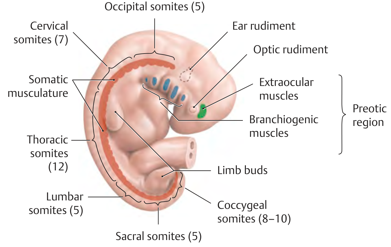
Figure: Right lateral view of a 5-week human embryo showing the regional distribution of somites (occipital, cervical, thoracic, lumbar, sacral, coccygeal) alongside the ear rudiment, optic rudiment, and limb buds. Preotic somites (blue/green, cranial to ear) give rise to extraocular and branchiogenic muscles innervated by cranial nerves. Postotic somites (red) give rise to somatic musculature. (Thieme Atlas of Anatomy)
5. The Molecular Segmentation Clock ("Clock and Wavefront" Model)
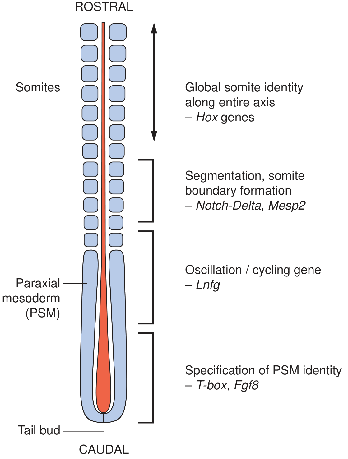
Figure: Somatogenesis and the molecular segmentation clock. From the tail bud (caudal) to formed somites (rostral), molecular control is organized in rostrocaudal zones: T-box/Fgf8 specify PSM identity; Lunatic fringe (Lnfg) is the oscillation/cycling gene; Notch-Delta and Mesp2 define somite boundary formation; HOX genes assign somite identity along the whole axis. (Emery's Elements of Medical Genetics and Genomics)
Somitogenesis is not random - it is controlled with extraordinary precision by a molecular clock:
The "Clock and Wavefront" Model (Cooke & Zeeman, 1970s)
- The clock is a wave of oscillating gene expression that sweeps from the caudal tail bud rostrally through the PSM. Each sweep "pinches off" one new pair of somites.
- The wavefront is a threshold of signaling activity (FGF8 and WNT gradients, high caudally) that determines where in the PSM the clock oscillation is interpreted as a boundary.
- As the embryo elongates caudally, the wavefront moves caudally too, allowing new somites to form progressively.
Key Molecular Players
| Gene/Pathway | Role |
|---|---|
| T-box genes (Tbx6) | Specification of PSM identity; Tbx6 mutations → failure to form somites |
| FGF8 (Fgf8) | High in caudal PSM; maintains PSM in undifferentiated state |
| WNT signaling | Interacts with FGF to maintain PSM and influence oscillation |
| Notch-Delta signaling (Notch receptor + Delta-like-1, Delta-like-3 ligands) | Core oscillation clock; establishes rostrocaudal polarity within each new somite; Mesoderm posterior-2 (Mesp2) defines somite boundaries |
| Lunatic fringe (Lnfg) | Modulates Notch signaling; key oscillation/cycling gene |
| Presenilin-1 | Component of Notch processing (γ-secretase complex) |
| HES genes (hes in mouse; c-hairy in chick) | Cycling genes downstream of Notch |
| FoxC1, FoxC2 | Forkhead transcription factors expressed in presomitic mesoderm before somite formation |
| HOX genes | Assign regional identity to each somite along the entire craniocaudal axis (which vertebral level it will form) |
(Emery's Elements of Medical Genetics and Genomics; The Developing Human)
6. Somite Structure: Epithelial-to-Mesenchymal Transitions
Newly formed somites are epithelial spheres - cuboidal cells arranged around a small lumen (somitocoele). They quickly undergo regionalization and differentiation:
SOMITE (epithelial sphere)
│
├─── SCLEROTOME (ventromedial) ──────► Vertebrae & ribs
│ (epithelial → mesenchymal)
│
└─── DERMOMYOTOME (dorsolateral) ────► remains epithelial
│
├── MYOTOME ─────────────────► Skeletal muscle
│
└── DERMATOME ───────────────► Dermis (back skin)
Signaling that Drives Differentiation
| Region | Signal | Source |
|---|---|---|
| Sclerotome induction | Sonic Hedgehog (SHH) | Notochord + floor plate of neural tube |
| Myotome induction | Wnt | Dorsal neural tube + surface ectoderm |
| Dermomyotome maintenance | Wnt + BMP | Surface ectoderm |
| Sclerotome also induced by | Noggin (BMP antagonist) | Notochord |
7. Somite Derivatives in Detail
A. Sclerotome → Axial Skeleton
The ventromedial cells of the somite undergo epithelial-to-mesenchymal transition (EMT) under SHH signaling and migrate medially to surround the notochord and neural tube. These sclerotomal cells form the vertebrae and ribs.
Resegmentation (von Ebner's concept):
Each sclerotome is divided into a cranial (rostral) and a caudal (dense) half. A vertebra forms from the caudal half of one sclerotome fusing with the cranial half of the next sclerotome below - not from a single somite. This means:
- Vertebral bodies are intersegmental - they bridge two adjacent somitic segments
- The spinal nerve (derived from the neural tube at the level of one somite) therefore exits between two vertebrae, running through the center of the original sclerotome
- The intervertebral disc forms at the junction between the cranial and caudal halves of a sclerotome
- The nucleus pulposus of the disc is a remnant of the notochord (which normally disappears as it is surrounded by vertebral bodies)
Structures formed from sclerotome:
- Vertebral bodies and arches
- Ribs (from thoracic sclerotomes)
- Sternum (from sternal bars of lateral plate mesoderm - not directly from somites)
- Intervertebral discs
B. Dermomyotome → Myotome and Dermatome
The dorsolateral portion of the somite - the dermomyotome - remains epithelial and then further divides:
Myotome (muscle precursors)
The myotome divides into:
| Division | Name | Innervation | Muscles Formed |
|---|---|---|---|
| Dorsal (medial) | Epimere / Epaxial | Dorsal (posterior) ramus of spinal nerve | Intrinsic (deep) back muscles (erector spinae, multifidus, rotatores, interspinales, intertransverse muscles); keep segmental arrangement |
| Ventral (lateral) | Hypomere / Hypaxial | Ventral (anterior) ramus of spinal nerve | Anterolateral chest wall, abdominal wall muscles (intercostals, external/internal oblique, transversus abdominis, rectus abdominis); diaphragm; limb musculature |
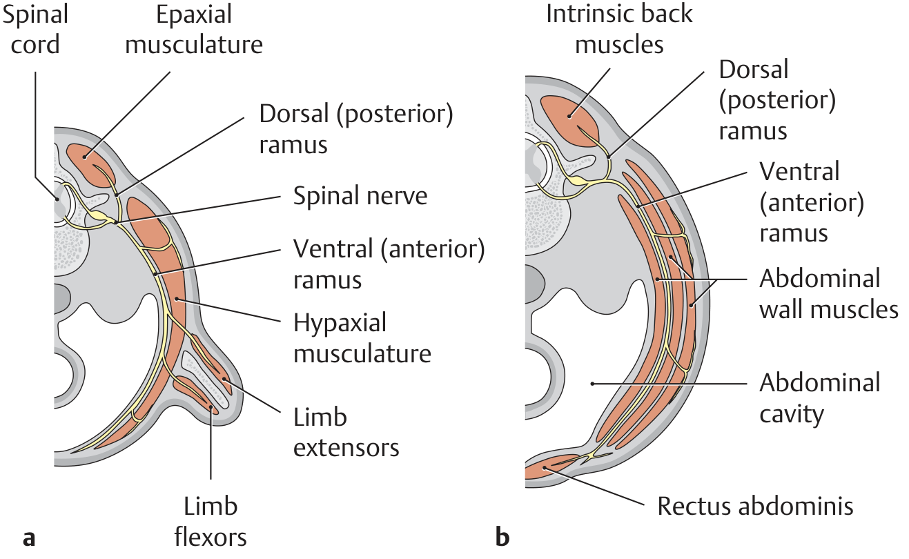
Figure: Transverse sections through a 6-week embryo showing the epaxial musculature (posterior, supplied by dorsal ramus) and hypaxial musculature (anterior/lateral, supplied by ventral ramus). At limb levels (a), myotome cells migrate into the limb bud to form limb muscles. At abdominal levels (b), hypaxial muscle forms the abdominal wall. (Thieme Atlas of Anatomy)
Limb muscles: At limb bud levels, myotome cells with replicative capacity migrate from the hypomere into the developing limb bud. They differentiate into a dorsal blastema (extensor muscles) and a ventral blastema (flexor muscles). Limb muscles are innervated by ventral rami through the brachial plexus (upper limb) and lumbosacral plexus (lower limb).
Fate of segmental arrangement: The original metameric (segmental) pattern:
- Persists in: deep intrinsic back muscles (rotatores, interspinales, intertransverse muscles), intercostal muscles
- Disappears (polymerization) in: superficial back muscles (which span multiple segments); but the segmental neurovascular supply remains as evidence of origin
Dermatome (dermis of back)
The dermatome gives rise to the dermis and subcutaneous tissue of the back. Note: the dermis of the ventral body wall and limbs is derived from lateral plate mesoderm, not somites. The dermis of the face and scalp is derived from neural crest cells.
8. Complete Summary of Somite Derivatives
PARAXIAL MESODERM (presomitic → somites)
│
├── SCLEROTOME (ventromedial)
│ ├── Vertebral bodies (via resegmentation)
│ ├── Vertebral arches (pedicles, laminae, spinous/transverse processes)
│ ├── Ribs (thoracic levels)
│ └── Nucleus pulposus contribution (notochordal remnant encased by sclerotome)
│
└── DERMOMYOTOME (dorsolateral)
│
├── MYOTOME
│ ├── Epaxial → intrinsic back muscles (dorsal ramus)
│ └── Hypaxial → body wall muscles, diaphragm, limb muscles (ventral ramus)
│
└── DERMATOME → dermis of the back
(The Developing Human; Fischer's Mastery of Surgery; Thieme Atlas of Anatomy)
9. Clinical Correlations
Spondylocostal Dysostosis (Jarcho-Levin Syndrome)
Mutations in the Notch-Delta segmentation clock genes disrupt orderly somite boundary formation → irregular, fused, "butterfly" vertebrae and rib fusions. Caused by recessive mutations in:
- DLL3 (Delta-like-3)
- MESP2 (Mesoderm posterior-2)
- LFNG (Lunatic fringe)
- HES7
- TBX6 (also dominant forms)
Hemivertebra
Failure of one half of a sclerotome to develop → asymmetric vertebra → congenital scoliosis.
Spina Bifida
Failure of the neural arches (vertebral arch components from sclerotome) to fuse posteriorly. Ranges from spina bifida occulta (hidden, no neural protrusion) to myelomeningocele (herniation of spinal cord + meninges).
Klippel-Feil Syndrome
Failure of normal segmentation of cervical somites → fusion of two or more cervical vertebrae. Clinically: short neck, limited neck movement, low posterior hairline.
Nucleus Pulposus Herniation (Disc Prolapse)
The nucleus pulposus is the notochordal remnant within each intervertebral disc. It retains high water content and viscoelastic properties. With age or trauma, the annulus fibrosus (sclerotomal fibrocartilage) can rupture, allowing the nucleus pulposus to herniate and compress spinal nerve roots.
Alagille Syndrome
Mutations in JAGGED1 (a Notch ligand) or NOTCH2 → variable features including butterfly vertebrae, congenital heart disease, cholestasis, and renal anomalies - demonstrating that Notch signaling operates far beyond somitogenesis.
Presenilin-1 Mutations
Presenilin-1 is a γ-secretase component essential for Notch processing during somitogenesis - but it is better known as the most common cause of early-onset familial Alzheimer's disease (autosomal dominant), illustrating the same molecular machinery is reused in adult neuronal function.
(Emery's Elements of Medical Genetics and Genomics)
10. Quick Reference Summary
| Feature | Detail |
|---|---|
| First somites appear | Day 20, caudal to otic placode |
| Final number | 42-44 pairs |
| Direction of formation | Craniocaudal |
| Origin | Paraxial mesoderm (flanking notochord) |
| Molecular clock | Notch-Delta oscillation; FGF8 wavefront; WNT |
| Somite boundary genes | Notch receptor, Delta-like-1/3, Mesp2, Lunatic fringe |
| Somite identity genes | HOX genes (along entire axis) |
| PSM specification | T-box (Tbx6), Fgf8 |
| Sclerotome inducer | Sonic Hedgehog (SHH) from notochord |
| Myotome inducer | Wnt from dorsal neural tube |
| Sclerotome → | Vertebrae, ribs (via resegmentation) |
| Myotome epaxial → | Intrinsic back muscles (dorsal ramus) |
| Myotome hypaxial → | Body wall, limb, diaphragm muscles (ventral ramus) |
| Dermatome → | Dermis of the back |
| Notochord remnant → | Nucleus pulposus of intervertebral disc |
Periodic Breathing
Searching sources...
Finding Sources
Finding Sources
Excellent leads across multiple high-quality sources. Let me read all key sections in parallel.
Reading File
Reading File
Reading File
Excellent content. Let me now get the Fishman's sleep & periodic breathing section (Cheyne-Stokes in heart failure), the high-altitude section, and fetch the alveolar gas diagram.
Reading File
Loading Image
Finding Sources
Let me now get the Fishman's detailed Cheyne-Stokes/heart failure section and the high-altitude periodic breathing section.
Reading File
Reading File
Excellent - very rich CSR and high-altitude content from Fishman's. Let me now get the rest of the CSR section and the management details.
Reading File
Finding Sources
Let me now get the Fishman's "loop gain" and main pathophysiology section, plus fetch the CSR waveform diagram.
Reading File
Loading Image
Now let me get the loop gain / pathophysiology of CSB section from Fishman's sleep chapter, plus neonatal/infancy periodic breathing details.
Finding Sources
Finding Sources
Reading File
Finding Sources
Reading File
I now have comprehensive, multi-source content. Let me compose the full answer.
Periodic Breathing
1. Definition
Periodic breathing is a cyclical pattern of breathing characterized by alternating episodes of hyperpnea (increased breathing) and apnea (absent breathing) or hypopnea (reduced breathing). The breathing waxes and wanes in a repetitive, oscillatory cycle rather than maintaining the steady rhythm of normal eupnea. It represents instability in the respiratory control system - a negative feedback loop that overshoots and undershoots, causing the system to oscillate rather than settle at a stable setpoint.
2. The Physiology: Why Breathing Oscillates
Normal Respiratory Control
Breathing is regulated by chemoreceptors that detect arterial PO₂, PCO₂, and pH:
- Central chemoreceptors (medullary) - primarily sense PCO₂/H⁺; high sensitivity, slow response
- Peripheral chemoreceptors (carotid and aortic bodies) - sense PO₂, PCO₂, pH; faster response, primarily hypoxia detectors
Normal breathing is stable because the feedback loop from lungs → bloodstream → chemoreceptors → brainstem → respiratory muscles → lungs is appropriately tuned: the response to a perturbation brings the system smoothly back to baseline.
The Concept of "Loop Gain"
Loop gain describes the overall gain of the respiratory control feedback loop:
$$\text{Loop Gain} = \frac{\text{Ventilatory response to a disturbance}}{\text{Original disturbance}}$$
- Loop gain < 1: System is stable; a perturbation is damped and breathing returns to normal
- Loop gain > 1: System is unstable; a perturbation is amplified, causing sustained oscillation → periodic breathing
Periodic breathing occurs when loop gain exceeds 1 due to:
- High controller gain (exaggerated chemoreceptor sensitivity to CO₂/O₂)
- Long circulation time (delayed feedback from lungs to chemoreceptors)
- Low lung/body CO₂ stores (small CO₂ buffer capacity → larger PCO₂ swings per breath)
- Proximity of PaCO₂ to the apneic threshold (the PCO₂ level below which breathing ceases)
3. The Experimental Model: Hyperventilation-Induced Periodic Breathing
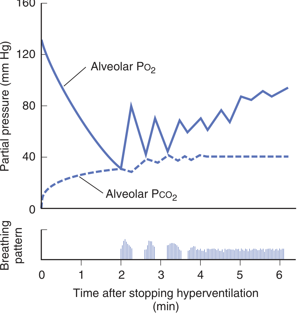
Figure: After forced hyperventilation for 2 minutes, alveolar PO₂ rises sharply (solid) and PCO₂ falls nearly to zero (dashed). When voluntary breathing stops, periodic breathing ensues - apneic pauses alternate with clusters of breaths. Gradually, PCO₂ returns to normal and breathing resumes. (Ganong's Review of Medical Physiology)
The sequence after hyperventilation:
- Forced hyperventilation → ↑ alveolar PO₂, ↓ alveolar PCO₂ (CO₂ blown off)
- Breathing stops voluntarily → PCO₂ is now below the apneic threshold → apnea
- During apnea: ↓ alveolar PO₂, ↑ alveolar PCO₂ (gas exchange continues without ventilation)
- Hypoxia stimulates carotid body chemoreceptors before PCO₂ returns to normal
- A few breaths occur (hyperpnea) → the hypoxic stimulus is eliminated → apnea again
- This cycle repeats, gradually dampening as PCO₂ normalizes
- Eventually PCO₂ returns to normal and stable breathing resumes
Key proof: This apnea does NOT occur if hyperventilation is done with a 5% CO₂ mixture (which prevents the fall in PCO₂), confirming that CO₂ depletion below the apneic threshold is the driver. (Ganong's Review of Medical Physiology)
4. The Apneic Threshold
The apneic threshold is the PaCO₂ level below which respiratory output from the brainstem ceases entirely. During wakefulness, a "wakefulness drive to breathe" keeps breathing going even when PaCO₂ falls (so true apnea from hypocapnia is rare). During sleep, this wakefulness drive disappears and ventilation comes under almost purely metabolic (chemical) control - making sleep the time when periodic breathing is most prominent and apnea is most easily triggered by hypocapnia.
At sleep onset, the apneic threshold rises by approximately 2-6 mmHg PCO₂ compared to wakefulness - meaning PaCO₂ must remain higher during sleep to sustain breathing. This narrows the gap between the normal sleeping PaCO₂ and the apneic threshold, predisposing to instability.
5. Cheyne-Stokes Respiration (CSR)
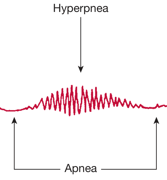
Figure: The characteristic waveform of Cheyne-Stokes respiration. Breathing waxes to a crescendo hyperpnea then wanes back to complete apnea in a repeating cycle. The hyperpnea phase (red, center) transitions into flat apnea. (Fishman's Pulmonary Diseases and Disorders)
Cheyne-Stokes respiration (CSR) is the classic clinical form of periodic breathing. It was first described by John Cheyne in 1818 (and William Stokes later elaborated on it; the pattern was actually first reported by John Hunter 37 years earlier - sometimes called Hunter-Cheyne-Stokes breathing).
Pattern
- Repetitive cycles of crescendo-decrescendo hyperpnea followed by central apnea
- Cycle length: 40-90 seconds (longer than altitude-related periodic breathing)
- Predominantly occurs during non-REM sleep (rare in REM sleep)
- Arousals occur at the peak of hyperpnea (not at apnea termination - this distinguishes it from OSA)
Conditions Associated with CSR
| Condition | Mechanism |
|---|---|
| Systolic heart failure (HFrEF) - most common | Prolonged circulation time + enhanced chemosensitivity |
| Uremia | Altered respiratory control |
| Stroke / brain disease | Disruption of inhibitory neural pathways |
| Atrial fibrillation (with/without LV dysfunction) | Cardiac output reduction → prolonged circulation time |
| Opioid use | Depressed respiratory drive; complex mechanism |
| Sleep in some normal individuals | Proximity to apneic threshold during sleep |
Two Mechanisms in Heart Failure
Mechanism 1 - Prolonged circulation time (the dominant mechanism):
- In CHF, reduced cardiac output → prolonged lung-to-brain circulation time (normally ~30-40 s; in CHF may be >60-90 s)
- When the lungs hyperventilate and lower PCO₂, the blood with low PCO₂ takes abnormally long to reach the medullary and carotid chemoreceptors
- During this delay, the lungs continue to over-ventilate, lowering PCO₂ even further
- When the hypocapnic blood finally arrives at the brainstem → apnea
- During apnea, PCO₂ builds up again and the cycle repeats
- The cycle length of CSR in heart failure is longer than altitude-related periodic breathing precisely because circulation time is prolonged
Mechanism 2 - Enhanced chemosensitivity:
- Some heart failure patients have increased responsiveness to CO₂ (disrupted inhibitory neural pathways)
- A small rise in PCO₂ triggers an exaggerated hyperpnea, which drives PCO₂ far below the apneic threshold → apnea
- When PCO₂ rises back to normal, the overly sensitive system again over-responds → cycling continues
(Ganong's Review of Medical Physiology; Fishman's Pulmonary Diseases and Disorders; Fuster and Hurst's The Heart)
CSR as a Prognostic Marker in Heart Failure
CSR is not merely a nuisance - it has important prognostic implications:
- CSR independently increases the risk of premature death by ~150% in HFrEF patients (observational data)
- Time with SaO₂ <90% (T90) predicts mortality in a dose-dependent manner (~16% increased risk per hour below 90%)
- Both CSA and OSA cause sustained sympathetic overactivation (hyperadrenergic state), worsening cardiac remodeling
- CSA is associated with hospital readmissions and worse outcomes
- Risk factors for CSR in HF: male sex, age >65, atrial fibrillation, awake hypocapnia (PaCO₂ <38 mmHg)
6. High-Altitude Periodic Breathing
At altitude above ~2000 m, virtually all healthy lowlanders develop periodic breathing, especially during sleep. This is a physiological response, not a pathological one.
Mechanism
- Hypoxia at altitude stimulates peripheral chemoreceptors → hyperventilation
- Hyperventilation lowers PaCO₂ → hypocapnia
- PaCO₂ falls below the apneic threshold (especially during sleep) → central apnea
- During apnea: PaO₂ falls further, PaCO₂ rises → peripheral chemoreceptors re-stimulated
- Burst of hyperventilation → PaCO₂ falls again below apneic threshold → next apnea
This is essentially the altitude equivalent of post-hyperventilation periodic breathing. The key driver is the narrow gap between sleeping PaCO₂ and the apneic threshold in the setting of baseline hypocapnia from altitude-induced hypoxic ventilatory response.
Features
- Cycle time: 12-34 seconds (short - because at altitude, peripheral chemoreceptor sensing of changes in PaCO₂/PaO₂ is rapid, unlike the prolonged circulation delay in CHF)
- Increases in severity and frequency with increasing altitude above 3000 m
- Gender differences: women have fewer events than men at equivalent altitudes (3400 m and 5400 m) and longer respiratory cycles
- At extreme altitude (>5500 m), paradoxically, apnea length and number decrease as high-frequency breathing dominates (greater central O₂-sensitive drive)
- Worsened by sleep (loss of wakefulness drive)
- Causes sleep fragmentation and arousals during the hyperpneic phase
- Oscillations in SaO₂ may increase susceptibility to HAPE and HACE
- Persists for >1 week even with acclimatization in many individuals; may gradually improve
Treatment
- Acetazolamide (carbonic anhydrase inhibitor): induces bicarbonate diuresis → metabolic acidosis → increases ventilatory drive → raises PaCO₂ above apneic threshold → reduces apneas. Standard first-line prevention. Dose: 125 mg at bedtime; also 125-250 mg q12h for AMS prevention.
- Supplemental oxygen: increases PaO₂ → reduces hypoxic drive → reduces the hyperpnea → raises PaCO₂ → narrows the oscillation
(Fishman's Pulmonary Diseases and Disorders)
7. Neonatal and Infant Periodic Breathing
Normal Periodic Breathing of Infancy
Periodic breathing is normal and expected in neonates, particularly premature infants. It differs from the pathological forms seen in adults:
- Pattern: cyclic episodes during sleep of brief apneic pauses (5-10 seconds) interrupting regular breathing, followed by a burst of rapid respiration
- Mechanism: immature central respiratory control in the brainstem; the respiratory oscillator has not yet achieved the stable gain characteristics of the mature system
- Clinical significance: generally benign; neurological complications do not occur unless hypoxic periods are prolonged and severe
Apnea of Prematurity (Pathological)
When apnea exceeds 20 seconds, or shorter apneas are accompanied by bradycardia (heart rate fall) or oxygen desaturation, the event is clinically significant:
Causes of pathological apnea in neonates:
- Idiopathic (apnea of prematurity) - most common
- Intraventricular hemorrhage
- Seizures
- Upper airway obstruction / atelectasis
- Pneumonia, sepsis, meningitis
- Phrenic nerve paralysis
- Gastroesophageal reflux
- Hypovolemia
- Heart failure
Treatment:
- Caffeine (methylxanthine): stimulates central respiratory drive by adenosine receptor antagonism; standard therapy for apnea of prematurity. Early caffeine therapy (<3 days of life) is associated with better neurodevelopmental outcomes in infants born <29 weeks gestation.
- Supplemental oxygen
- CPAP
(Bradley and Daroff's Neurology in Clinical Practice)
8. Summary: Types of Periodic Breathing
| Type | Setting | Cycle Length | Mechanism | Treatment |
|---|---|---|---|---|
| Post-hyperventilation | Experimental; after voluntary hyperventilation | Minutes | CO₂ depletion below apneic threshold; hypoxia restimulates | Self-resolving |
| Cheyne-Stokes (CSR) | Heart failure, uremia, stroke, normal sleep | 40-90 s | Prolonged circulation time + enhanced CO₂ sensitivity → oscillating feedback loop | Optimize HF therapy; O₂; CPAP; (ASV with caution) |
| High-altitude periodic breathing | Altitude >2000 m (virtually all lowlanders) | 12-34 s | Hypoxic hyperventilation → hypocapnia below apneic threshold during sleep | Acetazolamide; supplemental O₂ |
| Neonatal periodic breathing | Premature > term newborns; normal variant | 5-10 s apneic pauses | Immature central respiratory control | Usually benign; caffeine if pathological |
| Drug-induced CSA | Opioids | Variable | Suppressed respiratory drive + disrupted rhythmogenesis | Reduce opioid dose; CPAP/ASV |
9. Key Pathophysiological Concepts - Summary
| Concept | Significance |
|---|---|
| Apneic threshold | PaCO₂ below which breathing ceases; must be close to resting PaCO₂ for instability to develop |
| Loop gain | Overall feedback gain; must exceed 1 for oscillation; elevated by high chemosensitivity, long circulation time, low CO₂ stores |
| Circulation time | Delay between lung ventilation change and chemoreceptor detection; prolonged in HF → longer cycle |
| Wakefulness drive | Non-chemical drive that stabilizes breathing during waking; absent during sleep → instability unmasked |
| Hypocapnia | The final common pathway to apnea in most forms of periodic breathing |
| Arousals in CSR | Occur at peak hyperpnea; sustain oscillations (vs. OSA arousals at apnea termination) |
Periodic Breathing Physiology
Searching sources...
Finding Sources
Finding Sources
Your credits are exhausted. You can purchase a top-up or subscribe to a plan to continue using Orris. If you're already subscribed, your daily credits will reset tomorrow.
This is a shared conversation. Sign in to Orris to start your own chat.