
Functions of endorphins, dopamine and serotonin.
dopamine serotonin endorphin neurotransmitter pathways brain diagram
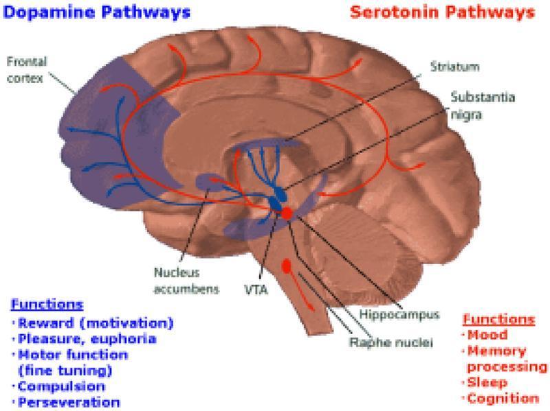
This anatomical diagram presents a sagittal view of the human brain, illustrating the major neurotransmitter pathways associated with the Brain Reward Cascade (BRC). The illustration distinguishes between the Dopamine and Serotonin systems using color-coded projections. The Dopamine pathways, highlighted in blue, originate in the Ventral Tegmental Area (VTA) and project to the Nucleus Accumbens and the Frontal Cortex. Labeled functions for this system include reward (motivation), pleasure, motor function, compulsion, and perseveration. The Serotonin pathways, highlighted in red, originate from the Raphe Nuclei and demonstrate a more diffuse distribution pattern, passing through the hippocampus, substantia nigra, and striatum before reaching the frontal cortex. Functional correlates for serotonin listed include mood, memory processing, sleep, and cognition. This educational graphic is designed to demonstrate the neuroanatomical circuitry underlying motivation and behavior, specifically highlighting the mesocorticolimbic system's role in reward and its interplay with wider serotonergic projections.
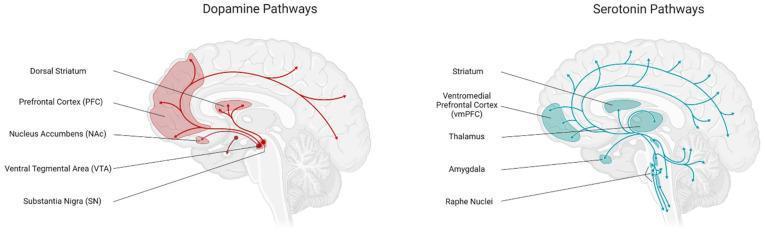
This medical illustration presents two sagittal sections of the human brain, side-by-side, detailing the central dopamine and serotonin neurotransmitter pathways involved in food intake regulation. The left panel depicts the Dopamine Pathways in red, showing projections originating from the Ventral Tegmental Area (VTA) and Substantia Nigra (SN) in the midbrain. Key innervated regions highlighted include the Dorsal Striatum, Nucleus Accumbens (NAc), and the Prefrontal Cortex (PFC), representing the mesolimbic and mesocortical reward circuits. The right panel illustrates the Serotonin Pathways in blue, originating from the Raphe Nuclei in the brainstem. This pathway shows a broader distribution, with projections reaching the Striatum, Thalamus, Amygdala, and the Ventromedial Prefrontal Cortex (vmPFC). Arrows indicate the directional flow of neural signaling. Both diagrams highlight the anatomical overlap in the striatum and prefrontal regions, emphasizing their integrated role in homeostatic and hedonic appetite control.
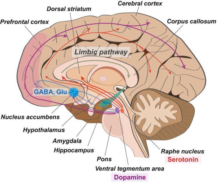
A medical schematic illustrating the neuroanatomical pathways and neurotransmitter circuits implicated in depression, depicted in a mid-sagittal section of the human brain. The diagram highlights key structural regions including the prefrontal cortex, dorsal striatum, nucleus accumbens, hypothalamus, amygdala, and hippocampus. The nucleus accumbens serves as a central hub, receiving GABAergic and glutamatergic (Glu) inputs from the prefrontal cortex. Two primary neurotransmitter pathways are color-coded: the serotonin pathway (red arrows) originates in the raphe nucleus and projects to the limbic pathway and hippocampus; the dopamine pathway (purple arrows) originates in the ventral tegmental area (VTA) and projects broadly to the prefrontal cortex and the cerebral cortex. The corpus callosum and brainstem structures (pons and raphe nucleus) provide anatomical orientation. This illustration is designed to teach the functional connectivity and chemical imbalances associated with depressive disorders, emphasizing the intersection of the reward system and the limbic system.
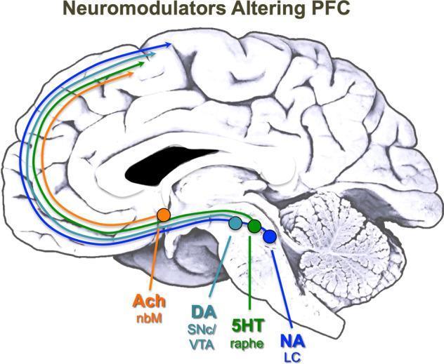
This medical illustration presents a midsagittal view of the human brain, serving as a neuroanatomical diagram of neuromodulator pathways. It identifies the origin and axonal projection routes of four key neurotransmitter systems to the prefrontal cortex (PFC). Visible anatomical landmarks include the cerebral cortex with prominent sulci and gyri, the C-shaped corpus callosum, the brainstem, and the cerebellum. The diagram uses color-coded circles to mark cell body locations and matching curved arrows to illustrate projection pathways. Starting anteriorly, the orange pathway represents Acetylcholine (Ach) from the nucleus basalis of Meynert (nbM). Within the brainstem, a light blue circle denotes Dopamine (DA) from the substantia nigra pars compacta (SNc) and ventral tegmental area (VTA); a green circle indicates Serotonin (5HT) from the dorsal raphe; and a dark blue circle signifies Noradrenaline (NA) from the locus coeruleus (LC). The illustration highlights the clinical significance of these long-range projections in modulating the PFC, which is essential for working memory, attention, and executive function.
Endorphins, Dopamine, and Serotonin: Functions
1. Endorphins
Key Functions
| Receptor | Endorphin | Effects |
|---|---|---|
| Mu 1 (μ1) | Beta-endorphin | Euphoria, supraspinal analgesia, confusion, dizziness, nausea |
| Mu 2 (μ2) | Beta-endorphin | Respiratory depression, cardiovascular and GI effects, miosis, urinary retention |
| Delta (δ) | Enkephalin | Spinal analgesia, cardiovascular depression, decreased brain and myocardial oxygen demand |
| Kappa (κ) | Dynorphin, beta-endorphin | Spinal analgesia, dysphoria, psychomimetic effects, feedback inhibition of the endorphin system |
2. Dopamine
Three Major Brain Systems
| Pathway | From → To | Key Functions |
|---|---|---|
| Nigrostriatal | Substantia nigra → Striatum | Motor control, movement initiation |
| Mesocorticolimbic | VTA → Nucleus accumbens, prefrontal cortex | Reward, motivation, pleasure, cognition |
| Tuberohypophyseal | Hypothalamus → Pituitary | Inhibits prolactin release |
Key Functions
- Reward and motivation: Dopamine's phasic (burst) firing signals when a reward occurs - particularly when it is better than expected. This reward prediction signal is central to learning, goal-directed behavior, and the experience of pleasure. (Kaplan & Sadock's Comprehensive Psychiatry)
- Motor control: The nigrostriatal pathway governs smooth, coordinated movement. Degeneration of these neurons causes Parkinson's disease, and dopamine replacement therapy restores motor function.
- Tonic enabling function: At baseline, steady-state (pacemaker) dopamine activity exerts a tonic enabling effect on motor, cognitive, and motivational processes - impaired in Parkinson's disease.
- Cognition and working memory: Dopamine in the prefrontal cortex is essential for executive function, attention, and working memory.
- Inhibitory neurotransmitter: In many brain regions, dopamine acts as an inhibitory neurotransmitter and a stabilizer. (Guyton & Hall Physiology)
- Drive, pleasure, sex: Dopamine is considered crucial for drive, pleasure, and sexual motivation. (Kaplan & Sadock)
- Peripheral roles:
- Kidney: Renal dopamine neurons regulate sodium homeostasis and produce vasodilation, diuresis, and natriuresis. Impairment may underlie some forms of hypertension.
- Retina: Dopamine-containing amacrine cells modulate high-acuity, light-adapted vision and photoreceptor coupling.
- Adrenal medulla / Periphery: Dopamine is the metabolic precursor of norepinephrine; it can activate alpha and beta adrenergic receptors at high concentrations.
3. Serotonin (5-Hydroxytryptamine / 5-HT)
Despite only 1 in a million neurons in the brain producing serotonin, this evolutionarily ancient molecule influences virtually all aspects of CNS function due to its extensive projection pattern. - Kaplan & Sadock's Comprehensive Psychiatry
Distribution
- >80% of the body's serotonin is in the GI tract, produced by enterochromaffin cells.
- Blood serotonin is concentrated in platelets.
- The brain serotonin supply is entirely derived from dietary tryptophan.
Key Functions
- Mood regulation: The most well-known role - low serotonergic function is strongly linked to depression. SSRIs work by enhancing serotonin availability, and their antidepressant efficacy is closely associated with serotonergic enhancement.
- Sleep-wake cycle: Serotonin regulates circadian rhythm and promotes wakefulness; it is a precursor to melatonin.
- Appetite and feeding behavior: Serotonin suppresses appetite; alterations are implicated in eating disorders and obesity.
- Anxiety and aggression: Low CNS serotonin is associated with increased aggression and impulsivity. (Kaplan & Sadock)
- Pain modulation: Descending serotonergic fibers from caudal raphe nuclei project into the spinal cord to inhibit pain signals.
- Cognition and memory: Serotonin modulates attention, memory processing, and other cognitive functions.
- Sexual behavior: Serotonin has an inhibitory effect on sexual behavior (explaining why SSRIs can cause sexual dysfunction).
- Temperature regulation: Serotonin influences thermoregulation.
- Blood pressure regulation: Serotonergic neurons modulate vascular tone and blood pressure.
- GI motility: Enterochromaffin cells release serotonin in response to luminal stimuli, triggering peristalsis via enteric neurons.
- Platelet aggregation: Serotonin released from activated platelets promotes further aggregation and vasoconstriction at wound sites, facilitating hemostasis.
- Vomiting reflex: Serotonin activates 5-HT3 receptors in the gut and brainstem, triggering the vomiting reflex (the basis for ondansetron as an antiemetic).
Neurotransmitter Pathways - Brain Diagram
Quick Comparison Summary
| Feature | Endorphins | Dopamine | Serotonin |
|---|---|---|---|
| Type | Endogenous opioid peptides | Catecholamine | Indoleamine |
| Origin | Scattered neurons (POMC-derived) | Substantia nigra, VTA, hypothalamus | Raphe nuclei (brainstem) |
| Primary role | Pain relief, stress modulation | Reward, motivation, motor control | Mood, sleep, GI motility |
| Mood effect | Euphoria, sense of well-being | Pleasure, drive | Emotional stability, well-being |
| Pain | Major analgesic system | Minor role | Descending pain inhibition |
| Motor | Minimal | Essential (Parkinson's when lost) | Minor |
| GI | Minor | Renal/peripheral only | Major (>80% of body serotonin) |
| Clinical relevance | Opioid drugs mimic/inhibit this system | Parkinson's, schizophrenia, addiction | Depression, anxiety, IBS, antiemetics |
Can u give me only functions
Endorphins
- Natural pain relief (analgesia)
- Produce feelings of euphoria and well-being
- Counter the stress response
- Modulate emotional reactions to pain
- Triggered by exercise, laughter, excitement
Dopamine
- Reward and pleasure signaling
- Motivation and goal-directed behavior
- Motor control and movement coordination
- Working memory and executive function
- Regulates attention and cognition
- Controls prolactin secretion (inhibits it)
- Renal sodium regulation and vasodilation (peripheral)
- Modulates vision in the retina
Serotonin
- Mood stabilization and sense of well-being
- Sleep-wake cycle regulation
- Appetite and satiety control
- Pain inhibition (descending spinal pathways)
- Regulates anxiety and aggression
- Cognitive functions - memory, attention
- Temperature regulation
- GI motility and digestion (majority of body serotonin is in the gut)
- Platelet aggregation and hemostasis
- Triggers the vomiting reflex (via 5-HT3 receptors)
- Precursor to melatonin (circadian rhythm)
Which txt book is this from ?
- Rosen's Emergency Medicine: Concepts and Clinical Practice (Endorphin System section)
- Tietz Textbook of Laboratory Medicine, 7th Edition
- Kaplan & Sadock's Comprehensive Textbook of Psychiatry
- Kaplan & Sadock's Comprehensive Textbook of Psychiatry
- Kaplan & Sadock's Synopsis of Psychiatry
- Guyton and Hall Textbook of Medical Physiology
- Lippincott Illustrated Reviews: Pharmacology
- Eric Kandel - Principles of Neural Science, 6th Edition
- Kaplan & Sadock's Comprehensive Textbook of Psychiatry
- Kaplan & Sadock's Synopsis of Psychiatry
- Biochemistry, 8th ed. - Lippincott Illustrated Reviews
- Katzung's Basic and Clinical Pharmacology, 16th Edition
- Histology: A Text and Atlas with Correlated Cell and Molecular Biology