A 12-year-old boy came to STAMCH with the complaints of fever and rapidly growing painful swelling on the right side of face. He also noticed that pain increases while taking food. On examination the physician found congestion (redness due to inflammation) in mucous membrane of vestibule of mouth, opposite the crown of upper second molar tooth on right side. 1+2+2=5 a) Name the organ which is most likely involved in this case? b) Why this condition is so much painful? c) Why does pain increase during eating?

Reading File
Finding Sources
Finding Sources
Reading File
Reading File
Finding Sources
Reading File
Loading Image
I now have everything needed for a thorough, well-sourced answer.

Clinical Case Analysis

Diagnosis: Acute Parotitis (inflammation of the parotid gland - most likely mumps/epidemic parotitis in a 12-year-old)

a) The Organ Involved: The Parotid Gland (1 mark)

The organ most likely involved is the right parotid gland - the largest of the three paired major salivary glands.
The key diagnostic clue is the location of congestion: redness in the mucous membrane of the vestibule of the mouth, opposite the crown of the upper second molar tooth on the right side. This is precisely where the parotid (Stensen's) duct opens into the oral cavity.
"The major duct of the parotid gland is called Stensen's duct, which opens into the vestibule of the mouth opposite the crown of the upper second molar tooth..."
  • Bailey and Love's Short Practice of Surgery, 28th Ed.
"The parotid duct leaves the anterior edge of the parotid gland midway between the zygomatic arch and the corner of the mouth. It crosses the face in a transverse direction and, after crossing the medial border of the masseter muscle, turns deeply into the buccal fat pad and pierces the buccinator muscle. It opens into the oral cavity near the second upper molar tooth."
  • Gray's Anatomy for Students
The anatomical diagram below shows the parotid gland, its duct, and the course it takes across the face:
Parotid gland lateral view and cross section showing Stensen's duct, facial nerve, and related structures
(Gray's Anatomy for Students - Fig. 8.65: Parotid Gland - lateral view (A) and cross section (B))

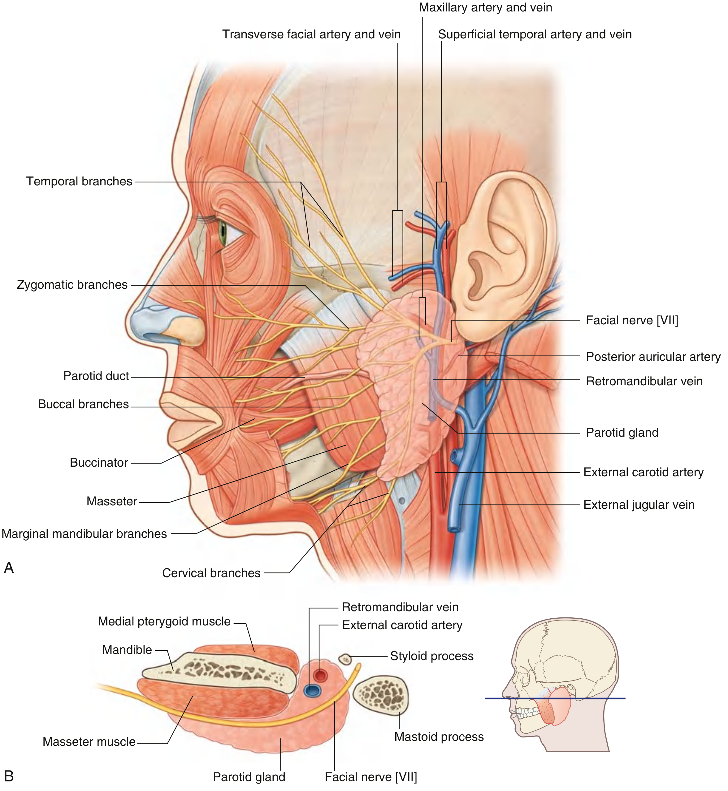
b) Why is this condition so painful? (2 marks)

The intense pain in parotitis is caused by the tough, inextensible fibrous capsule that tightly encloses the parotid gland.
When inflammation occurs (whether from mumps virus, bacterial infection, or duct obstruction), the gland swells due to edema, increased secretion, and cellular infiltration. However, unlike most swellings in the body where surrounding soft tissue can expand to accommodate the enlargement, the parotid gland is enclosed within a non-distensible capsule derived from the investing layer of the deep cervical fascia. This capsule cannot stretch.
As a result:
  • Inflammatory swelling increases the pressure within the confined space of the capsule
  • This raised intracapsular pressure compresses the nerve endings within and around the gland
  • The result is severe, throbbing pain - the harder the capsule, the more intense the pain
"Chewing movements are very painful because the tough capsule enclosing the parotid gland is unable to stretch."
  • Color Atlas of Human Anatomy Vol. 2, Internal Organs
This is the same principle that makes compartment syndrome so painful - a rigid boundary preventing expansion of inflamed tissue leads to ischemic and pressure-mediated pain.

c) Why does pain increase during eating? (2 marks)

Pain increases during eating for two interconnected reasons:
1. Stimulation of salivary secretion: Eating (especially the sight, smell, or taste of food) triggers parasympathetic stimulation of the parotid gland, causing the acini to actively secrete saliva. In an already inflamed and swollen gland, this additional secretory activity further increases pressure within the inextensible capsule, worsening pain.
2. Masseter muscle contraction compresses the gland: During chewing, the masseter muscle contracts powerfully. The parotid gland lies directly superficial and posterior to the masseter. As the masseter contracts with each chewing movement, it physically compresses the inflamed, swollen, and pressure-sensitive parotid gland, producing sharp exacerbations of pain.
Both mechanisms are captured here:
"Chewing movements are very painful because the tough capsule enclosing the parotid gland is unable to stretch."
  • Color Atlas of Human Anatomy Vol. 2, Internal Organs
The parotid duct also crosses the masseter before piercing the buccinator - so compression of the duct during chewing can further obstruct outflow and raise intraductal pressure, adding to discomfort.

Summary Table

FeatureExplanation
Organ involvedRight parotid gland (Stensen's duct opens opposite upper 2nd molar)
Why painfulTough fibrous capsule cannot stretch - raised intracapsular pressure compresses nerve endings
Why worse on eatingChewing compresses the gland via masseter; eating also reflexly stimulates salivary secretion, increasing intraglandular pressure further
Sources: Gray's Anatomy for Students; Bailey and Love's Short Practice of Surgery (28th Ed.); Color Atlas of Human Anatomy Vol. 2, Internal Organs

A 14-year-old boy came to STAMCH with the complaints of fever and rapidly growing painful swelling on the right side of face. On examination the physician found congestion in mucous membrane of vestibule of mouth opposite the crown of upper second molar tooth on right side. a) Name the organ which is involved in this case? b) Why this condition is painful? c) How does mucous membrane congested in this case?

Finding Sources
Finding Sources
Finding Sources
Finding Sources
Reading File
Reading File
I now have all the anatomical detail needed. I have the full answer from textbook sources.

Clinical Case Analysis

a) The Organ Involved: The Parotid Gland (Right Side)

The organ involved is the right parotid gland - the largest of the three paired major salivary glands.
The pathognomonic clue is the site of congestion: the vestibule of the mouth opposite the crown of the upper second molar tooth on the right side. This is the precise location of the opening (papilla) of the parotid duct (Stensen's duct) - the excretory duct of the parotid gland.
"The major duct of the parotid gland is called Stensen's duct, which opens into the vestibule of the mouth opposite the crown of the upper second molar tooth."
  • Bailey and Love's Short Practice of Surgery, 28th Ed.
"The duct then briefly runs caudally before exiting the papilla at the buccal mucosa opposite the second maxillary molar."
  • Cummings Otolaryngology Head and Neck Surgery
Parotid gland lateral view and cross section showing Stensen's duct course to the vestibule
(Gray's Anatomy for Students - Fig. 8.65: Parotid Gland and its duct)

b) Why is this condition so painful?

The severe pain is due to the tough, non-distensible fibrous capsule that tightly encloses the parotid gland.
When the gland becomes inflamed (e.g., from mumps virus or bacterial parotitis), it swells from edema, cellular infiltration, and increased secretory pressure. Unlike most body swellings that can expand into surrounding soft tissue, the parotid gland is bound within a rigid capsule derived from the investing layer of the deep cervical fascia - a tough layer that cannot stretch.
The result:
  • Inflammatory swelling rapidly builds raised intracapsular pressure
  • This pressure directly compresses the rich network of nerve endings (auriculotemporal nerve branches) within the gland
  • The confined expansion causes intense, throbbing pain that cannot be relieved until the inflammation subsides
"Chewing movements are very painful because the tough capsule enclosing the parotid gland is unable to stretch."
  • Color Atlas of Human Anatomy Vol. 2, Internal Organs
This is mechanistically identical to compartment syndrome - a rigid boundary trapping swelling and generating compressive pain. No other major salivary gland (submandibular, sublingual) has such a firm capsule, which is why parotitis is particularly painful.

c) How is the mucous membrane congested in this case?

This is an anatomical question about how the inflammatory process at the parotid gland leads to visible redness (congestion) of the mucous membrane in the vestibule of the mouth.
The answer lies in the precise anatomical course of Stensen's duct:
Step-by-step pathway of spread:
  1. Origin: The parotid duct (Stensen's duct) originates within the parotid gland, emerging from its anterior border. It is approximately 5 cm long with an internal calibre of ~0.6 mm.
  2. Course across the face: It travels horizontally across the masseter muscle, then turns medially at the anterior border of the masseter.
  3. Piercing the buccinator: The duct pierces the buccinator muscle and runs obliquely between the buccinator and the oral mucosa - this is the key segment.
  4. Opening at the papilla: It enters the oral cavity at the parotid papilla - a small elevation on the mucous membrane of the vestibule, directly opposite the crown of the upper second molar tooth.
"It pierces buccinator and runs obliquely between buccinator and the oral mucosa before entering the oral cavity at the parotid papilla opposite the second upper molar tooth."
  • Scott-Brown's Otorhinolaryngology Head & Neck Surgery
Mechanism of congestion:
When the parotid gland is inflamed, the inflammation tracks along Stensen's duct from the gland toward its opening. Because the distal portion of the duct runs between the buccinator muscle and the overlying oral mucosa, the inflammatory process (with its associated vasodilation and increased vascular permeability) directly involves the mucous membrane at the duct's opening. The result is:
  • Vasodilation of mucosal blood vessels around the papilla
  • Increased blood flow to the mucosa (hyperemia)
  • Visible redness and congestion of the vestibular mucosa at the exact site of the duct's opening - opposite the upper second molar
Additionally, in acute parotitis, pressure on the gland may cause purulent or cloudy secretions to discharge from the papilla, further inflaming the surrounding mucosa.

Summary

QuestionAnswer
Organ involvedRight parotid gland (Stensen's duct opens at upper 2nd molar)
Why painfulTough inextensible fibrous capsule cannot accommodate inflammatory swelling - raised intracapsular pressure compresses nerve endings
Why mucosa is congestedInflammation tracks along Stensen's duct to its papilla; the duct's distal segment runs between buccinator and oral mucosa, so peri-ductal inflammation directly congests the overlying vestibular mucosa opposite the upper 2nd molar
Sources: Scott-Brown's Otorhinolaryngology Head & Neck Surgery; Cummings Otolaryngology Head and Neck Surgery; Bailey and Love's Short Practice of Surgery (28th Ed.); Color Atlas of Human Anatomy Vol. 2
This is a shared conversation. Sign in to Orris to start your own chat.