
Minimal change disease
Reading File
Now I'll search the medical library for comprehensive content on Minimal Change Disease in parallel.
Finding Sources
Finding Sources
Searching PubMed
"minimal change disease" treatment
Excellent - multiple authoritative sources found. Now reading the key sections in parallel.
Reading File
Reading File
Reading File
Now let me read the treatment and management sections from Comprehensive Clinical Nephrology and Harrison's for more detail.
Reading File
Reading File
Reading File
Now let me get the prognosis/relapse details and fetch the key histology images:
Reading File
Loading Image
Loading Image
I now have comprehensive information from multiple authoritative sources. Let me compile the full answer.
Minimal Change Disease (MCD)
Overview
Minimal change disease (MCD) - also called "nil lesion" or historically "lipoid nephrosis" - is a podocytopathy that causes the nephrotic syndrome. It is the most common cause of nephrotic syndrome in children (70-90% of cases) but accounts for only 10-15% of idiopathic nephrotic syndrome in adults. It is most common between ages 1-7 years.
Etiology
Primary (idiopathic): The majority of cases are idiopathic.
Secondary causes:
- Drugs: NSAIDs, lithium
- Malignancies: Hodgkin's lymphoma (classically), non-Hodgkin's lymphoma
- Infections
- Biologic agents: Interferon-alpha, pamidronate, IL-2
- Allergies/atopy (40% in children, 30% in adults)
Pathogenesis
The pathogenesis of MCD is incompletely understood but involves immune dysregulation at the level of the podocyte:
-
T-cell dysfunction: Activated T cells may release circulating permeability factors (cytokines such as IL-13 and IL-4) that damage the glomerular capillary wall. T cell hybridomas from MCD patients secrete factors that provoke heavy proteinuria in rats.
-
CD80 overexpression: Podocytes in MCD show overexpression of CD80 (B7.1), normally expressed by dendritic cells/B cells. Urinary CD80 correlates with disease activity. Dysregulated interactions between CD80 on podocytes and CTLA-4 on T lymphocytes may perpetuate injury.
-
Angiopoietin-like-4 overexpression: Corticosteroids and N-acetyl-D-mannosamine can reduce this overexpression.
-
Anti-nephrin antibodies: A subset of MCD patients have autoantibodies against nephrin (a slit-diaphragm protein), pointing to an autoimmune mechanism. Immunofluorescence shows minimal IgG despite the antibodies being present.
-
Endothelial injury: The majority of MCD patients also show mild glomerular endothelial injury with circulating endothelial biomarkers, suggesting MCD is not purely a "podocytopathy."
-
B-cell involvement: Evidence includes successful treatment with anti-CD20 (rituximab).
Morphology / Histopathology
Light Microscopy
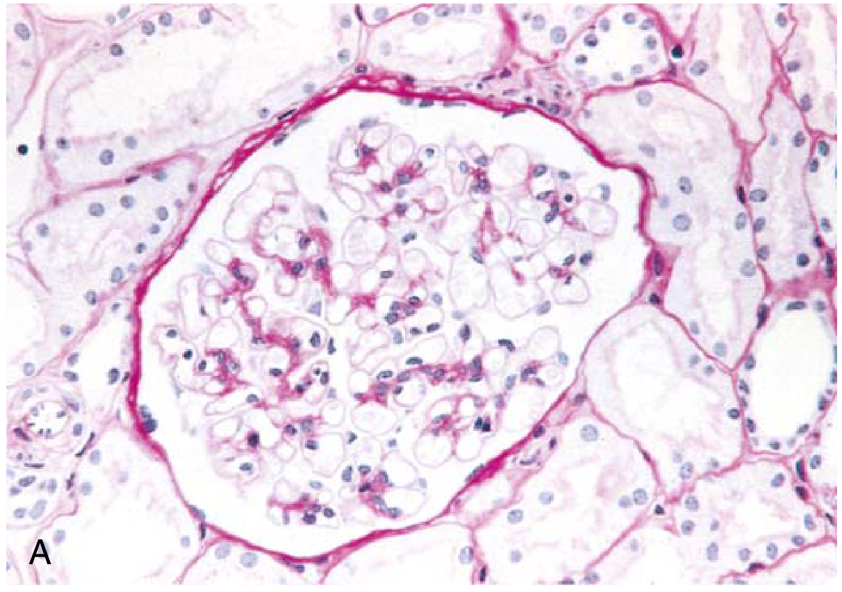
- Normal-appearing glomeruli - no proliferation, no sclerosis, no deposits (hence the name "minimal change")
- Tubules may show lipid droplet accumulation and protein reabsorption droplets from leaked proteins
Fig. A - PAS stain, Light Microscopy: Normal glomerular architecture with patent capillaries and no hypercellularity.
Immunofluorescence
- Negative for immune deposits (IgG, IgA, IgM, complement)
- Occasionally small amounts of IgM in the mesangium
Electron Microscopy (Diagnostic)
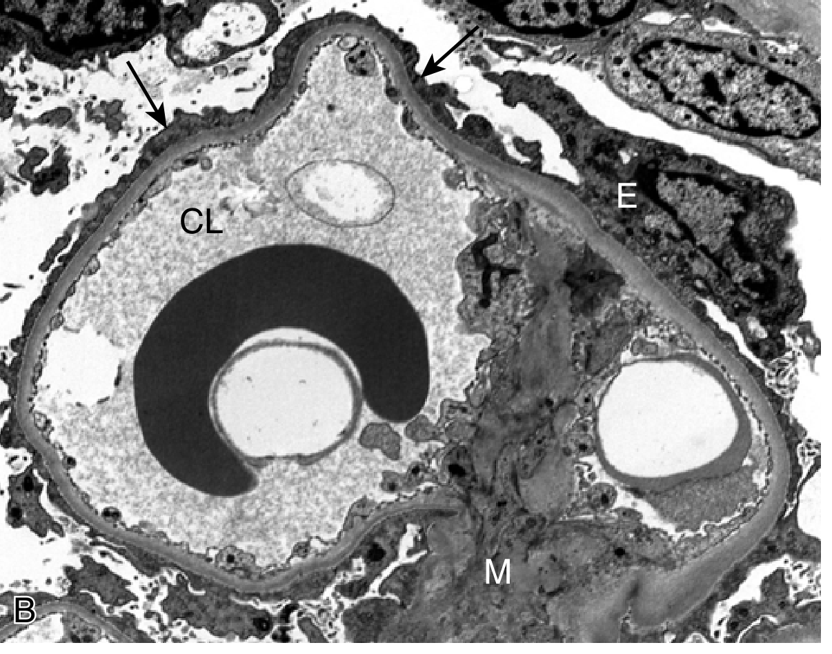
- Diffuse effacement (fusion) of podocyte foot processes along virtually every capillary loop - the hallmark finding
- Vacuolization and microvillus transformation of the visceral epithelium
- No electron-dense deposits
- Normal glomerular basement membrane
Fig. B - Electron Microscopy: Effacement of foot processes (arrows), CL = Capillary Lumen, E = Epithelial cell, M = Mesangium. No deposits visible.
Clinical Features
| Feature | Children | Adults |
|---|---|---|
| Onset | Abrupt | Abrupt |
| Edema | Periorbital + peripheral | Periorbital + peripheral |
| Proteinuria | ~10 g/24h (selective - mainly albumin) | ~10 g/24h |
| Hypertension | ~30% | 20-50% |
| Microscopic hematuria | ~20% | ~33% |
| Atopy/allergy | ~40% | ~30% |
| Decreased renal function | Uncommon | 25-40% (often reversible) |
- Urinary sediment: Acellular (no RBC casts)
- Selective proteinuria (mainly albumin, minimal high-molecular-weight proteins) - especially in children
- Serum complement levels and serology are normal
- Hypoalbuminemia, hyperlipidemia, edema - the classic nephrotic tetrad
Diagnosis
- Children: Clinical diagnosis (biopsy not usually required for initial presentation) - treated empirically with steroids. Biopsy done only in non-responders.
- Adults: Kidney biopsy required to confirm diagnosis
- EM showing foot process effacement with no deposits on IF is diagnostic
Treatment
Initial Therapy
Prednisone is first-line:
- Daily: 1 mg/kg/day (max 80 mg) OR
- Alternate-day: 2 mg/kg (max 120 mg)
- Taper after 2 months, total course 5-6 months
- Adults: Not considered steroid-resistant until failure after 16 weeks of treatment
- Children: >90% respond to a short course of corticosteroids
Relapse Management
- Infrequent relapses: Repeat prednisone course (similar to initial)
- Frequent relapses / Steroid-dependent / Steroid-resistant:
- Rituximab - 1000 mg IV, repeated in 2 weeks (anti-CD20)
- Cyclosporine - 3-5 mg/kg/day for 4 months (CNI; monitor trough levels)
- Tacrolimus - 0.05-0.1 mg/kg/day
- Mycophenolate mofetil (MMF) - 750-1000 mg twice daily
- Cyclophosphamide - up to 2 mg/kg/day for 8 weeks (less preferred due to gonadal toxicity and marrow suppression)
Supportive Therapy (All nephrotic patients)
- ACE inhibitors / ARBs + SGLT2 inhibitors - to reduce proteinuria and preserve renal function
- Lipid-lowering agents - for hypercholesterolemia (cardiovascular risk)
- Diuretics - for edema (avoiding intravascular volume depletion)
- Anticoagulation - for hypercoagulable state if indicated
Prognosis
- Generally excellent
- Children: >90% respond to steroids; relapses in >2/3 of initial responders; relapse frequency decreases after puberty
- Adults: 75-85% respond to steroids; ~40% relapse by 1 year; ~70-75% of children relapse after first remission
- 50%+ of all patients experience relapses overall
- 10-20% become steroid-dependent
- Patients with progressive renal decline usually demonstrate FSGS on subsequent biopsy (not true MCD)
- Long-term prognosis is less favorable in adults when acute kidney injury or steroid resistance occurs
Key Distinguishing Points vs. FSGS
| Feature | MCD | FSGS |
|---|---|---|
| LM | Normal | Segmental sclerosis (some glomeruli) |
| IF | Negative | Negative |
| EM | Diffuse foot process effacement, no deposits | Diffuse foot process effacement, no deposits |
| Steroid response | Excellent (>90% children) | Poor |
| Prognosis | Favorable | More progressive |
Both share diffuse foot process effacement on EM; FSGS is distinguished by segmental sclerotic lesions. Some consider MCD and FSGS part of a "podocytopathy spectrum."
Sources: Harrison's Principles of Internal Medicine 22E (2025), Goldman-Cecil Medicine, Robbins & Kumar Basic Pathology, Comprehensive Clinical Nephrology 7th Ed, National Kidney Foundation Primer on Kidney Diseases 8e.
This is a shared conversation. Sign in to Orris to start your own chat.