Molar pregnancy

Reading File
Finding Sources
Finding Sources
Searching PubMed

"hydatidiform mole"

Searching the Web

hydatidiform mole diagnosis management ACOG guideline hCG follow-up

Reading File
Reading File
Reading Web Page

https://www.cancer.gov/types/gestational-trophoblastic/patie…

Searching Images

Ultrasound complete hydatidiform mole snowstorm appearance
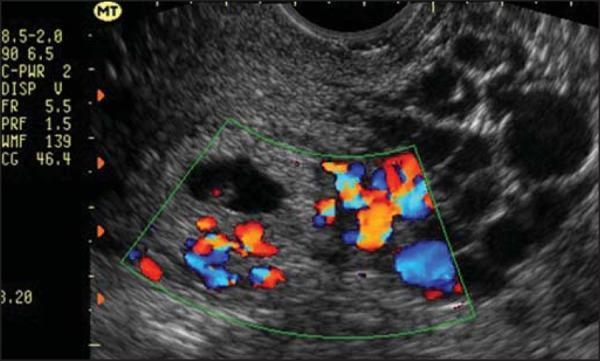
This diagnostic image is a transvaginal ultrasound featuring a sagittal view of the uterus, demonstrating a partial hydatidiform mole. The image shows a heterogeneous intrauterine mass with a classic 'snowstorm' or 'bunch of grapes' appearance, characterized by multiple small, anechoic (black) cystic vesicles representing hydropic villi. A distinct anechoic gestational sac containing a small fetal pole or embryo is visible, which is the key diagnostic feature distinguishing a partial mole from a complete hydatidiform mole. A Color Doppler overlay is applied to a central region of interest, showing prominent vascularity (red and blue signals) within the deciduous and placental tissue, indicating active blood flow. The surrounding myometrium appears intact. This image is used in obstetrics and gynecology to illustrate gestational trophoblastic disease (GTD) and the importance of high-resolution ultrasound in differentiating between complete and partial molar pregnancies based on the presence of fetal tissue and placental morphology.
This diagnostic image is a transvaginal ultrasound featuring a sagittal view of the uterus, demonstrating a partial hydatidiform mole. The image shows a heterogeneous intrauterine mass with a classic 'snowstorm' or 'bunch of grapes' appearance, characterized by multiple small, anechoic (black) cystic vesicles representing hydropic villi. A distinct anechoic gestational sac containing a small fetal pole or embryo is visible, which is the key diagnostic feature distinguishing a partial mole from a complete hydatidiform mole. A Color Doppler overlay is applied to a central region of interest, showing prominent vascularity (red and blue signals) within the deciduous and placental tissue, indicating active blood flow. The surrounding myometrium appears intact. This image is used in obstetrics and gynecology to illustrate gestational trophoblastic disease (GTD) and the importance of high-resolution ultrasound in differentiating between complete and partial molar pregnancies based on the presence of fetal tissue and placental morphology.

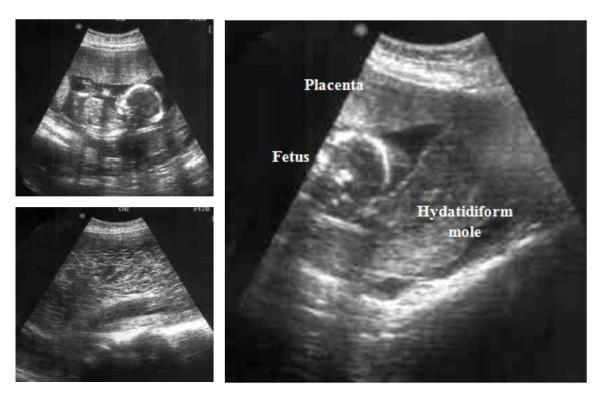
This obstetric ultrasound composite displays a rare twin pregnancy consisting of a complete hydatidiform mole and a coexisting live fetus. In the primary longitudinal view, the uterus contains two distinct compartments. One compartment shows a normal fetus with discernible anatomical landmarks and an adjacent granular, well-defined placenta. The secondary compartment is occupied by a large, heterogeneous, echogenic mass characteristic of a complete hydatidiform mole, often described as having a 'snowstorm' appearance due to complex internal cystic spaces and hydropic villi. The molar tissue is predominantly localized in the lower uterine pole. The separate smaller ultrasound frames highlight these findings individually: one focusing on the fetal anatomy and the other on the diffuse, amorphous texture of the gestational trophoblastic disease. This visual illustrates a high-risk obstetric scenario requiring careful monitoring for complications like pre-eclampsia and thyrotoxicosis due to extremely elevated beta-hCG levels.

This obstetric ultrasound composite displays a rare twin pregnancy consisting of a complete hydatidiform mole and a coexisting live fetus. In the primary longitudinal view, the uterus contains two distinct compartments. One compartment shows a normal fetus with discernible anatomical landmarks and an adjacent granular, well-defined placenta. The secondary compartment is occupied by a large, heterogeneous, echogenic mass characteristic of a complete hydatidiform mole, often described as having a 'snowstorm' appearance due to complex internal cystic spaces and hydropic villi. The molar tissue is predominantly localized in the lower uterine pole. The separate smaller ultrasound frames highlight these findings individually: one focusing on the fetal anatomy and the other on the diffuse, amorphous texture of the gestational trophoblastic disease. This visual illustrates a high-risk obstetric scenario requiring careful monitoring for complications like pre-eclampsia and thyrotoxicosis due to extremely elevated beta-hCG levels.

Molar pregnancy, also called a hydatidiform mole, is an abnormal pregnancy in which placental tissue grows abnormally. It is a form of gestational trophoblastic disease (GTD), and it is not a viable pregnancy.

Types

  • Complete mole: No fetus develops. The genetic material is usually entirely paternal, and the uterus contains swollen, cyst-like placental villi.
  • Partial mole: An abnormal fetus or fetal tissue may be present, usually with an abnormal triploid genetic pattern, alongside abnormal placental tissue.
Ultrasound appearance of a partial hydatidiform mole

Possible symptoms

Many are found early on ultrasound. Symptoms can include:
  • Vaginal bleeding in early pregnancy
  • Severe nausea and vomiting
  • Uterus measuring larger than expected for gestational age
  • Very high blood beta-hCG levels
  • Passage of grape-like tissue
  • Rarely, early pre-eclampsia, hyperthyroid symptoms, or ovarian cysts
Ultrasound may show a cystic, “snowstorm” appearance. A complete mole generally has no fetus, while a partial mole may show fetal tissue. NCI’s GTD overview explains the condition and treatment pathway.

Diagnosis

Clinicians use:
  1. Quantitative blood beta-hCG
  2. Pelvic/transvaginal ultrasound
  3. Histopathology of evacuated uterine tissue, sometimes supported by genetic testing

Treatment

The usual treatment is prompt uterine evacuation by suction curettage. Hysterectomy may be considered when fertility is not desired, but follow-up hCG testing is still needed.

Why follow-up is essential

After evacuation, beta-hCG is measured serially until it is negative, then monitored for a defined period according to the treating center’s protocol. A plateauing or rising hCG can indicate persistent gestational trophoblastic neoplasia (GTN), which is highly treatable, commonly with chemotherapy. Pregnancy should be avoided during surveillance because a new pregnancy raises hCG and makes interpretation difficult. The NCI describes weekly testing until normal followed by monthly follow-up for up to 6 months, though exact schedules vary by guideline and whether the mole was complete or partial.
Complete moles have a higher chance of persistent disease than partial moles. Textbook descriptions emphasize early bleeding, excessive vomiting, an enlarged uterus, elevated hCG, ultrasound findings, evacuation, and post-evacuation hCG surveillance. Textbook of Family Medicine, 9e, p. 20-6.
Seek urgent obstetric/gynecologic assessment now if there is heavy bleeding, fainting, severe abdominal pain, breathlessness, fever, or suspected molar pregnancy.
This is a shared conversation. Sign in to Orris to start your own chat.