
Give seminar on Synovial joint classification
Reading File
Now let me search the medical library for synovial joint classification content.
Finding Sources
Finding Sources
Finding Sources
Finding Sources
Excellent! Found the key section. Let me read the detailed content on synovial joint classification and also check the rheumatology textbook for the synovial tissue section.
Reading File
Reading File
Let me now read the specific section on types of synovial joints from the Imaging Anatomy Atlas:
Reading File
Finding Sources
Reading File
Reading File
Reading File
Reading File
I now have comprehensive content. Let me also fetch the key anatomical diagram showing the six types of synovial joints:
Loading Image
Loading Image
I have comprehensive material. Now let me deliver the full seminar on Synovial Joint Classification.
Seminar: Classification of Synovial Joints
1. Introduction - The Joint as a Functional Organ
A joint (articulation) is a point where two or more bones meet. Joints are classified in two broad ways:
- Functionally - by the degree of movement they permit
- Structurally - by the type of tissue connecting the bones
This dual classification framework is the foundation for understanding synovial joints.
2. Functional Classification of All Joints
| Term | Movement | Structural Basis | Examples |
|---|---|---|---|
| Synarthrosis | Immobile | Fibrous/cartilaginous | Skull sutures, sternomanubrial joint |
| Amphiarthrosis | Slightly mobile | Fibrocartilage/fibrous | Intervertebral discs, pubic symphysis |
| Diarthrosis | Freely mobile | Synovial joint cavity | Knee, hip, shoulder, elbow |
The diarthrosis (synovial joint) is the most common joint type in the body and the focus of this seminar.
- Imaging Anatomy: Bones, Joints, Vessels and Nerves, p. 46
3. Structural Classification of All Joints
| Type | How Bones Are United | Functional Class |
|---|---|---|
| Fibrous | Fibrous connective tissue | Synarthrosis / Amphiarthrosis |
| Cartilaginous | Hyaline or fibrocartilage | Synarthrosis / Amphiarthrosis |
| Synovial | Joint cavity filled with synovial fluid | Diarthrosis |
4. Anatomy of a Synovial Joint - Key Components
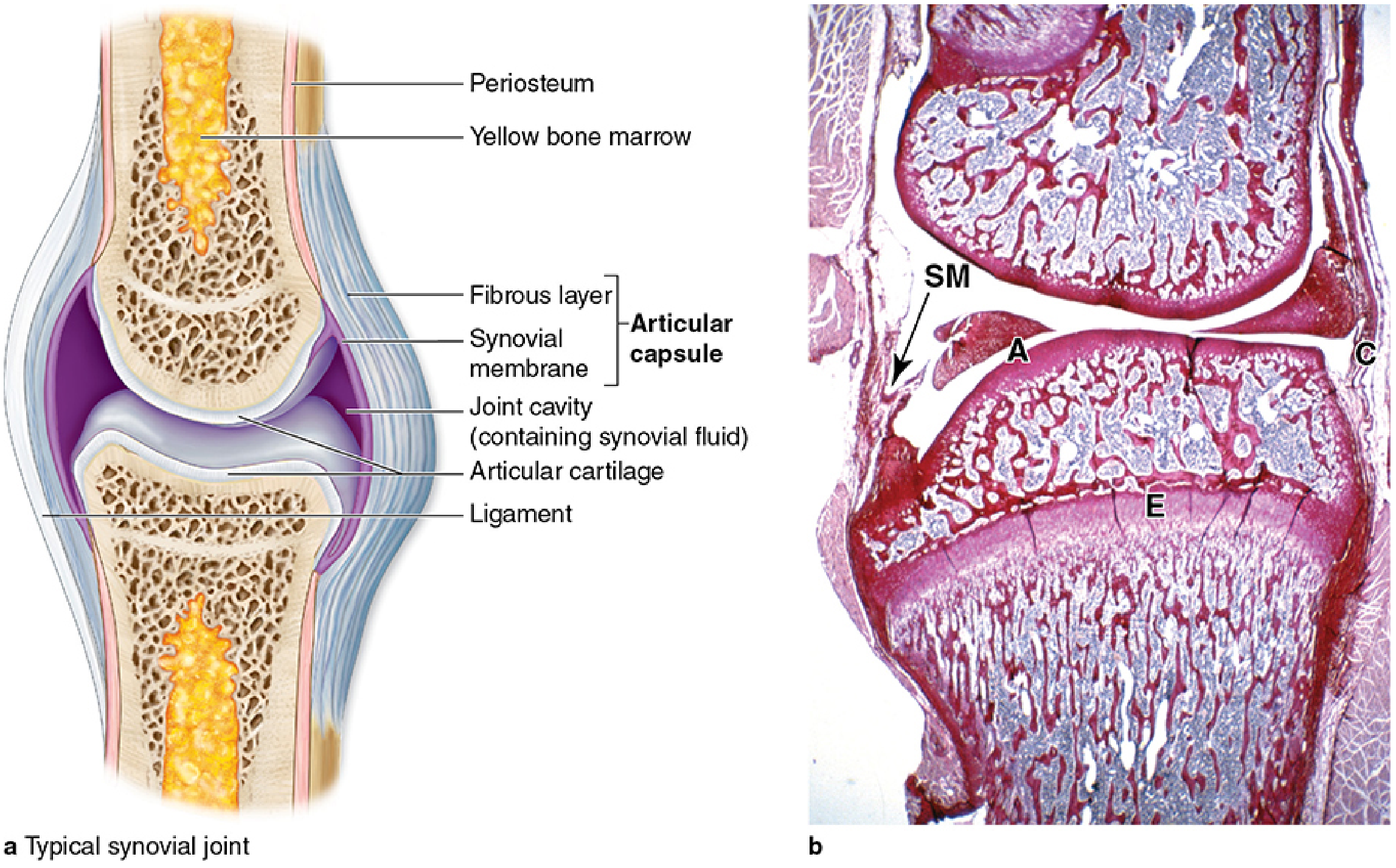
Fig. 1 - Typical synovial (diarthrotic) joint: (a) diagram showing articular capsule, synovial membrane, joint cavity with synovial fluid, articular cartilage, and ligaments; (b) longitudinal histological section of a mouse knee showing articular cartilage (A), synovial membrane (SM), epiphyseal plate (E), and capsule (C). - Junqueira's Basic Histology, p. 394
Essential Components
- Articular cartilage - Hyaline cartilage covers the articular surfaces of each bone; avascular and aneural, it reduces friction and absorbs compressive forces
- Joint (articular) capsule - Two layers:
- Outer fibrous layer - dense irregular connective tissue, continuous with periosteum
- Inner synovial membrane - highly vascular; lined by two specialised cell types
- Synovial membrane - Produces synovial fluid; contains:
- Type A (macrophage-like) synovial cells - derived from blood monocytes; remove wear-and-tear debris
- Type B (fibroblastic) synovial cells - produce hyaluronan and proteoglycans that form the lubricant synovial fluid
- Synovial fluid - Viscous, clear, lubricates all internal surfaces; supplies nutrients to avascular articular cartilage
- Ligaments - Classified as:
- Intracapsular - inside the joint capsule (e.g., cruciate ligaments of the knee)
- Extracapsular - outside the capsule (e.g., fibular collateral ligament)
- Bursae - Connective tissue sacs containing lubricating fluid; located outside the joint, subdendously, submuscularly, or subcutaneously
- Fibrocartilage discs/menisci - Present in some joints (knee, temporomandibular, sternoclavicular) to improve congruence and distribute load
- Imaging Anatomy: Bones, Joints, Vessels and Nerves, p. 49; Junqueira's Basic Histology, p. 394-395
5. Classification of Synovial Joints
Synovial joints are classified in two complementary ways:
A. Functional (Axes of Movement) Classification
| Class | Axes | Movement Planes | Examples |
|---|---|---|---|
| Uniaxial | 1 | 1 plane | Elbow (hinge), Atlantoaxial (pivot) |
| Biaxial | 2 | 2 planes | Metacarpophalangeal (condyloid), 1st CMC (saddle) |
| Multiaxial | 3+ | Multiple directions | Shoulder (ball-and-socket), Hip |
B. Morphological (Shape) Classification - The 6 Types
Morphologically, synovial joints are divided into six types: pivot, hinge, plane, saddle, condyloid, and ball-and-socket. (Imaging Anatomy, p. 49)
6. The Six Morphological Types in Detail
6.1 Pivot Joint (Trochoid Joint)
| Feature | Detail |
|---|---|
| Shape | Cylindrical bony process rotating within a ring of bone + ligament |
| Axes | Uniaxial |
| Movement | Rotation only |
| Examples | Atlantoaxial joint (C1-C2), Proximal radioulnar joint |
- At the atlantoaxial joint: the dens (odontoid process) of C2 rotates within the ring formed by the anterior arch of C1 and the transverse ligament. This allows approximately 50° of head rotation to each side.
- At the proximal radioulnar joint: the radial head rotates within the ring formed by the radial notch of the ulna and the annular ligament - enabling pronation/supination.
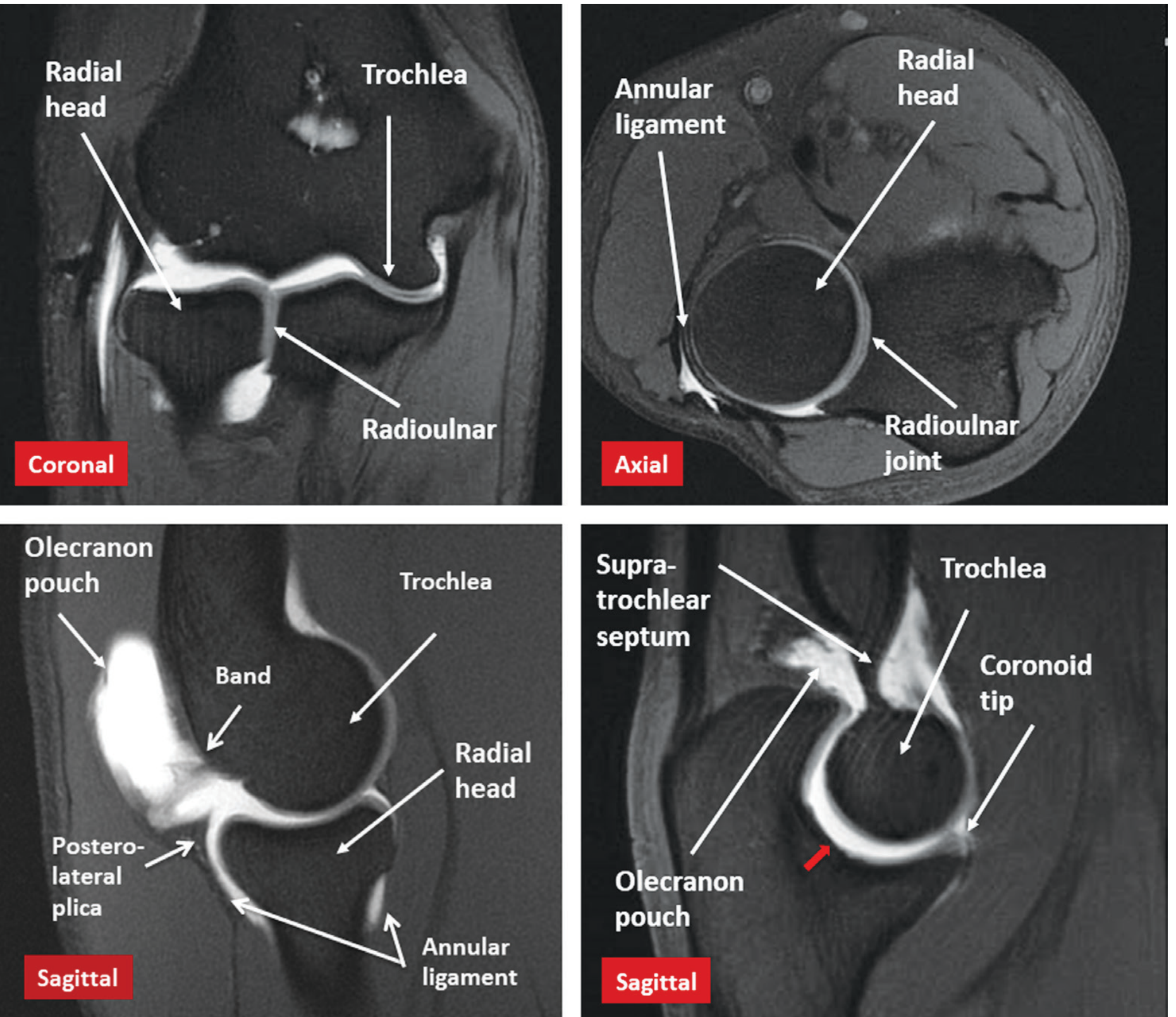
Fig. 2 - MRI arthrogram of the elbow showing the uniaxial hinge articulation and the pivot radioulnar joint. - Imaging Anatomy: Bones, Joints, Vessels and Nerves
6.2 Hinge Joint (Ginglymus)
| Feature | Detail |
|---|---|
| Shape | Convex surface fitting into a concave surface, like a mechanical door hinge |
| Axes | Uniaxial |
| Movement | Flexion and extension only |
| Examples | Elbow (humeroulnar), Ankle (talocrural), Knee (partially), Interphalangeal joints |
- The elbow (humeroulnar joint) is the classic example: the trochlea of the humerus articulates with the trochlear notch of the ulna. Movement is restricted to one plane.
- The ankle allows plantarflexion (~50°) and dorsiflexion (~20°).
- The interphalangeal joints (both proximal and distal) are the simplest and most numerous hinge joints in the body.
6.3 Plane Joint (Gliding Joint)
| Feature | Detail |
|---|---|
| Shape | Flat or slightly curved articular surfaces |
| Axes | Multiaxial (but limited amplitude) |
| Movement | Gliding / translatory motion in multiple directions |
| Examples | Intercarpal joints, Intertarsal joints, Acromioclavicular joint, Subtalar joint, Vertebral facet (zygapophysial) joints |
- These joints allow small-amplitude, gliding motion. Despite technically being multiaxial, the flat surfaces and strong ligaments limit the range significantly.
- The vertebral facet joints are the most numerous in the body (24 pairs) and permit the cumulative segmental movements of the spine.
- The acromioclavicular (AC) joint is a true diarthrodial synovial joint with a capsule and weak AC ligament.
6.4 Saddle Joint (Sellar Joint)
| Feature | Detail |
|---|---|
| Shape | Each surface is concave in one direction and convex in the other - like two saddles oriented at 90° to each other |
| Axes | Biaxial |
| Movement | Flexion/extension + abduction/adduction; circumduction; NO axial rotation |
| Examples | 1st carpometacarpal joint (trapezium + 1st metacarpal), Sternoclavicular joint |
- The 1st CMC joint (thumb basal joint) is the most important example - it gives the thumb its uniquely wide range of motion including opposition, making it critical for human hand function.
- The sternoclavicular joint is the only bony attachment of the upper limb to the axial skeleton. Despite its classification as a saddle joint, it functions like a ball-and-socket due to the presence of an intra-articular disc.
6.5 Condyloid Joint (Ellipsoid Joint)
| Feature | Detail |
|---|---|
| Shape | Oval/elliptical convex surface articulating with a shallow elliptical concavity |
| Axes | Biaxial |
| Movement | Flexion/extension + abduction/adduction; circumduction; limited/no axial rotation |
| Examples | Metacarpophalangeal (MCP) joints, Metatarsophalangeal (MTP) joints, Radiocarpal (wrist) joint |
- The wrist (radiocarpal) joint is a key condyloid joint: the distal radius articulates with an elliptical surface formed by the scaphoid and lunate bones together. This allows flexion-extension and radial-ulnar deviation.
- The MCP joints of the fingers allow the characteristic "knuckle" movements - flexion, extension, abduction, and adduction.
6.6 Ball-and-Socket Joint (Enarthrodial Joint / Spheroidal Joint)
| Feature | Detail |
|---|---|
| Shape | Spherical head fitting into a cup-shaped socket |
| Axes | Multiaxial |
| Movement | Flexion/extension, abduction/adduction, medial/lateral rotation, circumduction - greatest range of all joint types |
| Examples | Hip joint (coxofemoral), Glenohumeral (shoulder) joint |
- The hip joint: the spherical femoral head sits in the deep acetabulum. The deep socket gives stability at the cost of some mobility. Additional stability comes from the labrum, capsule, and ligamentum teres.
- The glenohumeral joint: the spherical humeral head articulates with the shallow glenoid cavity. The shallow socket provides greater range of motion compared to the hip, but sacrifices stability - making it the most commonly dislocated large joint in the body.
- Imaging Anatomy: Bones, Joints, Vessels and Nerves, p. 49-50
7. Summary Classification Table
| Type | Shape | Axes | Movements | Key Examples |
|---|---|---|---|---|
| Pivot | Cylindrical peg in ring | Uniaxial | Rotation | Atlantoaxial, proximal radioulnar |
| Hinge | Convex-concave (pulley) | Uniaxial | Flexion/Extension | Elbow, ankle, IPs |
| Plane | Flat/slightly curved | Multiaxial (limited) | Gliding | Facet joints, AC joint, intercarpal |
| Saddle | Reciprocal concave-convex | Biaxial | F/E, Abd/Add, Circumduction | 1st CMC, sternoclavicular |
| Condyloid | Oval convex in shallow concave | Biaxial | F/E, Abd/Add, Circumduction | Wrist, MCP, MTP |
| Ball-and-socket | Sphere in cup | Multiaxial | All movements | Hip, shoulder |
8. Special/Mixed Joints
Several clinically important joints do not fit neatly into a single category:
Temporomandibular Joint (TMJ)
- A mixed hinge and gliding joint between the concave mandibular condyle and the convex temporal articular surface
- Contains an intra-articular disc
- Allows both unipolar (rotational/hinge) and bipolar (gliding/translational) movements
- Lower compartment: hinge movement (mouth opening)
- Upper compartment: gliding movement (protrusion, lateral excursion)
Sacroiliac Joint
- The anterior portion is a true synovial (diarthrodial) joint
- The posterior third is a fibrous syndesmosis
- Unique: the sacral articular surface is covered by hyaline cartilage, while the iliac surface is covered by fibrocartilage
Knee Joint
- Primarily classified as a hinge but with a significant rotational component (tibial rotation occurs at the end of extension - the "screw-home mechanism")
- Contains intra-articular fibrocartilage menisci (medial and lateral) that improve congruence
9. Fibrous Joints (Context / Contrast)
Three sub-types for reference:
| Type | Description | Example |
|---|---|---|
| Sutures | Thin fibrous tissue between skull bones; may eventually ossify (synostosis) | Coronal, sagittal sutures |
| Syndesmosis | Bones joined by thick fibrous ligaments or interosseous membrane | Distal tibiofibular joint, interosseous membrane of forearm/leg |
| Gomphosis | Peg-in-socket fibrous joint | Tooth in alveolar socket |
10. Cartilaginous Joints (Context / Contrast)
| Type | Tissue | Examples |
|---|---|---|
| Synchondrosis (Primary) | Hyaline cartilage | Epiphyseal growth plates (temporary; ossify with age) |
| Symphysis (Secondary) | Fibrocartilage | Pubic symphysis, intervertebral discs, sacrococcygeal joint |
11. Clinical Relevance
- Hinge joints (IPs, elbow) are commonly involved in psoriatic arthritis - the distal IPs being characteristically affected.
- Ball-and-socket joints are the most common sites of osteoarthritis (hip, shoulder) due to high load-bearing demands over time.
- Condyloid joints (MCP, MTP) are hallmark targets of rheumatoid arthritis - MCP swelling and ulnar deviation are classic findings.
- Plane joints (facet joints) degenerate in spondylosis and are targeted in facet joint injections for back pain.
- Saddle joint (1st CMC) arthritis is the most common form of hand OA, causing the classic "squaring" deformity at the thumb base.
- Pivot joints: the atlantoaxial joint can be destabilized in rheumatoid arthritis and Down syndrome (atlanto-axial instability) due to erosion or laxity of the transverse ligament.
- Synovitis - inflammation of the synovial membrane - is detected on ultrasound as joint effusion (anechoic/hypoechoic, compressible, displaceable, no Doppler signal) or synovial thickening (hypoechoic, partially compressible, non-displaceable, may show Power Doppler signal). (Rheumatology, 2-Volume Set, 2022)
12. Key Mnemonics
"Please Help Some Crippled People Build"
- Pivot, Hinge, Saddle, Condyloid, Plane, Ball-and-socket
Axes of Movement:
- "Uniform Pivots Hang" = Uniaxial: Pivot, Hinge
- "Both Saddles Canter" = Biaxial: Saddle, Condyloid
- "Multiaxial Plane Balls" = Multiaxial: Plane, Ball-and-socket
13. Summary
Synovial (diarthrotic) joints are the predominant, freely mobile joint type of the body. They are defined structurally by a fluid-filled joint cavity, synovial membrane, articular cartilage, and a fibrous capsule. Functionally they are classified as uniaxial, biaxial, or multiaxial. Morphologically, six types are recognized - each shaped to serve a specific functional role, from the simple rotation of pivot joints to the full multiplanar freedom of ball-and-socket articulations.
Sources:
- Imaging Anatomy: Bones, Joints, Vessels and Nerves (Vol. 3) - Chapter 1: Joints and Cartilage
- Junqueira's Basic Histology: Text and Atlas, 17e - Chapter 8: Connective Tissue
- Rheumatology, 2-Volume Set (2022, Elsevier) - Chapter: Synovial Tissue and the Synovial Joint
- Firestein & Kelley's Textbook of Rheumatology - The Synovial Joint as an Organ
This is a shared conversation. Sign in to Orris to start your own chat.