
Bacterial Pneumoniae vs Viral Pneumoniae
Reading File
Now let me search the medical library and PubMed in parallel for comprehensive coverage.
Finding Sources
Searching PubMed
"bacterial pneumonia" AND "viral pneumonia" AND differentiation
Good. Now let me read the most relevant sections from Goldman-Cecil and Murray & Nadel in parallel:
Reading File
Reading File
Finding Sources
Now let me pull the dedicated pneumonia comparison sections from Goldman-Cecil and Harrison's:
Reading File
Reading File
Finding Sources
Good. Now let me get the dedicated CAP chapter from Goldman-Cecil and the microbiology section:
Reading File
Finding Sources
Excellent - found key resources. Let me read the Robbins pathology section and Frameworks for Internal Medicine sections in parallel:
Reading File
Reading File
Good - found the diagram image. Let me read more of the Frameworks section and the Robbins pathology content:
Reading File
Finding Sources
Now let me get the radiology patterns, Robbins pathology content, and the Goldman-Cecil CAP chapter:
Reading File
Finding Sources
Now let me get the Robbins pathology content on bacterial vs viral pneumonia morphology and the Goldman-Cecil CAP treatment section:
Reading File
Finding Sources
I now have comprehensive data. Let me get the viral pneumonia pathology from Robbins and the Goldman-Cecil lab section, then compose the answer:
Reading File
Reading File
I now have very comprehensive data from multiple authoritative textbooks. Let me fetch the key diagram image and then compose the full answer:
Loading Image
Loading Image
I now have all the material needed for a comprehensive, well-illustrated comparison. Here is the full answer:
Bacterial Pneumonia vs Viral Pneumonia
A comprehensive comparison drawn from Robbins & Cotran Pathologic Basis of Disease, Goldman-Cecil Medicine, Harrison's Principles, Murray & Nadel's Respiratory Medicine, Frameworks for Internal Medicine, and Grainger & Allison's Diagnostic Radiology.
1. Overview and Classification
Pneumonia is broadly classified by setting (community-acquired [CAP] vs. hospital-acquired [HAP]) and by causative agent. Within CAP, clinicians further split organisms into typical bacterial, atypical bacterial, and viral categories. The distinction matters because it drives empiric antibiotic choice.
"Why is it important to distinguish between typical and atypical community-acquired pneumonia? The usual pathogens differ between typical and atypical CAP, which informs the choice of empiric treatment." - Frameworks for Internal Medicine
2. Causative Organisms
Typical Bacterial CAP
| Pathogen | Key Association |
|---|---|
| Streptococcus pneumoniae | Most common overall (5-16% of CAP) |
| Haemophilus influenzae | COPD exacerbations; present in most adults' nasopharynx |
| Staphylococcus aureus | Post-influenza secondary infection; 3-5% |
| Klebsiella pneumoniae | Chronic alcoholics; cavitating |
| Moraxella catarrhalis | COPD, immunocompromised |
| Pseudomonas aeruginosa | Cystic fibrosis, neutropenia |
Atypical Bacterial CAP
| Pathogen | Key Points |
|---|---|
| Mycoplasma pneumoniae | Most common "atypical"; no cell wall; presents like a viral illness |
| Legionella pneumophila | Comorbid adults; urinary antigen test ~75% sensitive for serotype 1 |
| Chlamydia pneumoniae | Obligate intracellular; up to 1/5 of CAP cases |
| Coxiella burnetii | Q fever; zoonotic; nonproductive cough, severe headache |
Viral CAP
| Pathogen | Key Points |
|---|---|
| Influenza A/B | Most common viral; leads to primary viral or secondary bacterial pneumonia |
| SARS-CoV-2 | Newly emergent; bilateral ground-glass opacities |
| RSV, Parainfluenza | Especially in children and elderly |
| Human metapneumovirus, Adenovirus, Rhinovirus | Less common but significant |
"In the pre-COVID era, bacteria were identified in 15-30% of cases; respiratory viruses were detected in 20-27% of patients hospitalized with CAP, with rhinoviruses (5-13%) and influenza (1-9%) being most common." - Goldman-Cecil Medicine
3. Pathology and Morphology
This is where the most fundamental differences lie.
Bacterial pneumonia features predominantly intraalveolar neutrophilic inflammation. It comes in two anatomical patterns:
- Lobar pneumonia - consolidation of a large portion or entire lobe, classically from S. pneumoniae
- Bronchopneumonia - patchy, multifocal consolidation centered around bronchioles, typical of S. aureus, gram-negatives, anaerobes
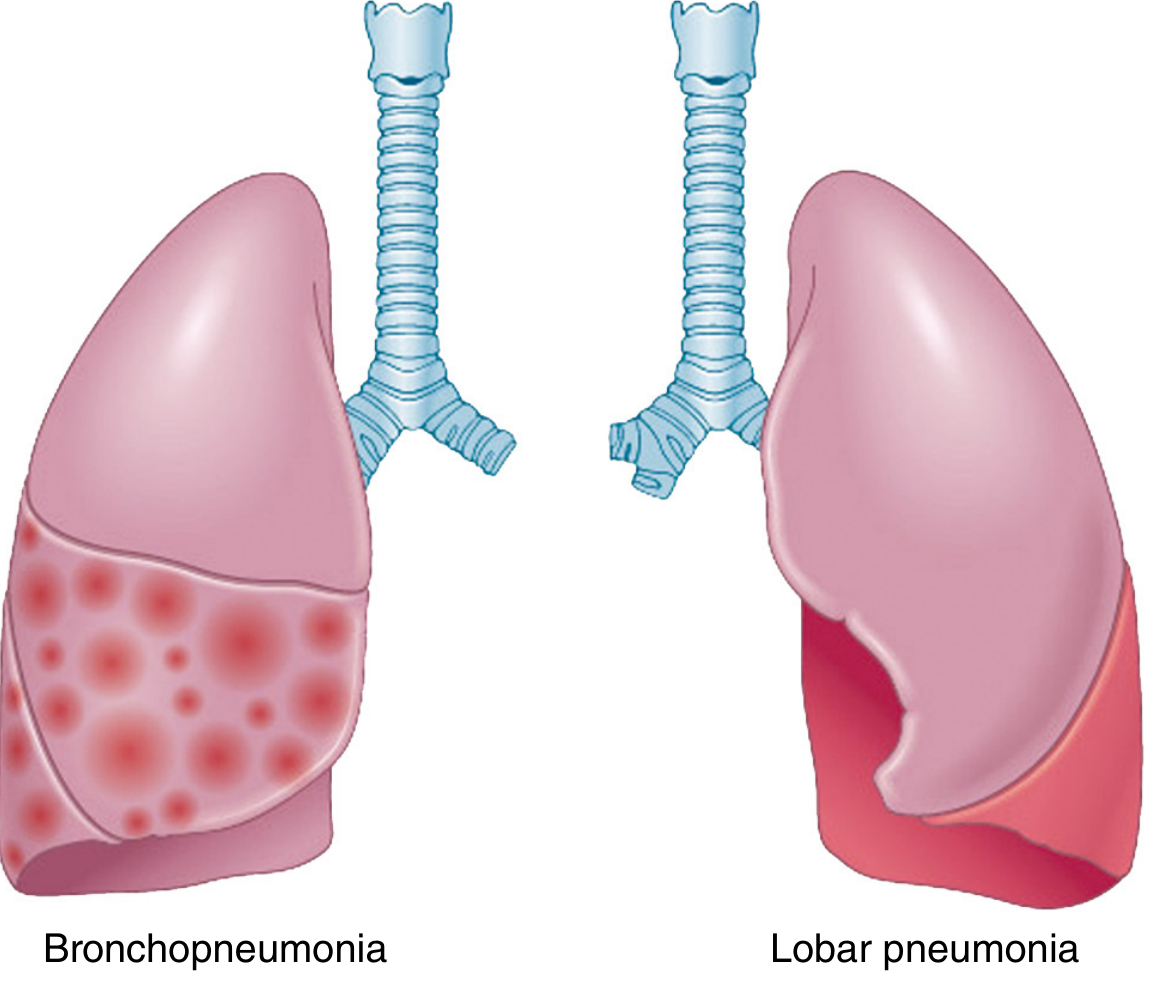
Comparison of bronchopneumonia (patchy foci) and lobar pneumonia (entire lobe consolidation) — Robbins, Cotran & Kumar Pathologic Basis of Disease
Four stages of lobar pneumonia (Robbins):
- Congestion - vascular engorgement, edema fluid, early neutrophils
- Red hepatization - massive neutrophils + red cells + fibrin in alveoli; lung feels liver-like
- Gray hepatization - RBCs disintegrate; fibrinopurulent exudate persists; gray-brown color
- Resolution - enzymatic digestion of exudate, macrophage clearance
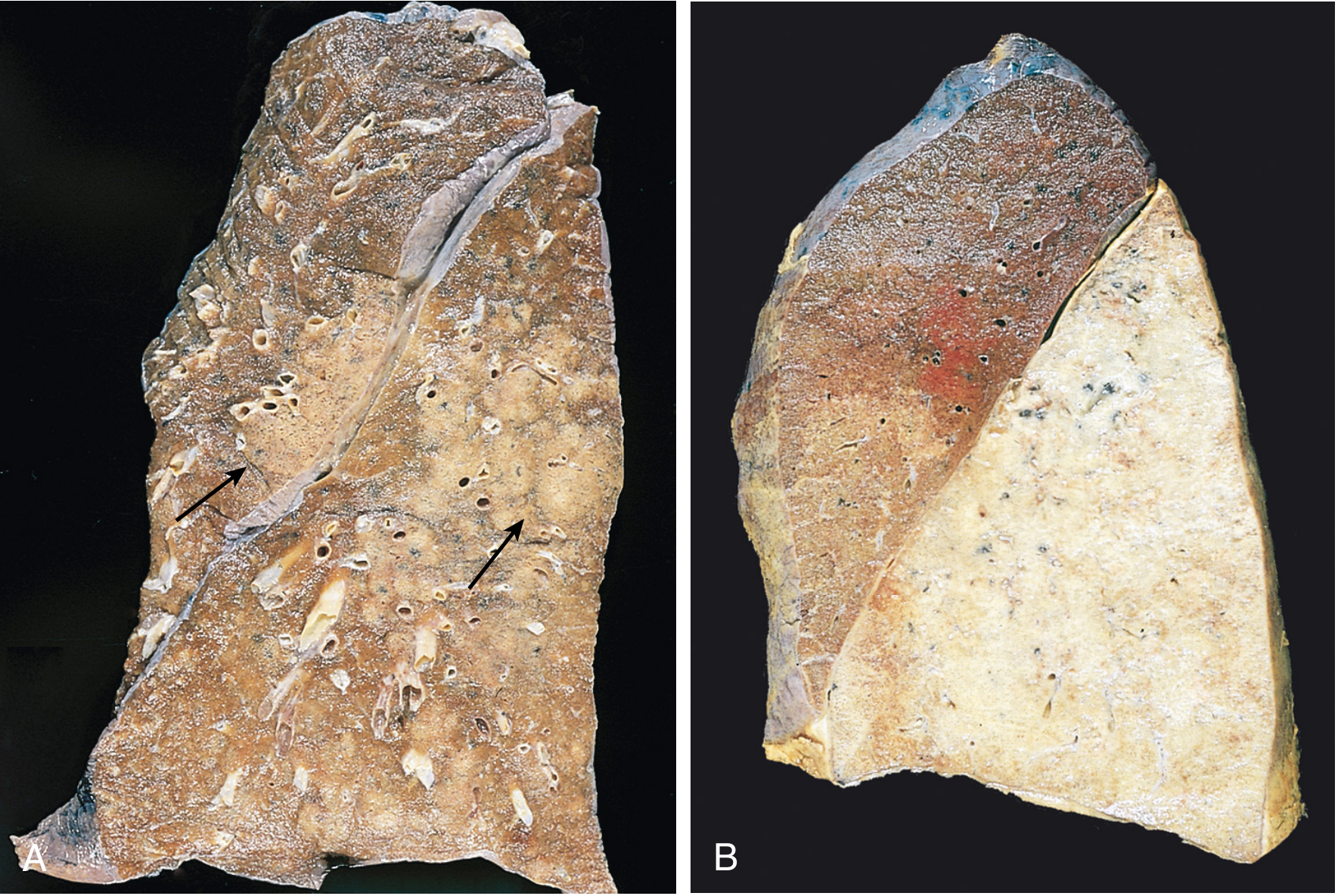
Gross pathology specimens: (A) Bronchopneumonia with patchy consolidation; (B) Lobar pneumonia with uniform gray hepatization — Robbins, Cotran & Kumar
Viral pneumonia is histologically distinct: it causes interstitial lymphocytic inflammation rather than alveolar exudate. Key features include:
- Infiltration of monocytes, lymphocytes, and macrophages in alveolar septa
- Destruction and hemorrhage in the respiratory epithelium
- Less or no intraalveolar neutrophilic exudate (at least early on)
- Large amounts of virus can be recovered from the lungs in primary viral pneumonia
"Bacterial pneumonias are characterized by predominantly intraalveolar neutrophilic inflammation, while viral pneumonia shows interstitial lymphocytic inflammation." - Robbins, Cotran & Kumar
4. Clinical Features
| Feature | Typical Bacterial | Viral / Atypical |
|---|---|---|
| Onset | Abrupt, acute | Gradual, insidious ("walking pneumonia") |
| Fever | High, with rigors/chills | Lower-grade, persistent |
| Cough | Productive, purulent/rusty sputum | Dry, non-productive |
| Pleuritic chest pain | Common (lobar) | Uncommon |
| Septic shock | Can occur | Rare |
| Auscultation | Localized crackles, bronchial breathing | Non-localizing scattered crackles, rhonchi, wheezes |
| Extrapulmonary features | Less common | Common (myalgias, headache, rash, myocarditis) |
"Typical bacterial pneumonia: acute-onset chills and fever, cough with sputum production, pleuritic chest pain. A smoldering onset with low-grade fever and fewer constitutional symptoms indicates atypical pneumonia caused by respiratory viruses or Mycoplasma, Chlamydia, or Legionella." - Frameworks for Internal Medicine / Textbook of Family Medicine
5. Radiology
| Pattern | Typical Organism(s) |
|---|---|
| Lobar consolidation | S. pneumoniae, K. pneumoniae |
| Bronchopneumonia | S. aureus, gram-negatives, anaerobes, S. pneumoniae |
| Interstitial / bilateral diffuse | Viruses, Mycoplasma pneumoniae |
| Round pneumonia | S. pneumoniae |
| Cavitation / necrotizing | S. aureus (including MRSA), K. pneumoniae, anaerobes, TB |
"Lobar pneumonia indicates a bacterial process, whereas interstitial infiltrates are more likely related to viruses and pneumocystis." - Fishman's Pulmonary Diseases and Disorders
The term "atypical pneumonia" is still used radiologically, but as Grainger & Allison note, it is technically imprecise - many atypical organisms (Mycoplasma, Legionella, Chlamydia) are bacteria, and they can present with dense consolidation, not only interstitial patterns.
6. Laboratory Diagnosis
| Test | Bacterial | Viral |
|---|---|---|
| WBC | Elevated neutrophilia (leukocytosis) | Often normal or leukopenia; lymphocytosis |
| Procalcitonin (PCT) | Elevated (≥0.25 µg/L) | Low / normal |
| CRP / ESR | Elevated | May be mildly elevated |
| Sputum Gram stain/culture | Useful if adequate sample | Not applicable |
| Respiratory PCR | For atypical bacteria (Mycoplasma, Legionella) | Method of choice for viral identification |
| Urinary antigen | Legionella, S. pneumoniae | Not applicable |
| Blood cultures | Low yield; reserve for severe CAP or MRSA/Pseudomonas risk | Not applicable |
"Laboratory findings include an elevated white blood cell count with neutrophilia in bacterial pneumonia, but viral infections and overwhelming bacterial infections can present with leukopenia. Procalcitonin is more likely to be elevated with bacterial than with viral infections." - Goldman-Cecil Medicine
7. The Overlap: Influenza and Secondary Bacterial Pneumonia
This is clinically important. After an influenza infection, three distinct pneumonic syndromes can occur:
- Primary viral pneumonia - progressive dyspnea, persistent fever, cyanosis in severe cases; marked inflammatory reaction in alveolar septa with monocytes, lymphocytes, macrophages; typical of 1918 pandemic and H5N1
- Secondary bacterial pneumonia - biphasic illness; patient appears to recover then fever recurs with localized pulmonary findings; caused by S. pneumoniae, S. aureus, H. influenzae
- Mixed viral + bacterial pneumonia
The mechanism: influenza de-epithelializes airways and destroys ciliary function, allowing bacterial colonization. Additionally, Staphylococcus and Pseudomonas produce proteases that enhance hemagglutinin cleavage, further promoting viral replication.
"Almost one-fifth of patients with bacterial-proven CAP are co-infected with a virus." - Frameworks for Internal Medicine
8. Treatment
Bacterial CAP
- Outpatient, no comorbidities: doxycycline or macrolide (if local resistance <25%)
- Outpatient, typical organism suspected: amoxicillin-clavulanate; alternatives: levofloxacin, moxifloxacin
- Hospitalized (non-ICU): β-lactam (ceftriaxone, cefotaxime) + azithromycin, or respiratory fluoroquinolone (levofloxacin/moxifloxacin)
- Severe / MRSA risk: add vancomycin or linezolid
- Duration: 5-7 days typically
- Atypical organisms: macrolides, doxycycline, or fluoroquinolones cover Mycoplasma and Chlamydia; doxycycline for Q fever
Viral CAP
- Influenza: neuraminidase inhibitor (oseltamivir); most effective when started early
- SARS-CoV-2: antivirals (nirmatrelvir/ritonavir), dexamethasone for severe disease
- Most other viral pneumonias: supportive care (oxygen, hydration, antipyretics)
- Antibiotics are NOT indicated for pure viral pneumonia but are given when secondary bacterial infection is present
9. Summary Comparison Table
| Feature | Bacterial Pneumonia | Viral Pneumonia |
|---|---|---|
| Key organisms | S. pneumoniae, S. aureus, H. influenzae, Legionella, Mycoplasma | Influenza, RSV, SARS-CoV-2, Adenovirus |
| Inflammation type | Intraalveolar - neutrophilic | Interstitial - lymphocytic |
| Radiographic pattern | Lobar or bronchopneumonia consolidation | Bilateral interstitial / ground-glass |
| Onset | Abrupt | Gradual |
| Sputum | Purulent, productive | Dry cough or scant |
| Procalcitonin | Elevated | Low |
| WBC | Neutrophilia | Lymphocytosis or normal |
| Treatment | Antibiotics (β-lactam ± macrolide/quinolone) | Antivirals (oseltamivir, etc.) or supportive |
| Complications | Empyema, bacteremia, lung abscess, ARDS | Secondary bacterial pneumonia, myocarditis, encephalitis |
| Pleuritis | Common (especially lobar) | Uncommon |
Sources: Robbins, Cotran & Kumar Pathologic Basis of Disease; Goldman-Cecil Medicine; Harrison's Principles of Internal Medicine 22E; Murray & Nadel's Textbook of Respiratory Medicine; Frameworks for Internal Medicine; Grainger & Allison's Diagnostic Radiology; Textbook of Family Medicine 9e; Fishman's Pulmonary Diseases and Disorders.
This is a shared conversation. Sign in to Orris to start your own chat.